
G.G. Akunna
Department of Anatomy, College of Medicine, Lagos State University, Ikeja Lagos, Nigeria
L.C. Saalu
Department of Anatomy, College of Medicine, Lagos State University, Ikeja Lagos, Nigeria
B. Ogunlade
Department of Anatomy, College of Medicine, Lagos State University, Ikeja Lagos, Nigeria
L.A. Enye
Department of Anatomy, College of Health Sciences, Afe Babalola University, Ado-Ekiti, Ekiti, Nigeria
Journal of Medical Sciences
Year: 2014 | Volume: 14 | Issue: 1 | Page No.: 46-50


ABSTRACT
Various commonly-used products have been reported to contain chemicals that could disrupt estrogen and testosterone hormone. As trade secrets, these chemicals are generally listed as fragrance to mask individual identity. In this study, the reproductive implications of two commonly used perfumes (designated as F1 and F2) in Nigeria were carried out. Sixty adult male wistar rats (12-13 weeks old) were randomly divided into four groups (A-F) of ten rats each. Group A and B rats were exposed to 5 mL kg-1 b.wt. of normal saline for 56 days and 112 days, respectively via whole body inhalation. Group C and D rats were exposed to 5 mL kg-1 b.wt. of a fragrance product designated as F1 for a period of 56 days and 112 days, respectively while Group E and Group F rats were exposed to 5 mL kg-1 b.wt. of a designated fragrance product F2 for a period of 56 days and 112 days, respectively. The results obtained from this study showed a significant (p<0.005) decrease in body weight and absolute testicular weight of the rat models exposed to fragrance when compared to the control groups. It was also observed that the concentration, mobility, livability and morphology of spermatozoa from groups C, D, E and F were significantly lower (p>0.005) when compared to values of the control group A and B. Based on the spermiographic evaluation from this study, fragrance materials could have an adverse effect on spermatozoa of the intact male wistar rats.
PDF Abstract XML References Citation
Received: March 30, 2013;
Accepted: May 14, 2013;
Published: January 15, 2014
How to cite this article
G.G. Akunna, L.C. Saalu, B. Ogunlade and L.A. Enye, 2014. Spermatotoxicity in Animal Models Exposed to Fragrance Components. Journal of Medical Sciences, 14: 46-50.
DOI: 10.3923/jms.2014.46.50
URL: https://scialert.net/abstract/?doi=jms.2014.46.50
DOI: 10.3923/jms.2014.46.50
URL: https://scialert.net/abstract/?doi=jms.2014.46.50
INTRODUCTION
Although researchers have severally reported the toxicity of fragrance exposure, most of the documentation spins around their dermatological implications. This is fairly acceptable as most fragrance-containing care products are applied directly on the skin (Vader and Christy, 2009). The exposure routes for industrial workers are mostly via the dermal and inhalation route while that of the consumers are via inhalation, dermal and oral routes (Caress and Steinemann, 2004; Vader and Christy, 2009).
It has been reported that through inhalation, ingestion and absorption, fragrance infiltrates the body and moves directly to the blood stream. Studies have shown that individual sensitivity to the effects of fragrance is relative (Elberling et al., 2005, 2007) and the fact that different fragrances cause different symptoms may be an indication of its varying toxicity (Environmental Health Network, 2002). Symptoms ranging from severe mucosal discharge, sinus problems, tremor, asthmatic attack, sneezing, migraine headache, convulsions, hyperactivity, nausea, sore throat, cough, chest tightness to shortness of breath after fragrance exposure have been vastly documented (Guin and Berry, 1980; De Groot, 1987; Schleuter et al., 1978).
Unswerving connection between memory and smell has been established (Rachel and Engen, 1996). This knowledge has resulted in placement of fragrance in the category of psychoactive drugs and highlighted the ability of fragrance to cross the brain barrier thereby resulting in potential damage to brain tissue (Andrea, 1997). Linalool, the most abundant fragrance substance has been indicated to cause lethargy, depression and severe respiratory difficulties after exposure (Dahom, 2011).
Research shows the ability of fragrance from citrus to efficiently boost metabolism, memory and alleviate food cravings and depression than were prescription anti-depressants, confirming the previous reports on its ability to permeate the brain barrier (Rachel and Engen, 1996).
Dose-dependent reduction in fecundity, median survival time and an increase in early life-stage mortality post administration of to musk xylene and musk ketone has been reported (Carlsson et al., 2000). Diethyl phthalate, a common fragrance substance in perfume and cologne has been shown to damage sperm DNA in adult, cause premature breast formation in developing baby girls and abnormal sexual development in male fetuses including cryptorchidsm (Duty et al., 2003).
Synthetic musk fragrance ingredients which are widely highly distributed in many consumer products have been examined in human blood, milk and fatty tissue. They represent a new group of human contaminants which are comparable with that of certain pesticides. Despite several reports on the toxic effect of fragrance, there is a dearth of literature ascertaining its effects on male fertility and testicular development (Thompson and Wansker, 1981).
Therefore, the solemnly aim here in is to evaluate the possible effect of fragrance exposure on male infertility in animal models.
MATERIALS AND METHODS
Materials: Two commonly used perfumes in Nigeria designated as F1 and F2 were obtained from Bayous Cosmetics in Lagos on 23rd of April, 2012 and were kept under standard temperature.
Experimental procedure: Sixty adult male wistar rat (12-13 weeks old) weighing 190-220 g were used for the study. The rats were randomly divided into four groups (A-F) of ten rats and the average weight difference between and within groups did not exceed±20% of the average weight of the sample population. Group A rats served as the first control and were exposed to 5 mL kg-1 b.wt. of normal saline for 56 days. Group B rats served as the chronic control group and were exposed to 5 mL kg-1 b.wt. of normal saline for a period of 112 days. Group C and Group D rats were exposed to 5 mL kg-1 b.wt. of F1 for a period of 56 days and 112 days respectively. Group E and Group F animals were exposed to 5 mL kg-1 b.wt. of F2 for a period of 56 days and 112 days, respectively. The study is consistent with the standard of the use of laboratory animals (WMA/APS, 2002; Akunna et al., 2011).
Method of exposure: The exposure was done via whole body inhalation. Small balls of cotton wool were soaked with the fragrance (Experimental Groups) and normal saline (Control Groups), respectively. The wools were rightly placed in a Petri dish inside the cages and covered with perforated plastic container to prevent direct contact for an exposed duration of at least six hours per day throughout the period of study.
Animal sacrifice and sample collection: The rats were first weighed and then subjected to cervical dislocation. The abdominal cavity was opened up through a midline abdominal incision to expose the reproductive organs. The testes were excised and trimmed of all fat. The rats were then anaesthetized by placing them into a glass chamber containing cotton wool soaked in chloroform till they lost consciousness. The testicles were then removed through a lower abdominal incision. The testes were then separated from the epididymis with the scalped blade. Sperm cells were sucked into a pre-warmed (37°C) Pasteur pipette from the caudal epididymis. It was flushed with 2-3 drops of 2.9% sodium citrate self kept at 37°C. Smears were prepared from these samples and strained with wells and Awa stain for morphological studies and eosin and nigrosin stain for live/dead ratio. Half of the spermatozoa samples collected were mixed with 0.5 mL of 2.9% sodium citrate solution for head forward unidirectional progressive motility. These were studied at x40 magnifications of the microscope (Zemjanis, 1970).
The mean percentages and standard error of mean were calculated for motility, live/dead ratio, Spermatozoa concentration and morphological studies. ANOVA (Analysis of Variance) was used to establish any significant difference in all these parameters (Snedecor, 1946).
RESULTS AND DISCUSSION
Changes in body weight, testis weight and volume of testes mean: As indicated in Fig. 1 and 2, the findings in our study showed a significant reduction in body weight, testicular weights and testicular volumes of the group of rats exposed to both F1 and F2. However, the group of rat exposed to F1 and F2 for a period of 112 days had a more significant (p<0.005) weight loss when compared to the group that were exposed to F1 and F2 for a period of 56 days, suggesting a time dependent nature of toxicity.
Our results are in conformity with previous reports of considerable decrease in body and testicular weight due to seminiferous tubular derangements (Sofikitis and Miyagawa, 1992; Saalu et al., 2010, 2009). Akunna et al. (2011) reported a significant loss in body weight of rats exposed to two Nigerian made perfumes. Although there was no significant difference between rats treated for the period of 154 days and rats treated for the period of 77 days. The testicular derangement indicated in the experimental model might have been as a result of active metabolites produced by the fragrance material (F1 and F2) which could have aided the production of lipid peroxides, resulting in oxidative degenerative changes in the cell and inhibition of mitochondrial action and eventually causing cell death (Akunna et al., 2013). This could be the rationale behind the significant reduction in testicular weight and volume of treated animals.
In a study done by Carlsson et al. (2000), to evaluate the impact of musk ketone (a major fragrance component in perfome) on reproduction, exposed models had a significant reduction in body weight and gonad somatic index which is an indication of the toxic effect of fragrance.
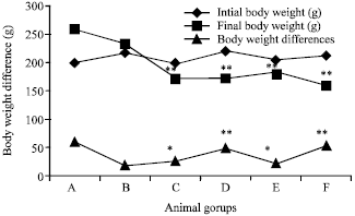 | |
| Fig. 1: | Effect of Fragrance exposure (5 mL kg-1 b.wt. for 56 days and 112 days) on the initial body weight, final body weight and body weight differences of experimental rats. *p<0.05, **p<0.005 significantly different from control. Values are expressed as Mean±SD for n = 10 in each group, A: 5 mL kg-1 b.wt. of normal saline for 56 days (Control), B: 5 mL kg-1 b.wt. of normal saline for 112 days (Control), C: 5 mL kg-1 b.wt. of Fragrance 1 (F1) for 56 days, D: 5 mL kg-1 b.wt. of Fragrance 1 (F1) for 112 days, E: 5 mL kg-1 b.wt. of Fragrance 2 (F2) for 56 days, F: 5 mL kg-1 b.wt. of Fragrance 2 (F2) for 112 days |
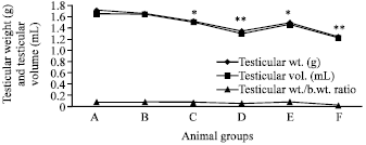 | |
| Fig. 2: | Effect of Fragrance exposure (5 mL kg-1 b.wt. for 56 days and 112 days) on testicular weight, testicular volume and testicular weight/body weight ratio of experimental rat, *p<0.05, **p<0.005 significantly different from control. Values are expressed as Mean±SD for n = 10 in each group, A: 5 mL kg-1 b.wt. of normal saline for 56 days (Control), B: 5 mL kg-1 b.wt. of normal saline for 112 days (Control), C: 5 mL kg-1 b.wt. of Fragrance 1 (F1) for 56 days, D: 5 mL kg-1 b.wt. of Fragrance 1 (F1) for 112 days, E: 5 mL kg-1 b.wt. of Fragrance 2 (F2) for 56 days, F: 5 mL kg-1 b.wt. of Fragrance 2 (F2) for 112 days |
| Table 1: | Effect of Fragrance exposure (5 mL kg-1 b.wt. for 56 days and 112 days) on sperm count, sperm motility and livability of experimental rats |
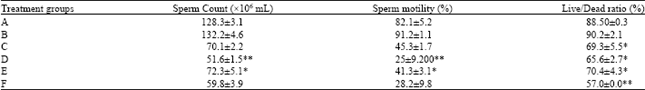 | |
| *p<0.05, **p<0.005 significantly different from control. Values are expressed as Mean±SD for n =10 in each group, A: 5 mL kg-1 b.wt. of normal saline for 56 days (Control), B: 5 mL kg-1 b.wt. of normal saline for 112 days (Control), C: 5 mL kg-1 b.wt. of Fragrance 1 (F1) for 56 days, D: 5 mL kg-1 b.wt. of Fragrance 1 (F1) for 112 days, E: 5 mL kg-1 b.wt. of Fragrance 2 (F2) for 56 days, F: 5 mL kg-1 b.wt. of Fragrance 2 (F2) for 112 days | |
| Table 2: | Effect of Fragrance exposure (5 mL kg-1 b.wt. for 56 days and 112 days) on sperm morphology of experimental rats |
 | |
| *p<0.05, **p<0.005 significantly different from control, Values are expressed as Man±SD for n =10 in each group, A: 5 mL kg-1 b.wt. of normal saline for 56 days (Control), B: 5 mL kg-1 b.wt. of normal saline for 112 days (Control), C: 5 mL kg-1 b.wt. of Fragrance 1 (F1) for 56 days, D: 5 mL kg-1 b.wt. of Fragrance 1 (F1) for 112 days, E: 5 mL kg-1 b.wt. of Fragrance 2 (F2) for 56 days, F: 5 mL kg-1 b.wt. of Fragrance 2 (F2) for 112 days | |
It should be noted however, that the control group of animal had a significant (p<0.005) increase in gross anatomical parameters which could mean that the control group of rat were still in their active growth phase during the study.
Sperm characteristics: As shown in Table 1, the groups of rat exposed to the two perfume for 112 days had a marked oligospermia (p<0.005), reduced sperm motility (p<0.005) and livability (p<0.005) when compared to the group exposed for 56 days which also had a significant decrease in sperm count (p<0.05), motility (p<0.05) and percentage amount of normal spermatozoa. These indicates a time dependent manner of fragrance toxicity. The morphological characteristic of the sperm cells in all the six groups of rats are as shown in Table 2, the following abnormalities were noticed both in the control group and the treated groups C, D, E, F; head less tail, rudimentary tail, curved mid-piece, curved tail. As shown in Table 2, there was a significant increase in percentage number of abnormal sperm of the exposed rat which was duration dependent. The observed decrease in sperm parameters could be as a result of the deleterious effect of fragrance on androgen-secreting capacity (Akunna et al., 2013). Diminution in spermatozoa concentration and mobility with an increase in the percentage of abnormal sperms is directly related to infertility (Mazzilli et al., 1994; Seed et al., 1996). Sperm membranes are very vulnerable to oxidative insult owed to a high concentration of polyunsaturated fatty acids and the need for Sertoli cell barrier protection (Winterbourn et al., 1975; Zorgniotti and Sealfon, 1988; Weese et al., 1993). Endocrine disruptors are chemical agents that have been severally documented as a possible cause of male infertility.
Currently, the process for deciding if a chemical is an environmental endocrine disruptor is to determine the effects of that chemical on the endocrine systems of humans and other animals. Fragrance has been indicated as a possible endocrine disrupting agent (Duty et al., 2003). The most likely effect of endocrine disruption in men may be a reduction in sperm concentration, motility and elevation in percentage abnormal sperm which was evident in our study (Carlsen et al., 1992; Sharpe, 1992).
CONCLUSION AND RECOMMENDATION
The overall results obtained in this study suggest that fragrance materials are toxic to spermatozoa of rat with the effect of a time dependent nature.
Despite the well established documentation on the effect of fragrance materials on human health, there is a need for further investigations in humans to determine its safety margin as far as fertility is concerned.
REFERENCES
- Akunna, G.G., L.C. Saalu, O.S. Ogunmodede, B. Ogunlade, G.A. Adefolaju and A.J. Bello, 2011. The effects of two Nigerian made perfume on the liver of adult Wistar rat. J. Med. Sci., 11: 220-225.
CrossRefDirect Link - Akunna, G.G., L.C. Saalu, B. Ogunlade, A.O. Ojewale and L.A. Enye, 2013. Consumption of bay leaf (a food spice) may be a safe and effective treatment for male infertility resulting from partial ligation of the left renal vein in wistar rat: Study suggest. Am. J. Res. Commun., 1: 123-142.
Direct Link - WMA and APS, 2002. Guiding principles for research involving animals and human beings. Am. J. Physiol.: Regul. Integr. Comp. Physiol., 283: R281-R283.
CrossRefPubMedDirect Link - Carlsen, E., A. Giwercman, N. Keiding and N.E. Skakkebaek, 1992. Evidence for decreasing quality of semen during past 50 years. Br. Med. J., 305: 609-613.
PubMedDirect Link - Carlsson, G., S. Orn, P.L. Andersson, H. Soderstrome and L. Norrgren, 2000. The impact of musk ketone on reproduction in zebrafish (Danio rerio). Mar. Environ. Res., 50: 237-241.
PubMedDirect Link - Vader, D. and L. Christy, 2009. Fragrance in the workplace is the new second-hand smoke. Proceedings of the Annual Conference on American Society of Business and Behavioral Sciences. Volume 16. Febrary 19-20, 2009, Las Vegas,.
Direct Link - Duty, S.M., N.P. Signh, M.J. Silva, D.B. Barr and J.W. Brock et al., 2003. The relationship between environmental exposure to phthalates and DNA damage in human sperm using the neutral comet assay. Environ. Health. Perspect., 111: 1164-1169.
Direct Link - Elberling, J., A. Linneberg, A. Dirksen, J.D. Johansen and L. Frolund et al., 2005. Mucosal symptoms elicited by fragrance products in a population-based sample in relation to atopy and bronchial hyper-reactivity. Clin. Exp. Allergy, 35: 75-81.
PubMed - Elberling, J., P. Skov, H. Mosbech, A. Dirkson and J. Johansen, 2007. Increased release of histamine in patients with respiratory symptoms related to perfume. Clin. Exp. Allergy, 37: 1676-1680.
PubMedDirect Link - Guin, J.D. and V.K. Berry, 1980. Perfume sensitivity in adult females. A study of contact sensitivity to a perfume mix in two groups of student nurses. J. Am. Acad. Dermatol., 3: 299-302.
PubMedDirect Link - Rachel, S.H. and T. Engen, 1996. Odor memory: Review and analysis. Psychon. Bull. Rev., 3: 300-313.
CrossRefDirect Link - Saalu, L.C., A.A. Osinubi, P.I. Jewo, A.O. Oyewopo and G.O. Ajayi, 2010. An evaluation of influence of Citrus paradisi seed extract on doxorubicin-induced testicular oxidative stress and impaired spermatogenesis. Asian J. Scient. Res., 3: 51-61.
CrossRefDirect Link - Saalu, L.C., G.O. Ajayi, A.A. Adeneye, I.O. Imosemi and A.A. Osinubi, 2009. Ethanolic seed extract of grapefruit (Citrus paradisi Macfad) as an effective attenuator of doxorubicin-induced oxidative stress in the rat heart. Int. J. Cancer Res., 5: 44-52.
CrossRefDirect Link - Schleuter, D.P., R.J. Soto, E.D. Baretta, A.A. Herrmann and L.E. Ostrander et al., 1978. Airway response to hair spray in normal subjects and subjects with hyperactive airways. Chest, 75: 544-548.
PubMedDirect Link - Seed, J., R.E. Chapin, E.D. Clegg, L.A. Dostal, R.H. Foote and M.E. Hurtt et al., 1996. Methods for assessing sperm motility, morphology and counts in the rat, rabbit and dog: A consensus report. ILSI risk science institute expert working group on sperm evaluation. Reprod. Toxicol., 10: 237-244.
Direct Link - Caress, S.M. and A.C. Steinemann, 2004. Prevalence of multiple chemical sensitivities. A population-based study in the Southeastern United States. Am. J. Pub. Health., 94: 746-747.
PubMedDirect Link - Thompson, J.A., Jr. and B.A. Wansker, 1981. A case of contact dermatitis, erythema multiforme and toxic epidermal necolysis. J. Am. Acad. Dermatol., 5: 666-669.
CrossRefDirect Link - Weese, D.L., M.L. Peaster, K.K. Himsl, G.E. Leach, P.M. Lad and P.E. Zimmern, 1993. Stimulated reactive oxygen species generation in the spermatozoa of infertile men. J. Urol., 149: 64-67.
Direct Link - Winterbourn, C.C., R.E. Hawkins, M. Brian and R.W. Carrell, 1975. The estimation of red cell superoxide dismutase activity. J. Lab. Clin. Med., 85: 337-341.
PubMedDirect Link