
M.B. Tijjani
Department of Veterinary Physiology, Pharmacology and Biochemistry, University of Maiduguri, P.M.B 1069, Borno State, Nigeria
A.A. Barkindo
Department of Animal Science and Rangeland Management, Modibbo Adama University, Yola, P.M.B 2076, Adamawa State, Nigeria
S. I. Ngulde
Department of Veterinary Physiology, Pharmacology and Biochemistry, University of Maiduguri, P.M.B 1069, Borno State, Nigeria
B. Wampana
Department of Veterinary Physiology, Pharmacology and Biochemistry, University of Maiduguri, P.M.B 1069, Borno State, Nigeria
K.A. Sanda
Department of Veterinary Physiology, Pharmacology and Biochemistry, University of Maiduguri, P.M.B 1069, Borno State, Nigeria
Journal of Medical Sciences
Year: 2013 | Volume: 13 | Issue: 6 | Page No.: 477-482


ABSTRACT
Decoction of Detarium microcarpum, used by traditional herbalist as antidiarrheal agents, was investigated for its efficacy. Determination of phytochemical constituents of the aqueous extract was carried qualitatively using standard laboratory procedures. Experimentally-induced diarrhoea and intraluminal pooling and charcoal transit time using castor oil was used as a model for assessing antidiarrheal efficacy of the plant. The result showed that the plant contain alkaloid, tannins, flavonoids and carbohydrates. The extract gave a protection against castor oil induced diarrhoea of 53% compared with standard drug loperamide that gave 91% protection. Intraluminal fluid volume was significantly (p≤0.05) decreased compared negative control. Distance travelled by charcoal meal was found to be significantly (p≤0.05) lower than the negative control and is comparable to with the positive control group that received atropine. The results of this study suggest that the plant was found to possess antidiarrheal effect as claimed by herbalist.
PDF Abstract XML References Citation
Received: March 31, 2013;
Accepted: April 29, 2013;
Published: July 04, 2013
How to cite this article
M.B. Tijjani, A.A. Barkindo, S. I. Ngulde, B. Wampana and K.A. Sanda, 2013. Evaluation of Antidiarrheal Efficacy of Detarium microcarpum Stem Bark Aqueous Extract in Albino Rats. Journal of Medical Sciences, 13: 477-482.
DOI: 10.3923/jms.2013.477.482
URL: https://scialert.net/abstract/?doi=jms.2013.477.482
DOI: 10.3923/jms.2013.477.482
URL: https://scialert.net/abstract/?doi=jms.2013.477.482
INTRODUCTION
Since time immemorial, humans have been using plants for the management of various conditions. Many of such plants have well been scientifically proven to be effective through experimental bioassay and the active principle have isolated and developed into drugs used conventional orthodox medicine. Some of the plants used in ethno medicine which was not found effective are discouraged of their continual usage especially in acute and life threatening conditions such as diarrhoea.
Diarrhoea is ranked high among the serious health problems claiming lives of children and immune compromised patients in the developing countries accounting for more than 5 million deaths worldwide each year in infants and children of less than 5 years. These could be attributed to lack of access to potable water and constitute a medical emergency primary health care units are not readily available (Shoba and Thomas, 2001). In order to reduce the impact of diarrhoea in developing countries, international organizations such as WHO have recommended the use of traditional remedies of proven efficacy in remote communities as a tentative approach before accessing standardized medical treatment (Atta and Mouneir, 2004).
This study attempts to determine the efficacy of the plant Detarium microcarpum stem bark in an attempt to further encourage their use if found effective.
MATERIALS AND METHODS
Plant material: The stem bark of D. microcarpum was collected in suburb of Yola town in Nigeria. It was identified and authenticated by a taxonomist in the Department of Biological Sciences, University of Maiduguri. It was then air dried to a constant weight, pulverized using mortar and pestle. The resulting powder was stored in an airtight container and kept in a cool, dark and dry place prior to extraction process.
Preparation of extracts: The plant was extracted using soxhlet extractor, where 250 mL of distilled water was added to 500 g of the powder. After 60 min, the mixture was cooled and decanted. The decoction was filtered with a filter and the filtrate was dried using digital heating drying oven (DHG-9030A) preset at 50°C. The air-dried extract was secured in air-tight material until use. A concentration of 200 mg mL-1 solution was constituted for use in the experiment.
Phytochemical analysis: The crude aqueous extract of D. microcarpum stem-bark was subjected to qualitative chemical screening for the identification of the various classes of active chemical constituents. The phytochemical analyses were carried out according to standard methods described by Trease and Evans (1997) and Sofowora, 2006. 80 mg mL-1 of the stem-bark extract was constituted for the analysis.
Test for carbohydrate
General test Molisch’s test: Two drops of Molisch’s reagent was added to 2 mL of the aqueous solution of the extract in a test tube, then a small volume of concentrated tetraoxosulphate VI acid was allowed to run down the side of the inclined test tube to form a layer without shaking. The presence of purple colour at the interface was indicative of the presence of carbohydrate.
General Test for Monosaccharide Barfoed’s Test: One milliliter of the aqueous solution and 1 mL of Barfoed’s reagent were mixed in a test tube and placed in a water bath for about 1 min. The presence of red precipitate was indicative of monosaccharide.
Standard test for free reducing sugar fehling’s test: To 2 mL of the aqueous solution of the extract was added 5 mL mixture of equal volumes of Fehling’s solutions I and II and boiled in a water bath for about 2 min. The brick red precipitate observed indicates the presence of reducing sugars.
Standard test for combined reducing sugars: One milliliter (1 mL) of the aqueous solution of the extract was hydrolysed by boiling with 5 mL of 10% hydrochloric acid solution. Then the above procedure for Fehling’s test was carried out and the presence of brick red precipitate was indicative of combined reducing sugars.
Test for tannins
Ferric chloride test: To 2 mL of the aqueous solution of the extract was added few drops of 10% ferric chloride solution. The occurrence of bluish black colour indicates the presence of gallic tannins and greenish-black colour indicates the presence of catechol tannins. Control tests were done by repeating the procedure using distilled water and wild cherry bark separately in place of the extract as standards.
Formaldehyde test: To 2 mL of the aqueous solution of the extract in a test tube was added a drop of 10% formaldehyde solution and 3 drops of 10% hydrochloric acid. The mixture was then observed for the presence of precipitate indicative of tannin. Control tests were done by repeating the procedure using distilled water and catechol separately in place of the extract as standards.
Test for saponins (froth test): Three milliliter of the aqueous solution of the extract was mixed with 10 mL of distilled water in a test tube. The tube was stoppered and shaken vigorously for about 5 min. It was then allowed to stand for 30 min and observed for honey comb froth for positive result.
Test of cardiac glycosides: To about 2 mL of the aqueous solution of the extract was added 3 drops of strong solution of lead acetate. This was mixed thoroughly and filtered. The filtrate was shaken with 5 mL of chloroform in a separating funnel. The chloroform layer was evaporated to dryness in a small evaporating dish. The residue was dissolved in glacial acetic acid containing a trace of ferric chloride. This was then transferred to the surface of 2 mL concentrated tetraoxosulphate (vi) acid in a test tube. The upper layer and interface of the two layers were observed for bluish-green and reddish-brown colouration respectively which are positive for cardiac glycosides. The control study was carried out in the absence of the extract.
Test for flavonoids: Two grams of the powdered stem bark was detanned with acetone. The residue was extracted in water after evaporating the acetone on a water bath. The mixture was filtered while hot and used for the following tests for flavonoids.
Lead acetate test: To 5 mL of detanned extract was added 10% lead acetate solution and observed for coloured precipitates indicative of flavonoid. Control test was run by repeating the procedure using distilled water in place of the extract.
Sodium hydroxide test: Five milliliters of 20% sodium hydroxide was added an equal volume of detanned extract and observed for yellow coloration which was indicative of flavonoid. Control test was run by repeating the procedure using distilled water in place of the extract.
Test for alkaloids: One gram (1 g) of the extract was dissolved in 5 mL of 10% ammonia solution and extracted with 15 mL of chloroform. The chloroform portion was evaporated to dryness and the resultant residue dissolved in 15 mL of dilute tatraoxosulphate (vi) acid. This was used for the following tests.
Mayer’s test: Three drops of Mayer’s reagent was added to 2 mL of the acidic solution in a test tube and observed for an opalescence or yellowish white precipitate which was indicative of the presence of alkaloids. Control test was carried out by repeating the procedure using water in placed of the extract.
Dragendorff’s test: Two milliliters of acidic solution in a test tube was neutralized with 10% ammonia solution. Dragendorrff’s reagent was added and turbidity or precipitate was observed indicative of presence of alkaloids. Control test was carried out by repeating the procedure using 10% ammonia in place of the extract as standard.
Test for anthraquinones
Borntragers reaction for free anthraquinones: One gram of powdered stem bark was placed in a dry test tube and 20 mL of chloroform was added. It was heated in a steam bath for five minutes. The extract was filtered while hot and allowed to cool. To the filtrate was added equal volume of 10% ammonia solution. It was then shaken and the upper layer was observed for bright pink coloration indicative of anthraquinones. The control test was performed by adding 10 mL of 10% ammonia solution in 5 mL of chloroform in a test tube.
Test for combined form of anthraquinone: One gram of powdered stem bark was boiled with 10 mL of ferric chloride and 5 mL of dilute hydrochloric acid for 5 min. The mixture was filtered while hot, allowed to cool and the filtrate was shaken with equal volume of chloroform. The layers were allowed to separate in a separating funnel. The chloroform layer was transferred into another tube containing 5 mL of 10% ammonia solution and the upper aqueous layer was observed for a bright-pink colour. Control test was performed by adding 10 mL of 10% ammonia solution in 5 mL of chloroform in a test tube.
Experimental animals: Albino rats of both sexes weighing between 150-200 g were obtained from laboratory animal house of the Faculty of Pharmacy; University of Maiduguri, Nigeria, was used for the study. They were allowed to acclimate for two weeks in the Department of Veterinary Physiology Laboratory, University of Maiduguri. The rats were fed with standard laboratory diet and allowed access to water ad-libitum. Assessment of anti-diarrheal effect of D. microcarpum by protection of induced diarrhoea.
Twenty-five rats divided into five groups of five rats each and are designated as groups A to E. Group A was orally given distilled water, groups B and C were given graded (400 and 600 mg kg-1) doses of the extract. Group D serving as the positive control received the standard drug loperamide. After 1 h, 1 mL of castor oil was administered to all the rats to induce diarrhoea and was then kept in individual cages with a clean white sheet of paper placed at its bottom. Numbers of dry and wet faeces were counted after 4 hour and percentage protection from diarrhoea was deduced. Group E were not administered castor oil and they served as the negative control.
Assessment of anti-diarrheal effect of D. microcarpum by intestinal secretory inhibition: Total of twenty-five was used for studying the anti-diarrheal effect of the extract using castor induced enteropooling by administering 0.2 mL. of castor oil orally. The rats were divided into five groups of five animals per group designated as A, B C, D and E. Group A serve as negative control and were administered castor oil only while groups B, C and D were given graded doses (400, 600 and 800 mg kg-1) while group E served as the positive control which were given atropine only.
Two hours after administration of the castor oil, animals in each group were sacrificed and volume of the content of the small intestine was measured by milking it into graduated tube (Robert et al., 1976).
Assessment of antidiarrheal effect by intestinal motility: Twenty five rats were fasted for 18 hours and grouped into five groups (A-E) of five rats each. Group A served as the negative control and were given distilled water. Groups B, C, D and E were first given castor oil an hour before treatment and then administered 10% charcoal solution 1 hour after treatment. Treatments given to group B was distilled water only, groups C and D were administered graded doses (400 and 800 mg kg-1, respectively) of the extract while group E served as the positive control and was administered standard drug, atropine. Animals were sacrificed after 1 h and intestinal motility was assessed by measuring the distance travelled by charcoal along the intestinal length with respect to the total length of the intestine as described by Mascolo et al. (1994).
Statistical analysis: Data obtained from this experiment was expressed as Mean±standard deviation and were analysed using one way analysis of variance. Statistical software, Graphpad InStat (2003) was used for the analysis and p≤0.05 was considered significant.
RESULTS
Phytochemical analysis: The presence of carbohydrate, alkaloids, tannins and flavonoids were detected in the aqueous extract of D. microcarpum as shown in Table 1.
| Table 1: | Phytochemical constituents of D. microcarpum aqueous extract |
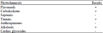 | |
| +: Present, -: Not detected | |
| Table 2: | Effect of D. microcarpum aqueous extract on castor-oil induced diarrhoea |
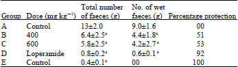 | |
| Values are expressed as Mean±SD, N = 5, Values with superscript a in the given column are significantly (p≤0.01) lower than the values in control group | |
Protection from induced diarrhoea test: Inhibition of castor induced diarrhoea was presented in Table 2. It showed that, compared to the control that had mean total number of faeces of 13±2.0, groups treated with 400 and 600 mg/kg of the aqueous extract of D. microcarpum stem bark showed significantly (p≤0.05) lower count of 6.4±2.5 and 5.8±2.5, respectively. Similarly, out of these totals, wet faecal counts for groups treated with the extracts at 400 and 600 mg kg-11 were 4.4±1.8 and 4.2±2.7, respectively. These values were significantly (p≤0.05) low in comparison to control that had a value of 9.0±1.6. Positive control group that received a standard drug Loperamide showed remarkable resistance to castor-induced diarrhoea because the value of the mean number of wet faeces was 0.6±0.1 out of 0.8±0.2 mean total. Negative control which received distilled water had no wet faceces. The percentage protection offered by the extract to the castor-oil induced diarrhoea was deduced to be 51 and 53% for 400 and 600 mg kg-1 while a very high value of 92% was obtained for the group treated with loperamide. This result signifies that the protective effect of extract was not dose dependant.
Enteropooling test: Result of enteropooling test (Table 3) showed that in the groups which received the aqueous extract of D. microcarpum stem bark at a dose rate of 400, 600 and 800 mg kg-1 had a volume of intraluminal fluid content weighing 1.8±0.4, 1.3±0.7 and 1.1±0.3, respectively. When these values were compared with the control group that had 2.4±1.0, statistically significant (p<0.05) reduction was observed in 600 and 800 mg kg-1 extract treated group. However, in the lowest dose of 400 mg kg-1, the value obtained was 1.8±0.4 and there was no significant (p<0.05) difference compared to the control.
| Table 3: | Effect of D. microcarpum aqueous extract on castor-oil induced enteropooling in albino rats |
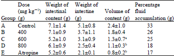 | |
| Values are expressed as Mean±SD, Values with superscript, a in the given column are significantly (p≤0.05) lower than the values in control group, Values with superscript, b in the given column are significantly (p≤0.01) lower than the values in control group | |
| Table 4: | Effect of D. microcarpum aqueous extract on charcoal transit time in albino rats |
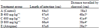 | |
| Values are expressed as Mean±SD, Values with superscript ain the given column are significantly (p≤0.01) lower than the values in control group | |
The calculated value of percentage fluid accumulation was lowest in atropine control group having 17% which was comparable to 18% recorded in the groups that received the extract at 800 mg kg-1. The values for percentage fluid accumulation for 400, 600 mg kg-1 were 26 and 27%, respectively.
Intestinal motility test: Results from the gastrointestinal motility tests (Table 4) showed that the average distance (58±3 cm) moved by the charcoal marker was greatest for the control (group A). These distances were least (28±5 cm) for the group which received atropine. In the groups treated with the aqueous extract of D. microcarpum stem bark at 400, 600 and 800 mg/kg, the values for distance travelled by charcoal were 48±5, 41±4 and 38±4 cm, respectively which were all significantly (p<0.05) lower compared to the negative control group.
DISCUSSION
In line with the objective of WHO’s objectives (Sofowora, 2006) of ethno-medicinal research of evaluating the efficacy of herbs used in managing disease condition and disease states to encourage or discourage its use, D. microcarpum was evaluated for antidiarrheal efficacy in experimental animals.
Ricinoleic acid released from castor oil when hydrolysed by pancreatic lipase causes irritation and inflammation of the intestinal mucosa leading to the release of prostaglandin that stimulate motility and secretion (Pierce et al., 1971). This agent was used in inducing functional diarrhoea in this experiment.
The presence of tannins in the extract may explain the antidiarrheal activity of the as they are known to have astringent action. Tannins are referred to as gastrointestinal modifiers which following ingestion and consequent hydrolysis precipitate proteins and other large molecules, thereby altering fluidity of the intestinal content and hence used as an antidiarrheal agent (Bone, 1998). Flavonoids have been ascribed to have antidiarrheal activity of due to their ability to inhibit intestinal motility and hydro electrolytic secretion (Di Carlo et al., 1993).
Modulators of intestinal motility such as antimuscarinics and opiates have been categorized as antidiarrheal agent due to their ability to relax gastrointestinal smooth muscle by inhibiting the effect of endogenous acetyl choline (Akubue, 2006). Atropine also inhibits secretion of intestinal mucosa. D. microcarpum stem aqueous extract was also observed in this study to reduce significantly the intestinal motility in rats that were fed with charcoal meal similar to that obtained in atropine treated group.
In the pathophysiology of diarrhoea, there is an increase in frequency of defecation associated with watery stool. The significant reduction of frequency of defecation and number of wet faecal dropping observed in this study demonstrates the efficacy of D. microcarpum stem aqueous extract as antidiarrheal agent.
Gastrointestinal Protestants and astringent such as kaolin are also in symptomatic control of diarrhoea because of their ability to coat the intestinal mucosa thereby preventing irritation and erosion of potentially harmful substances (Nicholas and McDonald, 2001). The percentage protection offered by D. microcarpum stem aqueous extract to diarrhoea induced by castor oil was about 55%. This signifies the extract possess some marginal coating effect on the intestinal mucosa.
CONCLUSION
The claim by traditional herbalists on use of the plant D. microcarpum stem bark decoction in the management of diarrhoea can be said to be scientifically justified since in this study, its aqueous extract was found to contain tannins, a known astringent and was able to reduce intestinal motility and secretion. These properties are attributes of some conventional antidiarrheal agents of orthodox medicine and therefore its usage for this purpose in traditional medicine should be encouraged. Further studies need to be carried out to identify the active principle responsible for this activity as well as toxicological studies to determine its safety margin.
REFERENCES
- Atta, A.H. and S.M. Mouneir, 2004. Antidiarrhoeal activity of some Egyptian medicinal plant extracts. J. Ethnopharmacol., 92: 303-309.
CrossRefPubMedDirect Link - Di Carlo, G., G. Autore, A.A. Izzo, P. Maiolino and N. Mascolo et al., 1993. Inhibition of intestinal motility and secretion by flavonoids in mice and Rats: Structure-activity relationships. J. Pharm. Pharmacol., 45: 1054-1059.
CrossRefPubMedDirect Link - Mascolo, N., A.A. Izzo, G. Autore, F. Barbato and F. Capasso, 1994. Nitric oxide and castor oil-induced diarrhea. J. Pharmacol. Exp. Ther., 263: 291-295.
PubMedDirect Link - Pierce, N.F., C.C. Carpenter Jr, H.L. Elliot and W.B. Greenough, 1971. Effects of prostaglandins, theophylline and cholera exotoxin upon transmucosal water and electrolyte movement in the canine jejunum. Gastroenterology, 60: 22-32.
Direct Link - Robert, A., J.E. Nezamis, C. Lancaster, A.J. Hanchar and M.S. Klepper, 1976. Enteropooling assay: A test for diarrhea produced by prostaglandins. Prostaglandins, 11: 809-828.
CrossRefPubMedDirect Link - Shoba, F.G. and M. Thomas, 2001. Study of antidiarrhoeal activity of four medicinal plants in castor-oil induced diarrhoea. J. Ethnopharmacol., 76: 73-76.
CrossRefDirect Link