
Anthony C.C. Egbuonu
Department of Biochemistry, University of Nigeria Nsukka, Enugu State, Nigeria
Ifeoma I. Ijeh
Department of Biochemistry, Michael Okpara University of Agriculture Umudike, Abia State, Nigeri
Lawrence U.S. Ezeanyika
Department of Biochemistry, University of Nigeria Nsukka, Enugu State, Nigeria
Onyechi O. Obidoa
Department of Biochemistry, University of Nigeria Nsukka, Enugu State, Nigeria
Journal of Medical Sciences
Year: 2013 | Volume: 13 | Issue: 4 | Page No.: 276-282


ABSTRACT
High blood pressure (a condition associated with vascular constriction) is a major feature of metabolic syndrome (MES). MES, a constellation of metabolic disorders, is prevalently higher in females and was associated with a reduced concentration of a vasodilator molecule, Nitric Oxide (NO). L-arginine (ARG), a precursor of NO may improve MES, warranting this study. Two groups (n = 8) of female Wistar albino rats were (per orally for twenty eight days) exposed to a single dose of 60 mg kg-1 b.wt. of ARG and 3 mL kg-1 b.wt. of distilled water, DW, respectively as treated and control groups. Significant differences in means were separated by student’s t-test (p<0.05; p<0.01) and results expressed as Mean±Standard deviation. ARG exposure caused a significant reduction (p<0.01) in sodium ion (Na+) concentration (136.42±1.66 mmol L-1; 6.54%), but a non-significant decrease (p>0.05) in potassium ion (K+) concentration (4.54±0.66 mmol L-1; 14.01%) in the rats’ serum, suggesting improved/reduced blood pressure. ARG treatment in the rats had a significant increase (p<0.01) in Alkaline Phosphatase (ALP) activity (8.30±0.23 IU L-1; 196.43%) in the rats’ serum, indicating adverse influence on high metabolic organs, including the brain. Sodium ion had a significant negative correlation (r = 0.01) with potassium ion, whereas the heart histomorphology revealed degenerations in the ARG-fed rats, apparently confirming the observations and suggestions thereto. Thus, ARG may improve blood pressure in the rats, perhaps at the expense of compromised heart function and histology of the rats. These may be pointing to a new arginine phenomenon, hence warrant follow up.
PDF Abstract XML References Citation
Received: January 10, 2013;
Accepted: February 23, 2013;
Published: May 21, 2013
How to cite this article
Anthony C.C. Egbuonu, Ifeoma I. Ijeh, Lawrence U.S. Ezeanyika and Onyechi O. Obidoa, 2013. Influence of L-arginine on the Heart Histology and Function Markers of Metabolic Syndrome in Female Wistar Albino Rats. Journal of Medical Sciences, 13: 276-282.
DOI: 10.3923/jms.2013.276.282
URL: https://scialert.net/abstract/?doi=jms.2013.276.282
DOI: 10.3923/jms.2013.276.282
URL: https://scialert.net/abstract/?doi=jms.2013.276.282
INTRODUCTION
High blood pressure (a condition associated with vascular constriction) is a major feature of metabolic syndrome (MES). It was associated with a reduced concentration of nitric oxide, NO (a vasodilator molecule) and characterized by a cluster of cardiovascular risk factors, including high blood pressure (Deedwania and Gupta, 2006; Gallagher et al., 2010) and increased health challenges (Pelucchi et al., 2010; Siddiqui, 2011). Metabolic syndrome scourge is pandemic (Gotto et al., 2006; Grundy, 2008) and is prevalent in children (Pedrosa et al., 2011). The syndrome afflicts about 20-30% of the adult population the world over (Grundy, 2008; Chaabo et al., 2010), including Nigeria (30.7%) as reported by Ijeh et al. (2010). The increasing prevalence of MES (Bakoma et al., 2011) may be higher among the female gender, with huge implications.
Garlichs et al. (2000) had associated reduced NO concentration with the pathophysiology of MES. NO regulated cardiovascular function (McGrowder and Brown, 2007) and at abnormal concentration, had elicited pathological condition (Lokhande et al., 2006) in animals. A reduction in the concentration of ARG affected the biological activity of NO (Subratty et al., 2007; Harisa, 2011), suggesting exogenous supply of ARG my influence the activity of NO in animals. Indeed, possible role of ARG in metabolic processes that could improve MES features in animals was suggested (Sepehri et al., 2006; Van Waardenburg et al., 2007; Ezeanyika and Egbuonu, 2011; Harisa, 2011) and appear to be supported by recent studies (Egbuonu and Ezeanyika, 2012a, b; Egbuonu and Ezeanyika, 2013).
Since high blood pressure is a major feature of MES (Expert Panel on Detection, Evaluation and Treatment of High Blood Cholesterol in Adults, 2001 (NCEP) (USA); Gallagher et al., 2010), this study aimed to ascertain the effect of ARG on the heart histology and function markers of MES in female rats. Objectives set to achieve the stated aim include the study of the effect of ARG on serum alkaline phosphatase activity, sodium ion and potassium ion concentrations as well as on the heart histology of female Wistar albino rats. In particular, potassium ion contributes to the optimal functioning of the cells and the organs and its deficiency (hypokalaemia) was associated with cardiac dysfunction (Bush, 1991) and high blood pressure (Siani et al., 1987; Strazzullo et al., 1990). On the other hand, elevated sodium ion concentration (hypernatraemia) indicated high blood pressure (Jaitovich and Bertorello, 2010).
MATERIALS AND METHODS
Chemicals: The chemicals (analytical grade) used in this study were products of reputable companies based in Europe and America.
Concentration determination/justification: Based on the WHO reported daily ARG oral intake (Marshal, 1994) and the concentration used in earlier studies (Alexander et al., 2004; Egbuonu et al., 2010a-c), the concentration of ARG used in this study was calculated and adjusted to 60 mg kg-1 b.wt.
Animals and treatment: The female Wistar rats (60-80 g) used in this study were procured from the animal house of the Faculty of Biological Sciences University of Nigeria. The animal study was according to International guidelines for the care and use of laboratory animals in Biomedical Research (CCAC, 1985; WMA/APS, 2002). The animal study was conducted between August and September, 2010.
The rats were acclimatized for a week and randomized into two groups (sample size of eight rats each) based on their body weight. Group B rats were exposed to ARG (60 mg kg-1 b.wt.) whereas Group A rats were given Distilled Water (DW) (3 mL kg-1 b.wt.). The rats were exposed orally for 28 consecutive days.
The rats were kept in a well-ventilated stainless steel cages at room temperature (28±2°C) and tropical humid condition. They were maintained under twelve hours of light alternating with twelve hours of darkness. The rats were allowed free access to tap water and standard rat chow (Grand Cereals and Oil Mills Limited, Jos, Nigeria) throughout the experimental period.
Sample collection and preparation: After 28 days oral intubation, the animals were fasted overnight and sacrificed on day 29. The respective blood sample of the animals was collected by ophthalmic venous plexus or retro orbital sinus venipuncture, using sterile capillary tube to direct blood into clean non-anticoagulated glass tubes.
Clotted blood in each tube was centrifuged (at 3000 rpm for 10 min) to yield the serum. The respective serum was aspirated separately into stoppered polystyrene tube and stored in a deep freezer for subsequent use in determining the biochemical markers of MES related to heart function. Organ specimen (heart) excised from the sacrificed rats for histology were fixed in 10% formaldehyde buffered saline (formal saline) until used.
Our choice for using female rats in this study was based on the recent listing of female gender as an independent risk factor for the development of MES (Ravikiran et al., 2010) and reports that MES was prevalently higher in females (Mangat et al., 2010; Kilic et al., 2010).
Parameters determined
Serum alkaline phosphatase (ALP) activity: The serum alkaline phosphatase (ALP) activity was assayed by the method of Walter and Schutt (1974). This is based on the principle that alkaline phosphatase could hydrolyze colorless phosphate esters of various alcohols and phenols yielding p-nitrophenol as the yellow nitrophenolate ion in alkaline solution that was measured colorimetrically at 405 nm.
Photometric estimation of sodium (Na+) and potassium (K+) ions concentrations: The estimation of sodium and potassium ions in serum was with flame emission photometer. This is based on the principle that passing a liquid sample through a nebuliser (atomizer) could excite the atoms which on falling back to the ground state, emit light of characteristic wavelength, color and intensity. On passing through a suitable filter, a photosensitive detector measures the emitted light as the amount (concentration) of the atom (metallic ion) present.
Organ histology: Organ specimen (heart) for histological examination was promptly excised from the sacrificed rats and fixed in 10% formaldehyde buffered saline (formal saline) as reported by Egbuonu et al. (2010c). Briefly, after dehydration (in graded levels (70-100%) of alcohol), clearing (in xylene impregnated with paraffin wax) and sectioning (at 5 microns thickness using rotary microtome) the sections were floated on a water bath maintained at a temperature of 2-3°C below melting point of the paraffin wax. The sections were dried on a hot plate maintained at a temperature of 2-3°C above the melting point of the paraffin and stained. Then, the sections were mounted, using haematoxylin and eosin.
Statistical analysis: Data were analyzed by Student’s t-test to determine the significant differences in means, using the Statistical Package for the Social Sciences (SPSS) for Windows version 16.0 (SPSS Inc., Chicago, IL., USA). Results were expressed as mean and standard deviation (Mean±SD) of eight rats per group at significance levels of p<0.05 and p<0.01. Furthermore, correlation of the results for possible association among the studied parameters was by Pearson’s bivarate methods (r = 0.01).
RESULTS
Serum alkaline phosphatase (ALP) activity: Results of this study reveal that rats exposed to ARG had a significant (p<0.01) increase in their serum ALP activity (8.30±0.23 IU L-1) compared with those exposed to DW (2.80±0.10 IU L-1). This is an increase of over one-fold (196.43%) in ARG-fed group relative to the control (Fig. 1).
Serum sodium ion (Na+) concentration: As depicted in Fig. 2, the sodium ion concentration in serum decreased in the ARG-treated rats (136.42±1.66 mmol L-1) relative to control (145.97±1.22 mmol L-1). The observation, representing a decrease of 6.54% was statistically significant (p<0.01).
Serum potassium ion (K+) concentration: The results of this study as presented in Fig. 3 show that ingesting ARG by rats non-siginificantly decreased (p>0.05) the serum K+ ion concentration (4.54±0.66 mmol L-1) in comparison with the control (5.28±0.44 mmol L-1). This observation represents a decrease of 14.01% relative to the control value.
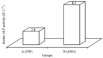 | |
| Fig. 1: | Effect of DW and ARG on serum ALP activity of rats |
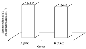 | |
| Fig. 2: | Effect of DW and ARG on serum sodium ion concentration of rats |
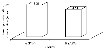 | |
| Fig. 3: | Effect of DW and ARG on potassium ion concentration in serum of rats |
 | |
| Fig. 4: | Heart section of untreated, control (Group A) rats showing normal papillary muscles (arrow heads). H and E stains, x400 |
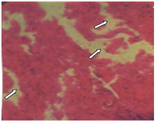 | |
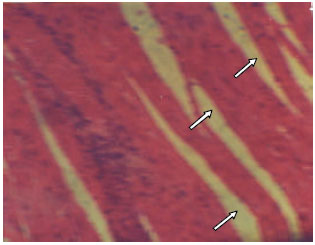
| Fig. 5: | Heart section of rats treated with high ARG (Group B) showing degeneration of the papillary muscles (arrow heads). H and E stains x400 |
Histomorphology of the heart: Heart sections of the control (Group A) showed typical histology with normal papillary muscles (Fig. 4).
| Table 1: | Correlations output of serum potassium ion and sodium ion |
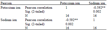 | |
| **Correlation is significant at the 0.01 level (2-tailed) | |
Sections collected from rats treated with ARG (Group B) showed moderate degeneration of the papillary muscles (Fig. 5).
Correlation of results: The results of Pearson’s correlation analysis showed that sodium ion concentration had a significant negative correlation (r = 0.01) with potassium (Table 1).
DISCUSSION
The female gender is an independent risk factor for the development of metabolic syndrome, MES (Ravikiran et al., 2010). MES, a constellation of metabolic disorders, is prevalently higher in the females. It was associated with a reduced concentration of NO (a vasodilator molecule) and characterized by a cluster of cardiovascular risk factors, including hypertension (high blood pressure) (Deedwania and Gupta, 2006; Gallagher et al., 2010). Possible role of ARG in metabolic processes that could improve MES in animals was suggested (Van Waardenburg et al., 2007; Ezeanyika and Egbuonu, 2011) and appear to be supported by recent studies (Egbuonu and Ezeanyika, 2012a, b; Egbuonu and Ezeanyika, 2013). High blood pressure (a condition associated with vascular constriction and heart function) is a major feature of MES. These therefore warranted this study.
ARG ingestion by rats elicited a significant increase (p<0.01) in ALP activity in the rats’ serum, indicating alterations of pancreatic function (Fraulob et al., 2010) and other high metabolic organs, possibly the heart and brain. The ALP enzyme is present in various high metabolic organs hence is not a specific marker of toxic effect. In similar studies, we demonstrated that L-arginine exposure improved the kidney/renal function (Egbuonu and Ezeanyika, 2013) and liver function (Egbuonu et al., 2013), but damaged the brain function (unpublished).
Sodium ion accumulation caused high blood pressure in animals (Jaitovich and Bertorello, 2010). ARG ingestion to the female rats decreased (p<0.01) the sodium ion (Na+) concentration, implying lowered blood pressure and possible benefit on MES in the rats. This is consistent with earlier reports of ARG-induced vasodilation (Rang et al., 2003) and reduction of hypertension or high blood pressure (Alexander et al., 2004) in animals. The possible vasodilatory activity of ARG via its metabolite, NO, could enhance the increase in the wall of the blood vessels resulting in lowered blood pressure. On the other hand, ARG treated rats had a decrease in serum potassium ion (K+) concentration. Decreased potassium ion (hypokalaemia) resulted to neurological and cardiac dysfunctions in animals (Bush, 1991), but the observation was not statistically significant (p>0.05) hence may not be treatment related.
Furthermore, correlation of the results showed that sodium ion had a significant negative correlation (r = 0.01) with potassium ion, suggesting overriding effect of ARG on sodium ion over potassium ion in relation to blood pressure reduction in this study. Generally, degenerations observed in the heart sections of the rats appear to indicate ARG-induced adverse influence on their heart histomorphology, apparently confirming the observation on the serum ALP activity and the suggestion thereto. Agent-induced histomorphologic alterations in organs was reported by Egbuonu et al. (2010c). It appears that the possible blood pressure lowering potential of ARG elicited adverse influence on the heart histology. We did neither know nor study how this could have occurred but L-arginine-induced complex effect had been reported in animals and attributed to the multiple catabolic pathways of ARG (Schriek et al., 2007). ‘Arginine paradox’ (a phenomenon referring to the dependence of cellular NO production on exogenous L-arginine concentration despite the theoretical saturation of nitric oxide synthase enzymes with intracellular L-arginine) may be fundamental to this complexity.
CONCLUSION
In conclusion, this study suggests that exposure to ARG may improve blood pressure (a major feature of MES) in the female rats, perhaps at the expense of compromised heart histology and function of the rats. This may be pointing to a an entirely new arginine phenomenon that warrants further investigation and caution in the use of ARG against MES associated with heart function in female rats.
REFERENCES
- Alexander, B.T., M.T. Llinas, W.C. Kruckeberg and J.P. Granger, 2004. L-Arginine attenuates hypertension in pregnant rats with reduced uterine perfusion pressure. Hypertension, 43: 832-836.
CrossRefPubMedDirect Link - WMA and APS, 2002. Guiding principles for research involving animals and human beings. Am. J. Physiol.: Regul. Integr. Comp. Physiol., 283: R281-R283.
CrossRefPubMedDirect Link - Bakoma, B., K. Eklu-Gadegkeku, A. Agbonon, K. Aklikokou, E. Bassene and M. Gbeassor, 2011. Preventive effect of Bridelia ferruginea against high-fructose diet induced glucose intolerance, oxidative stress and hyperlipidemia in male wistar rats. J. Pharmacol. Toxicol., 6: 249-257.
CrossRefDirect Link - Chaabo, F., A. Pronczuk, E. Maslova and K.C. Hayes, 2010. Nutritional correlates and dynamics of diabetes in the Nile rat (Arvicanthis niloticus): A novel model for diet-induced type 2 diabetes and the metabolic syndrome. Nutr. Metab. (Lond)., Vol. 7.
CrossRefDirect Link - Deedwania, P.C. and R. Gupta, 2006. Management issues in the metabolic syndrome. J. Assoc. Physicians. India, 54: 797-810.
PubMed - Egbuonu, A.C.C. and L.U.S. Ezeanyika, 2012. Effect of L-arginine on selected markers of metabolic syndrome related to oxidative stress, glucose metabolism and nitric oxide synthesis in female Wistar Albino rats. Int. Res. J. Biochem. Bioinf., 2: 186-192.
Direct Link - Egbuonu, A.C.C. and L.U.S. Ezeanyika, 2012. Effect of L-arginine on markers of metabolic syndrome related to abdominal obesity and disorder of lipid metabolism in female Wistar Albino rats. Am. J. Biochem., 2: 7-13.
CrossRef - Egbuonu, A.C.C., O. Obidoa, C.A. Ezeokonkwo, P.M. Ejikeme and L.U.S. Ezeanyika, 2010. Some biochemical effects of sub-acute oral administration of L-arginine on monosodium glutamate-fed Wistar albino rats 1: Body weight changes, serum cholesterol, creatinine, and sodium ion concentrations. Toxicol. Environ. Chem., 92: 1331-1337.
CrossRefDirect Link - Egbuonu, A.C.C., C.A. Ezeokonkwo, P.M. Ejikeme, O. Obidoa and L.U.S. Ezeanyika, 2010. Some biochemical effects of sub-acute oral administration of L-arginine on monosodium glutamate-fed wistar albino rats 2: Serum alkaline phosphatase, total acid phosphatase and aspartate aminotransferase activities. Asian J. Biochem., 5: 89-95.
CrossRefDirect Link - Egbuonu, A.C.C., L.U.S. Ezeanyika, P.M. Ejikeme and O. Obidoa, 2010. Histomorphologic alterations in the liver of male wistar rats treated with l-arginine glutamate and monosodium glutamate. Res. J. Environ. Toxicol., 4: 205-213.
CrossRefDirect Link - Egbuonu, A.C.C. and L.U.S. Ezeanyika, 2013. L-arginine exposure improves renal function markers of metabolic syndrome in female rats. Am. J. Biochem. Mol. Biol., 3: 50-60.
CrossRefDirect Link - Ezeanyika, L.U.S. and A.C.C. Egbuonu, 2011. Impact of nitric oxide and insulin resistance on the pathophysiology of the metabolic syndrome: Possible role of L-arginine and glutamate. J. Med. Med. Sci., 2: 657-662.
Direct Link - Fraulob, J.C., R. Ogg-Diamantino, C. Fernandes-Santos, M.B. Aguila and C.A. Mandarim-de-Lacerda, 2010. A mouse model of metabolic syndrome: Insulin resistance, fatty liver and Non-Alcoholic Fatty Pancreas Disease (NAFPD) in C57BL/6 mice fed a high fat diet. J. Clin. Biochem. Nutr., 46: 212-223.
CrossRef - Gallagher, E.J., D. Leroith and E. Karnieli, 2010. Insulin resistance in obesity as the underlying cause for the metabolic syndrome. Mt. Sinai J. Med., 77: 511-523.
CrossRefPubMedDirect Link - Garlichs, C.D., J. Beyer, H. Zhang, A. Schmeisser and K. Plotze et al., 2000. Decreased plasma concentrations of L-hydroxy-arginine as a marker of reduced NO formation in patients with combined cardiovascular risk factors. J. Lab. Clin. Med., 135: 419-425.
CrossRefPubMedDirect Link - Gotto, A.M. Jr., G.L. Blackburn, G.E. Dailey, A.J. Garber, S.M. Grundy, B.E. Sobel and M.R. Weir, 2006. The metabolic syndrome: A call to action. Coron. Artery Dis., 17: 77-80.
PubMed - Grundy, S.M., 2008. Metabolic syndrome pandemic. Arterioscler. Thromb. Vasc. Biol., 28: 629-636.
CrossRefPubMedDirect Link - Harisa, G.E.D.I., 2011. L-arginine ameliorates arylesterase/paraoxonase activity of paraoxonase-1 in hypercholesterolemic rats. Asian J. Biochem., 6: 263-272.
CrossRefDirect Link - Ijeh, I.I., U. Okorie and C.E.C.C. Ejike, 2010. Obesity, metabolic syndrome and BMI-metabolic-risk sub-phenotypes: A study of an adult Nigerian population. J. Med. Med. Sci., 1: 254-260.
Direct Link - Jaitovich, A. and A.M. Bertorello, 2010. Intracellular sodium sensing: SIK1 network, hormone action and high blood pressure. Biochimica Biophysica Acta, 1802: 1140-1149.
CrossRefPubMedDirect Link - Kilic, S., N. Yilmaz, G. Erdogan, M. Aydin, N. Tasdemir, M. Doganay and S. Batioglu, 2010. Effect of non-oral estrogen on risk markers for metabolic syndrome in early surgically menopausal women. Climacteric, 13: 55-62.
CrossRefPubMedDirect Link - Lokhande, P.D., B.S. Kuchekar, A.R. Chabukswar and S.C. Jagdale, 2006. Nitric oxide: Role in biological system. Asian J. Biochem., 1: 1-17.
CrossRefDirect Link - Mangat, C., N.K. Goel, D.K. Walia, N. Agarwal and M.K. Sharma et al., 2010. Metabolic syndrome: A challenging health issue in highly urbanized union territory of North India. Diabetol. Metab. Syndrome, Vol. 2.
CrossRefDirect Link - McGrowder, D. and P.D. Brown, 2007. Effects of nitric oxide on glucose transport: In vivo and in vitro studies. Asian J. Biochem., 2: 1-18.
CrossRefDirect Link - Expert Panel on Detection, Evaluation and Treatment of High Blood Cholesterol in Adults, 2001. Executive summary of the third report of the National Cholesterol Education Program (NCEP) expert panel on detection, evaluation and treatment of high blood cholesterol in adults (Adult Treatment Panel III). J. Am. Med. Assoc., 285: 2486-2497.
CrossRefPubMedDirect Link - Pelucchi, C., E. Negri, R. Talamini, F. Levi and A. Giacosa et al., 2010. Metabolic syndrome is associated with colorectal cancer in men. Eur. J. Cancer, 46: 1866-1872.
CrossRef - Ravikiran, M., A. Bhansali, P. Ravikumar, S. Bhansali and P. Dutta et al., 2010. Prevalence and risk factors of metabolic syndrome among Asian Indians: A community survey. Diabetes Res. Clin. Pract., 89: 181-188.
CrossRefPubMedDirect Link - Schriek, S., C. Ruckert, D. Staiger, E.K. Pistorius and K.P. Michel, 2007. Bioinformatic evaluation of L-arginine catabolic pathways in 24 cyanobacteria and transcriptional analysis of genes encoding enzymes of L-arginine catabolism in the cyanobacterium Synechocystis sp. PCC 6803. BMC Genomics, Vol. 8.
CrossRefDirect Link - Sepehri, G., S. Vahid, B. Fariba and F. Rasoul, 2006. Effect of L-NAME/L-arginine microinjection into nucleus accumbens shell on morphine withdrawal signs in male rats. Int. J. Pharmacol., 2: 171-176.
CrossRefDirect Link - Siani, A., P. Strazzullo, L. Russo, S. Guglielmi, L. Iacoviello, L.A. Ferrara and M. Mancini, 1987. Controlled trial of long term oral potassium supplements in patients with mild hypertension. Br. Med. J., 294: 1453-1456.
Direct Link - Siddiqui, A.A., 2011. Metabolic syndrome and its association with colorectal cancer: A review. Am. J. Med. Sci., 341: 227-231.
CrossRefPubMedDirect Link - Subratty, A.H., L.H. Semfa and M.D. Manraj, 2007. TAME-esterase and oxidative stress contribute to dysmetabolic syndrome in dyslipidaemia. Asian J. Biochem., 2: 323-329.
CrossRefDirect Link - Van Waardenburg, D.A., C.T. de Betue, Y.C. Luiking, M. Engel and N.E. Deutz, 2007. Plasma arginine and citrulline concentrations in critically ill children: Strong relation with inflammation. Am. J. Clin. Nutr., 86: 1438-1444.
PubMedDirect Link