
G. G. Akunna
Department of Anatomy, College of Medicine (LASUCOM) Ikeja, Lagos State University, Lagos, Nigeria
L. C. Saalu
Department of Anatomy, College of Medicine (LASUCOM) Ikeja, Lagos State University, Lagos, Nigeria
O. S. Ogunmodede
Department of Anatomy, College of Medicine (LASUCOM) Ikeja, Lagos State University, Lagos, Nigeria
B. Ogunlade
Department of Anatomy, College of Medicine (LASUCOM) Ikeja, Lagos State University, Lagos, Nigeria
G. A. Adefolaju
Department of Anatomy, College of Medicine, University of Ilorin, Kwara, Nigeria
A. J. Bello
Department of Anatomy, College of Medicine (LASUCOM) Ikeja, Lagos State University, Lagos, Nigeria
Journal of Medical Sciences
Year: 2011 | Volume: 11 | Issue: 5 | Page No.: 220-225


ABSTRACT
The use of perfumes is becoming increasingly popular in our environment. Attention is therefore understandably being focused on the safety of these perfumes. Hence, this study aimed to determine changes in the anatomical parameters of the liver and the activities of the biomarker enzymes of the liver (alanine transaminase, aspartate transaminase, alkaline phosphatase and gamma-glutamyl transpeptidase) following the exposure of the rats to two popularly used Nigerian made perfumes. Thirty-six Wistar rats were allocated into groups: A, B, C, D, E and F with each group consisting of six rats. Animals in groups C and D were exposed to the first and second perfumes by inhalation respectively for 77 days; animals in groups E and F were exposed to the first and second perfumes by inhalation respectively for a period of 154 days, while groups A and B animals served as the control groups for the periods of 77 days and 154 days, respectively. The rats were sacrificed at the end of each period of exposure after which blood was obtained for enzyme assay and the liver weights, liver volumes, liver weight/body weight ratio were evaluated. The results showed a significant decrease in the animals body weights, liver weights, liver volumes and liver weight/bodyweight ratios in the experimental groups of rats as compared to the control groups. There were also increases in the activities of alanine transaminase, aspartate transaminase, alkaline phosphatase, gamma-glutamyl transpeptidase. It is concluded that these perfumes have a deleterious effect on the rat liver.
PDF Abstract XML References Citation
Received: July 12, 2011;
Accepted: September 28, 2011;
Published: December 07, 2011
How to cite this article
G. G. Akunna, L. C. Saalu, O. S. Ogunmodede, B. Ogunlade, G. A. Adefolaju and A. J. Bello, 2011. The Effects of Two Nigerian Made Perfumes on the Liver of Adult Wistar Rat. Journal of Medical Sciences, 11: 220-225.
DOI: 10.3923/jms.2011.220.225
URL: https://scialert.net/abstract/?doi=jms.2011.220.225
DOI: 10.3923/jms.2011.220.225
URL: https://scialert.net/abstract/?doi=jms.2011.220.225
INTRODUCTION
The frequent use of fragranced cosmetic products is alarming amid the populace in Nigeria. Among these products are the two perfumes, herein referred to Perfume One (P1) and Perfume Two (P2). Like most perfumes, constituents of P1 and P2 are not always listed in the ingredients because perfume formulae are considered trade secrets. Instead the ingredients are listed as fragrance.
Fragrance materials are not only used in manufacturing colognes or perfumes but also in producing household domestic and food products (where they are used as flavours). Most sweet smelling personal care products ranging from cosmetics, drugs, deodorants, detergent, air fresheners, plastics, industrial greases to other household products contain fragrance as constituents (Rastogi et al., 2001; Calkin and Jellinek, 1994; Andrea, 1997). The safety of these fragrances, particularly in perfumes or colognes has been a concern and several questions arising from perfume uses are due to its ability to cause discomforts ranging from mucosal symptoms, asthmatic attack, headache, nausea to other types of allergies in certain individuals (Kumar et al., 1995; Millqvist and Lowhagen, 1996; Johansen, 2003; Caress and Steinemann, 2004; Kelman, 2004; Guiraud-Pons and Vigan, 2004; Elberling et al., 2005; Nardelli et al., 2008). The materials used in perfume manufacturing are mainly petroleum-based synthetic compounds.
Ven though the toxicity of these synthetic compounds in fragrance has been severally documented, most of the works reported on their toxicity revolve around their dermatological effects which is understandable due to the fact that most personal care products are applied directly on the skin (Ortiz and Yiannias, 2004; Api et al., 2008; Nigam, 2009). There are, however, various documented studies that evaluate the safety of fragrance components other than its dermatological effects (Cadby et al., 2002; Smith et al., 2004). Caress and Steinemann (2005) showed that common fragranced products emit volatile organic compounds that are hazardous or toxic. Andrea (1997), documented that the emissions of fragrance products cause various combinations of sensory and pulmonary irritations, decrease in expiratory airflow velocity and neurotoxicity which was indicated by the alterations of functional observational battery used in the course of the study.
With respects to the two perfumes under study, there is a documented report that they contain the following potentially toxic substances; linalool, polycyclic synthetic musk (galaxolide and tonalide), oakmoss absolutes, coumarin and furanocoumarin (Burr, 2008). Linalool is known to cause cellular damage by free radical generation pathway and lipid peroxidation (Zhou, 1998). Polycyclic synthetic muskhas has been variously shown to disrupt the balance of hormones in the human body, contaminate human blood and breast milk (Rimkus and Wolf, 1996; Bitsch et al., 2002; Schreurs et al., 2002; Peters, 2005). Furthermore, oakmoss absolutes have also been demonstrated by Bridges (2002) to contain allergens and carcinogenic compounds. Couramin has been reported to be a fragrance ingredient present in natural extracts of grapefruit (Saalu et al., 2007, 2009; Born et al., 2000) documented coumarin as a rat liver toxicant when they administered a toxic dosage of coumarin to rats with a resultant increase in the incidence of rat cholangiocarcinomas and parenchymal liver-cell tumors in a chronic bioassay. Finally, furanocoumarin, has been shown to cause severe allergic reactions and increase sensitivity to ultraviolet radiation (Polanska et al., 2010).Certain enzymes in the body are used as biomarkers in testing for liver health. When the liver is intoxicated or diseased, the level of these enzymes in the blood increases above the stipulated reference range thereby indicating hepatoxicity. These enzymes are Alanine Transaminase (ALT), Aspartate Transaminase (AST), Alkaline Phosphatase (ALP) and Gamma-Glutamyl Transpeptidase (GGT) (Schiele et al., 1998; Bruck et al., 2001). Hence, Liver damage is indicated when, ALT rise above the reference level of 10-40 U L-1 in males and 7-35 IU L-1 in females, AST rises above 14-20 IU L-1 in males and 10-36 IU L-1 in females, when ALP rise above 20-140 U L-1 and GGT rises above 0-51 UI L-1 (Schiele et al., 1998; Bruck et al., 2001). The references values however are largely laboratory specific.
The phenomenal rise in the usage of the perfumes in Nigeria and especially those reported in this study means that exposure to their ingredients has increased as well. However, despite apparent harmful nature of these perfume constituents, there is a dearth of information in the literature on the accumulative effects of these compounds in the perfumes on the liver of even laboratory animals.
The aim of this study was therefore to investigate the effects of two commonly- used Nigeria made perfumes (P1 and P2) on the gross the anatomical parameters and activities of liver biomarker enzymes ALT, AST, ALP and GGT in the rat.
MATERIALS AND METHODS
Animals: Thirty-six healthy male Wistar rats (about 16 weeks old), weighing between 130 and 170 g were obtained from the animal house of Anatomy department, University of Ilorin, Nigeria. The animals were housed in well ventilated wire wooden cages in the animal facility of the department of Anatomy, University of Ilorin, Nigeria. The rats were maintained under standard natural photoperiodic condition of twelve hours of light alternating with twelve hours of darkness (i.e., L:D;12 h:12 h photoperiod) at room temperature (25-26°C).
The rats were allowed unrestricted access to water and rat chow and were acclimatized for 20 days before the commencement of the experiment. The weights of the animals were estimated at procurement, during acclimatization, at commencement of the experiments and after the experiment was completed using an electronic analytical and precision balance (BA210S, d = 0.0001 g). Experimental procedures involving the animals and their care were conducted in conformity with international national and institutional guidelines for the care of laboratory animals in biomedical research and use of laboratory animals in bio-medical research promulgated by the Canadian Council of Animal Care (1985).
Chemicals: The perfumes P1 and P2 were obtained from cosmetics shop in Lagos, Nigeria on 11th January, 2011 and kept under room temperature throughout the experimental period.
Animals grouping and treatment: The rats were randomly divided into six groups as stated below:
Group A which served as the early control group were not exposed to the perfumes. They were sacrificed on day 77 of the experiment. Group B served as the chronic control group and were treated as group A, except that they were autopsied on day 154. Animals in group C were exposed to P1 for a period of 77 days after which they were sacrificed. The rats that were allocated to group D were exposed to P2 for a period of 77 days and sacrificed thereafter. Groups E and F were exposed to P1 and P2 perfumes, respectively and sacrificed on day 154.
As a means of exposing the animals to the perfumes, small balls of cotton wool were soaked in 5 mL of the perfumes for the fragrance-exposed groups while small balls of cotton wool were soaked in 5 mL water for the control groups. The wools were placed in a Petri dish inside the cages and covered with perforated plastic for an exposed duration of at least six hours per day for 77 days (Groups A, C and E) and 154 days (Groups B, D and F).
Animal sacrifice and sample collection: The rats were at the time of sacrifice first weighed and then subjected to cervical dislocation. The abdominal cavity of each rat was opened up through a midline abdominal incision to expose the liver. Each liver was excised and weighed; the liver was weighed with an electronic analytical and precision balance (BA210S, d = 0.0001 g). The liver volumes were measured using water displacement method. Blood was collected from the rat heart in potassium-EDTA containing test tubes using 10 mL syringes for biochemical investigations.
The blood samples were centrifuged at 5000 rmp for 5 min in a refrigerated centrifuge and serum was obtained for determination of liver biomarker enzymes activities.
Biochemical investigations: The alanine transaminase, aspartate transaminase, alkaline phosphatase and Gamma-Glutamyl transpeptidase activities were all determined according to the methods of Rutman and Frankel (1957).
Statistical analysis: All data were expressed as Mean±SD of number of experiments (n = 6). The level of homogeneity among the groups was tested using Analysis of Variance (ANOVA) as done by Snedecor and Cochran (1980). Where heterogeneity occurred, the groups were separated using Duncan Multiple Range Test (DMRT). A value of p<0.05 was considered to indicate a significant difference between groups (Duncan, 1957).
All analyses were performed using Genstat (2007) for windows.
RESULTS
Body weight changes: Table 1 shows that rats in experimental groups lost body weight significantly compared to the rats in control groups. Though there was no significant difference in loss of body weight between rats treated for the period of 154 days and rats treated for the period of 77 days.
Changes in liver weights and liver volume: The liver weight, liver volume and liver weight/body weight ratios of the rats in experimental groups were the least, being significantly lower, at 154 days (p<0.001) and 77 days (p<0.05) than the control groups (Table 1).
Liver biomarker enzymes: Serum level of the biomarker enzymes; ALT, AST, ALP and GGT was significantly elevated in the experimental rats (hence Group E: 160.5±7.2, 230±9.1, 390±16.4, 220±19.2, respectively compared to Group B- chronic control: 48.2±4.5 8, 6.05±5.01, 200±16.03, 44.05±4.5) (Table 2).
The elevation in the level of the marker enzyme increased significantly just as the duration of the exposure was increase from 77 days (p<0.05) to 154 days (p<0.001).
| Table 1: | Effect of Coolriver and Miyaki on the body weight, liver weight and liver volume in rats |
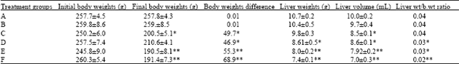 | |
| Values are given as Mean±SD, *p<0.05 **p<0.001, significant from control groups. Group A: Control group (day 77); Group B: Control (group 154); Group C: P1 (77days); Group D: P2 (77 days); Group E: P1 (154 days); Group E: P2 (154 days) | |
| Table 2: | Effect of P1 and P2 on the liver biomarker enzymes in rats |
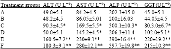 | |
| Value are given as Mean±SD, *p<0.05, **p<0.001, significance from control group. Group A: Control group (day 77); Group B: Control (group 154); Group C: P1 (77days); Group D: P2 (77 days); Group E: P1 (154 days); Group E: P2 (154 days) | |
DISCUSSION
The findings from the present study demonstrated that P1 and P2 constituents inhibited growth of male Wister rats. The gain in live weights of the control rats means that the rats were still in the active growth phase. The significant reduction of the anatomical parameters of live weights, liver weights and volumes of the experimental groups indicates that the chemical components of P1 and P2 have a negative effect on the body metabolic process. These findings agree with our earlier reports using other organotoxic models (Saalu et al., 2010, 2011).
The results of this study suggest that ingredients in P1 and P2 induce cellular damage in rat liver progressively with time. This toxicity is in accordance with the study done by Andrea (1997) in which neurotoxicity of fragrance in mice was shown to be more severe after repeated exposure. In this study, the hepatotoxic effect of P1 and P2 on rat liver over time was evidenced by decrease in body weight, liver weight, liver volume, liver weight/body weight ratio as well as substantial increase in the serum activities of ALT, AST, ALP and GGT which are indicators of cellular leakage and loss of functional integrity of cell membrane in the liver (Szymonik-Lesiuk et al., 2003; Yuan et al., 2008).
Among other toxic compounds in these perfumes is linalool, known to cause cellular damage by free radical generation pathway and lipid peroxidation (Zhou, 1998).
In accordance with Halliwell and Whiteman (2004), the hepatotoxicity experienced by the liver during exposure to the perfume could be as a result of the production of active metabolite (reactive oxygen species) and failure of the antioxidant defense mechanism. Active metabolite has been shown by Comporti (1989) to bind to macromolecules inducing lipid peroxidative degradation of poly unsaturated fatty acids. Saalu (2010) has shown the various pathways by which this toxicity could be accomplished. In line with the studies done by Evans and Cooke (2004) and Muller et al. (2007), the active metabolite could have lead to formation of lipid peroxides that might have caused hepatotoxicity resulting in changes in the cell permeability and inhibition of mitochondrial activity which is followed by cell death. The cell death could be an explanation as to why there was decrease in liver weight and liver volume of groups of animals treated with P1 and P2.
The active metabolites and its triggered lipid peroxidation might be involved in the main mechanisms by which fragrance materials injured hepatocytes. Normally, most of these oxygen-derived species are produced at a low level by normal aerobic metabolism and the damage they cause to cells is constantly repaired by antioxidant defense mechanism (Saalu, 2010). However, under severe levels of oxidative stress, the damage causes ATP depletion, distorting antioxidant defense mechanism and causing the cell to simply fall apart (Schmeiser et al., 2001; Apostolidis et al., 2002). This lipid peroxidative degradation of biomembrane could be the main mechanism by which fragrance compositions injure the liver.
An obvious sign of hepatic injury is a leakage of cellular enzyme into plasma (Schafer et al., 2001). When the liver cell plasma membrane is damaged, a variety of enzymes normally located in the cytosol are released into blood stream. Their estimation in the serum is a useful quantitative marker for the extent and type of hepatocellular damage (Schmidt et al., 1975). ALT and AST are the most often used and most specific indicators of hepatic injury and represent markers of hepatocellular necrosis. These liver enzymes catalyze transfer of alpha- amino group aspartate and alanine to the alpha-ketoglutaric acid. Whereas ALT is primarily localized to the liver, AST is present in a wide variety of tissue, including heart, skeletal, kidney, brain and liver. AST is present in both the mitochondria and cytosol of hepatocytes but ALT is found only in the cytosol. In an asymptomatic person with isolated elevation of AST or ALT activities, diagnostic clues can be garnered from the degree of elevation (Ansari et al., 1991). In agreement with the study done by Zhou (1998). which the health risks caused by musk ketone was evaluated, these perfumes (P1 and P2) also caused a significant increase in the activities of ALT, ALT, ALP and GGT enzymes which are biomarkers for liver health.
CONCLUSION
It could be concluded based on these research findings that the ingredients in these two popularly used Nigerian made perfumes induce cellular damage in rat liver which is progressive with time. Despite this glaring evidence of their toxicity in laboratory animals, we can not directly extrapolate these findings for the human. It is therefore recommend that more studies be done to elucidate the actual concentration needed to cause toxicity in humans.
REFERENCES
- Polanska, A., W. Silny, M. Czarnecka-Operacz and D. Jenerowicz, 2010. Allergic and toxic reaction caused by fragrances: A case report. Dermatol. Alergol., 27: 511-514.
Direct Link - Ansari, R.A., S.C. Tripathi, G.K. Patnaik and B.N. Dhawan, 1991. Antihepatotoxic properties of picroliv: An active fraction from rhizomes of Picrorhiza kurroa. J. Ethnopharmacol., 34: 61-68.
PubMed - Api, A.M., D.A. Basketter, P.A. Cadby, M.F. Cano and G. Ellis et al., 2008. Dermal sensitization Quantitative Risk Assessment (QRA) for fragrance ingredients. Toxicol. Pharmacol., 52: 3-23.
PubMedDirect Link - Apostolidis, S., T. Chandra, I. Demirhan, J. Cinat, H.W. Doerr and A. Chandra, 2002. Evaluation of carcinogenic potential of two nitro-musk derivatives, musk xylene and musk tibetene in a host-mediated in vivo/in vitro assay system. Anticancer Res., 22: 2657-2662.
PubMed - Bitsch, N., C. Dudas,W. Korner, K. Failing, S. Biselli, G. Rimkus and H. Brunn, 2002. Estrogenic activity of musk fragrances detected by the e-screen assay using human MCF-7 cells. Arch. Environ. Contaminat. Toxicol., 43: 257-264.
PubMed - Born, S.L., D. Caudill, B.J. Smith and L.D. Lehman-McKeeman, 2000. In vitro kinetics of coumarin 3, 4-epoxidation: application to species differences in toxicity and carcinogenicity. Toxicol. Sci., 58: 23-31.
CrossRefDirect Link - Bridges, B., 2002. Fragrance emerging health and environmental concerns. Flavour Fragrances J., 17: 361-371.
Direct Link - Bruck, R., H. Shirin, H. Aeed, Z. Matas, A. Hochman, M. Pines and Y. Avni, 2001. Prevention of hepatic cirrhosis inrats by hydroxyl radical scavengers. J. Hepatol., 35: 457-464.
PubMed - Cadby, P.A., W.R. Troy and M.G.H. Vey, 2002. Consumer exposure to fragrance ingredients: Providing estimates for safety evaluation. Regul. Toxicol. Pharmacol., 36: 246-252.
PubMed - Caress, S.M. and A.C. Steinemann, 2004. A national population study of the prevalence of multiple chemical sensitivity. Arch. Environ. Health, 59: 300-305.
PubMed - Caress, S.M. and A.C. Steinemann, 2005. National prevalence of asthma and chemical hypersensitivity: An examination of potential overlap. J. Occup. Environ. Med., 47: 518-522.
CrossRefPubMedDirect Link - Comporti, M., 1989. Three models of free radical-induced cell injury. Chem. Biol. Interactions, 72: 1-56.
CrossRef - Elberling, J., A. Linneberg, A. Dirksen, J.D. Johansen and L. Frolund et al., 2005. Mucosal symptoms elicited by fragrance products in a population-based sample in relation to atopy and bronchial hyper-reactivity. Clin. Exp. Allergy, 35: 75-81.
PubMed - Evans, M.D. and M.S. Cooke, 2004. Factors contributing to the outcome of oxidative damage to nucleic acids. Bioessays, 26: 533-542.
CrossRefPubMedDirect Link - Halliwell, B. and M. Whiteman, 2004. Measuring reactive species and oxidative damage in vivo and in cell culture: How should you do it and what do the results mean? Br. J. Pharmacol., 142: 231-255.
CrossRefPubMedDirect Link - Johansen, J.D., 2003. Fragrance contact allergy: A clinical review. Am. J. Clin. Dermatol., 4: 789-798.
PubMed - Kumar, P., V.M. Caradonna-Graham, S. Gupta, X. Cai X, P.N. Rao and J. Thompson, 1995. Inhalation challenge effects of perfume scent strips in patients with asthma. Ann. Allergy Asthma Immunol., 75: 429-433.
PubMed - Millqvist, E. and O. Lowhagen, 1996. Placebo-controlled challenges with perfume in patients with asthma-like symptoms. Allergy, 51: 434-439.
PubMedDirect Link - Muller, F.L., M.S. Lustgarten, Y. Jang, A. Richardson and H. Van Remmen, 2007. Trends in oxidative aging theories. Free Radical Biol. Med., 43: 477-503.
CrossRefPubMedDirect Link - Nardelli, A., A. Carbonez, W. Ottoy, J. Drieghe and A. Goossens, 2008. Frequency of and trends in fragrance allergy over a 15-year period. Contact Dermatitis, 58: 134-141.
CrossRefPubMedDirect Link - Nigam, P.K., 2009. Adverse reactions to cosmetics and methods of testing. Indian J. Dermatol. Venereol. Leprol., 75: 10-19.
CrossRefDirect Link - Ortiz, K. and J. Yiannias, 2004. Contact dermatitis to cosmetics, fragrances and botanicals. Dermatol. Ther., 17: 264-271.
CrossRefDirect Link - Rastogi, S.C., S. Heydorn, J.D. Johansen and D.A. Basketter, 2001. Fragrance chemicals in domestic and occupational products. Contact Dermatitis, 45: 221-225.
CrossRefDirect Link - Reitman, S. and S. Frankel, 1957. A colorimetric method for the determination of serum glutamic oxalacetic and glutamic pyruvic transaminases. Am. J. Clin. Pathol., 28: 56-63.
CrossRefPubMedDirect Link - Rimkus, G.G. and M. Wolf, 1996. Polycyclic musk fragrances in human adipose tissue and human milk. Chemosphere, 33: 2033-2043.
CrossRefPubMedDirect Link - Saalu, L.C., T. Kpela, L.A.J. Shittu and O.A. Ashiru, 2007. Grapefruit seed extract moderates morphologic, functional and biochemical evidences of epidoxorubicin-induced testicular toxicity. J. Med. Sci., 7: 650-654.
CrossRefDirect Link - Saalu, L.C., A.A. Osinubi, P.I. Jewo, A.O. Oyewopo and G.O. Ajayi, 2010. An evaluation of influence of Citrus paradisi seed extract on doxorubicin-induced testicular oxidative stress and impaired spermatogenesis. Asian J. Scient. Res., 3: 51-61.
CrossRefDirect Link - Saalu, L.C., 2010. The incriminating role of reactive oxygen species in idiopathic male infertility: An evidence based evaluation. Pak. J. Biol. Sci., 13: 413-422.
CrossRefDirect Link - Saalu, L.C., P.I. Jewo, O.E. Yama and J.A. Oguntola, 2011. Evaluation of the histomorphometric evidences of hydroxyurea-induced testicular cytotoxicity in sprague-dawley rat. J. Pharmacol. Toxicol., 6: 409-417.
CrossRefDirect Link - Schafer, T., E. Bohler, S. Ruhdorfer, L. Weigl and D. Wessner et al., 2001. Epidemiology of contact allergy in adults. Allergy, 56: 1192-1196.
CrossRefPubMedDirect Link - Schiele, F., M. Vincent-Viry, B. Fournier, M. Starck and G. Siest, 1998. Biological effects of eleven combined oral contraceptives on serum triglycerides, γ-glutamyltransferase, alkaline phosphatase, bilirubin and other biochemical variables. Clin. Chem. Lab. Med., 36: 871-878.
CrossRefPubMedDirect Link - Schmeiser, H.H., R. Gminski and V. Mersch-Sundermann, 2001. Evaluation of health risks caused by musk ketone. Int. J. Hyg. Environ. Health, 203: 293-299.
CrossRefPubMedDirect Link - Schmidt, E., F.W. Schmidt, J. Mohr, P. Otto, I. Vido, K. Wrogeman and C. Herfarth, 1975. Liver Morphology and Enzyme Release: Further Studies in the Isolated Perfused Rat Liver. In: Pathogenesis and Mechanisms of Liver Cell Necrosis, Keppler, D. (Ed.). Medical and Technical Publishing Co. Ltd., Lancaster, UK., ISBN: 978-94-011-6620-1, pp: 147-162.
- Szymonik-Lesiuk, S., G. Czechowska, M. Stryjecka-Zimmer, M. Slomka, A. Madro, K. Celiński and M. Wielosz, 2003. Catalase, superoxide dismutase, and glutathione peroxidase activities in various rat tissues after carbon tetrachloride intoxication. J. Hepatobiliary Pancreat. Surg., 10: 309-315.
PubMed - Yuan, L.P., F.H. Chen, L. Ling, P.F. Dou, H. Bo, M.M. Zhong and L.J. Xia, 2008. Protective effects of total flavonoids of Bidens pilosa L. (TFB) on animal liver injury and liver fibrosis. J. Ethnopharmacol., 116: 539-546.
PubMed - Duncan, B.D., 1957. Multiple range tests for correlated and heteroscedastic means. Biometrics, 13: 164-176.
Direct Link - Kelman, L., 2004. Osmophobia and taste abnormality in migraineurs: A tertiary care study. Headache: J. Head Face Pain 44: 1019-1023.
CrossRefPubMedDirect Link - Snedecor, G.W. and W.G. Cochran, 1980. Statistical Methods. 7th Edn., Iowa State University Press, Iowa, USA., ISBN-10: 0813815606, Pages: 507.
Direct Link - Saalu, L.C., G.O. Ajayi, A.A. Adeneye, I.O. Imosemi and A.A. Osinubi, 2009. Ethanolic seed extract of grapefruit (Citrus paradisi Macfad) as an effective attenuator of doxorubicin-induced oxidative stress in the rat heart. Int. J. Cancer Res., 5: 44-52.
CrossRefDirect Link
Evelyn Ezih Reply
Nice piece of work...its high time scientist questions some of these products.....but then can i do without perfumes....lol
bayo ade Reply
As much as i like this article,its only exposing the negative deeds of Nigeria...
jane ogunfadewo Reply
why didnt you give us the name of the perfumes.....so we can avoid it...hahaa....but then are you sure it can affect human as the study is on rats...................