
R. Muralinaidu
Department of Oral and Maxillofacial Pathology, Rajah Muthiah Dental College and Hospital, Annamalai University, Annamalainagar-608 002, Tamil Nadu, India
S. Jayalakshmi
CAS in Marine Biology, Annamalai University, Parangipettai-608 502, Tamil Nadu, India
C.R. Ramachandran
Department of Oral and Maxillofacial Pathology, Rajah Muthiah Dental College and Hospital, Annamalai University, Annamalainagar-608 002, Tamil Nadu, India
Journal of Medical Sciences
Year: 2008 | Volume: 8 | Issue: 6 | Page No.: 559-563


ABSTRACT
The patient suffering from oral cancer usually comes to the dental surgeon when the lesion is in its advanced stages leading to poor treatment outcome and prognosis. Hence early detection is the key to success. Autofluorescence spectroscopy is a new non invasive real time technique which is explored for the early detection of oral cancer. This study has been designed to analyze the fluorescent property of tissue as it progresses from normal to malignancy when it is excited at 405 nm of light. This study uses a fiber optic-based fluorescence spectroscopy system to measure the autofluorescence spectra of six each in normal control, premalignant and malignant group of hamster`s buccal cheek pouch carcinogenesis model. The tissues were excited at 405 nm and the emission scan was obtained over a range of wavelengths. A prominent peak at 635 nm and a small peak at 700 nm in the malignant tissues when excited at 405 nm was observed which was absent in the normal and premalignant group. An addition small peak at 490 nm was observed in all the three groups. Mean intensity ratio parameters I490/635, I490/700 and I635/700 were introduced and found that the difference between the groups and within the groups was statistically significant. Further ratio parameters were able to differentiate between the normal-malignant and premalignant-malignant group. The results obtained indicate that this technique would help the surgeons to early diagnose the lesion at the early stages.
PDF Abstract XML References Citation
How to cite this article
R. Muralinaidu, S. Jayalakshmi and C.R. Ramachandran, 2008. Autofluorescence Spectroscopy of Oral Squamous Cell Carcinoma. Journal of Medical Sciences, 8: 559-563.
DOI: 10.3923/jms.2008.559.563
URL: https://scialert.net/abstract/?doi=jms.2008.559.563
DOI: 10.3923/jms.2008.559.563
URL: https://scialert.net/abstract/?doi=jms.2008.559.563
INTRODUCTION
Oral cancer is one of the most prevalent disease in developing nations. In India, the prevalence of oral cancer is from 50 to 70% among all cancers occurring in the body compared with 2-3% only in the UK and USA (Gupte et al., 2001). The incidence of oral cancer is more in males than in females. This is attributed to the indiscriminate abuse of tobacco both in smoke and smokeless form added along with alcohol consumption.
The cancer in the oral cavity usually presents itself initially as a non healing ulcer or an exophytic growth. When the patient suffering from oral cancer seeks medical help, the cancer would have been in the advanced stages, which might compromise the treatment output, leading to poor prognosis. Presently the treatment modality for oral cancer at the advanced stages involves extensive surgeries, chemotherapeutic agents, radiation therapy or combination of these together, which have their own side effects. In this scenario it would be prudent to confront this disease at it early stages. Many researchers are extensively working upon techniques, which would help in early detection of the disease at its premalignant and early invasive stages.
Presently many non invasive techniques are available like local application of vital stains such as lugols iodine (Epstein et al., 1992), toluidine blue (Epstein et al., 1992; Martin et al., 1998) and performing exfoliative cytology (Ogden and Cowpe, 1989) for early diagnosis of the disease. These techniques are very sensitive and the results depend upon the experience of the person who performs and interprets it. On the other hand there is a Gold standard technique called tissue biopsy which is an invasive technique and is considered to be the confirmatory test in the diagnosis of the lesion. Yet this technique has its own drawbacks. It is contraindicated in certain disease conditions, it has to be performed by an experienced dental surgeon and the right area of biopsy has to be determined. To bridge the gap between the non invasive and invasive technique, a new technique is needed to serve the purpose.
Autofluorescence spectroscopy is a new optical real time technique which is under research for the early diagnosis of oral cancer. Here, the term Autofluorescence means that the biological tissue has an inherent property to fluoresce due to the presence of biomolecules called fluorophores when suitably excited by ultra violet or visible (UV/VIS) light. This technique is based upon the principle of fluorescence. One of the tissue metabolic products is porphyrin which are formed during the hematogenic process and also during cellular metabolism. It is well established that porhyrin has a property to fluoresce when excited suitably (Vengadesan et al., 1998). During carcinogenesis, there is increased cellular proliferation and cellular metabolism associated with increased vascularity.
In this in vivo study, an attempt has been made to analyze the fluorescent property of the tissue as it progresses from normal to malignant stage.
MATERIALS AND METHODS
Golden Syrian hamster cheek pouch model is a well established animal model to study DMBA induced oral carcinogenesis. Male golden Syrian hamsters (Mesocricetus auratus, retired breeders 150-200 g) were procured from National Institute of Nutrition, Hyderabad and maintained at the Central Animal House, Rajah Muthiah Institute of Medical Science. The animals were housed in plastic cages under controlled environmental conditions with a 12 h light/dark cycle and had free access to water and standard food. Totally, eighteen animals were taken up for the study. They were grouped as control (n = 6), premalignant group (n = 6) and malignant group (n = 6). A 0.5% solution of 7,12-dimethylbenz(a)anthracene (DMBA, Sigma) in liquid paraffin oil was painted in the right buccal cheek pouch and the left buccal cheek pouch was left untreated which acted as internal control. The DMBA application was done two times a week for first two weeks and then three times a week for 12 weeks. As the painting was started there was increased inflammation with purulent discharge in the buccal pouch. Thus time was given for the inflammation to subside and for the animal to get conditioned to DMBA painting. For the premalignant group animals, as soon as the clinically white patch (leukoplakia) was observed some where around four to six weeks the application of the DMBA was stopped. For the malignant group the DMBA was applied till a clinically apparent tumor was observed at about the end of 14 weeks. Rajah Muthiah Medical College, Annamalai University Institutional Animal Ethics Committee Clearance was obtained.
Fluorescence measurement: The animals from the control, premalignant and malignant group were subjected to fluorescence spectroscopy analysis under ketaminium hydrochloride anesthesia. A fiber optic probe connected to the Varian Fluorescent Spectrophotometer, which was, in turn, connected to the computer, was used for the analysis. The right buccal pouch was pulled out and the fiber optic probe was gently placed over the mucosa of the animal in control group and over the lesion proper in premalignant (leukoplakia) and malignant group. The tissue was excited at 405 nm and was scanned over 420 to 750 nm range of wavelength for the fluorescence emission by the tissue. The scan thus obtained was emission spectra.
Statistical analysis: The emission curve of the control group, premalignant (leukoplakia) and malignant group were averaged to get individual average emission spectra of each group. To find the statistical difference between the average spectrum ratio parameters were introduced. With the obtained ratio parameters through ANOVA the statistical significance was seen between the groups.
RESULTS AND DISCUSSION
Figure 1 shows the typical autofluorescence spectra of the normal, premalignant (leukoplakia) and oral squamous cell carcinoma. The fluorescence intensities gradually decreased with the progression of the scan in normal and premalignant group. There is a sharp increase in the fluorescence intensity forming a prominent peak at around 635 nm and a small peak at around 700 nm seen in the malignant group. No such peaks were observed in the normal and premalignant group. In the normal, premalignant and malignant group a small additional peak around 490 nm was also observed. Mean ratio parameters between 490, 635 and 700 nm viz., I490/635, I490/700 and I635/700 were introduced. An ANOVA test showed significant difference at p<0.05 in the ratio value among all categories. To further identify the relationship between groups, LSD post hoc test was performed (Table 1).
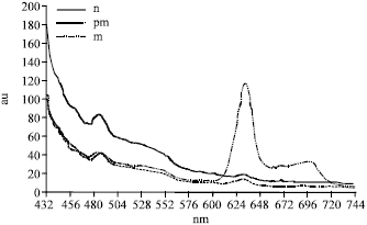 | |
| Fig. 1: | Average emission at 405 nm excitation |
| Table 1: | Results of LSD post hoc test |
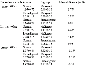 | |
| *Indicates statistically significant with p<0.05 | |
The patient suffering from initial stages of oral cancer usually does not suffer from grave symptoms except for white or red patch with or without ulcerations. The patient presents himself to the dental office usually when the oral cancer is in the advanced stages, thus making it very difficult to treat and have a very poor prognosis. To reduce the mortality rate associated with this disease, it would be prudent to confront this disease at its premalignant or early stages of invasion. Autofluorescence spectroscopy is one such real time optical technique, which is under research for the early diagnosis of cancer. This technique exploits the usage of biomolecules called fluorophores, tissue metabolite like porphyrin which has the property to fluoresce when excited between 400-450 nm of light (Coghlan et al., 2000, 2001). When light of certain wavelength is absorbed by an atom or molecule, an electron is excited to the higher energy level. When these displaced electrons return to the original ground state it may emit a quantum of light, which is known as fluorescence. This technique has a many advantages compared with the non invasive procedures currently available. (1) It can detect subtle changes in the tissue based upon tissue metabolites (porphyrin), (2) both quantitative and qualitative analysis is possible thus avoiding bias and (3) it is simple and very easy to perform. The only disadvantage is that this technique cannot be used to differentiate different pathological types of oral cancers.
In the present in vivo study, the Syrian golden hamsters buccal cheek pouch were used for the induction of tumor by local application of 0.5% solution of 7,12-dimethylbenz(a)anthracene in liquid paraffin. This carcinogenesis animal model is a well established and is used in various chemoprevention experiments due to its advantages. Induction of tumor though various stages of occurrence such as inflammatory reaction, hyperkeratosis, papillary stage seen in human is possible in this animal model. Clinical distinction can be made between premalignant lesion (leukoplakic patch) and malignant oral cancer tumor. The duration of the induction of tumor is relatively less 14 weeks.
Bottiroli et al. (1995), Dhingra et al. (1996) and Vengadesan et al. (1998) worked in this area and have concluded that there is localization of porphyrins in the tumor tissue. But not many in vivo studies are available where autofluorescent property of the oral tissue has been studied comparing the normal, premalignant lesion (leukoplakia) and oral cancer tissue. This has led us to take up the study to compare the fluorescent characteristics of the tissue from normal to malignant transformation. Porphyrin is a metabolic product in the heme biosynthesis. It leads to the synthesis of protoporphyrin IX (PpIX), which acquires an atom form heme. δ-Aminolevulinate synthetase, the enzyme catalyzing the committed step in this pathway, is feedback inhibited by heme. Porphyrin produces a red fluorescence, with peaks at 590, 630 and 690 nm when excited in the blue spectral region between 400 and 450 nm. In this study, the results revealed that, when the cancer tissue was excited at 405 nm there was a prominent peak at 635 nm and another small peak at 700 nm. These two peaks can be attributed to porphyrin fluorescence. As stated above, there is an accumulation of the porphyrin due to the increase in the cellular metabolism, breakdown of RBC`s and increased neovascularization (Onizawa et al., 2002; Dhingra et al., 1996). When compared with the normal control and premalignant group no such peak was found. Vengadesan et al. (1998) and Onizawa et al. (2002) in their in vitro study of malignant animal tissue found prominent porphyrin peak at 630 nm when excited at 405 nm.
The peak around 490 nm which was observed in all the three groups may be attributed to nicotinamide adenine dinucleotide of reduced form (NADH) (Wu et al., 2006). When mean ratio parameters between the NADH and porphyrin peaks were introduced, statistical significance of p-value less than 0.05 was observed between groups and within groups. In the LSD post hoc test, it was evident that all the ratio parameter, I490/635, I490/700 and I635/700 showed significant difference between the normal-malignant groups and premalignant-malignant groups. But the normal-premalignant group showed no difference. Probably some other excitation wavelengths have to be studied to show the difference between the normal-premalignant groups. However, from the results obtained from the present study it is evident that autofluorescence technique shows significant difference between the normal and diseased (malignant) tissue.
CONCLUSION
Autofluorescence spectroscopic analysis of clinically subtle lesions, which occurs long before the apparent lesions, has to be explored. Since this technique is noninvasive, it can help the dental surgeon to detect early, choose the site for biopsy and to mark the boundaries of oral squamous cell carcinoma lesion. Further in vivo studies have to be done in order to exploit the potentiality of this technique for mass screening at the primary clinics.
ACKNOWLEDGMENT
The Authors thank the University Grants Commission, New Delhi, India to support this study under Major Research Project No. F.30-163/2004(SR).
REFERENCES
- Bottiroli, G., A.C. Croce, D. Locatelli, R. Marchesini and E. Pignoli et al., 1995. Natural fluorescence of normal and neoplastic human colon: A comprehensive ex vivo study. Lasers Surg. Med., 16: 48-60.
CrossRefPubMedDirect Link - Coghlan, L., U. Utzinger, R. Drezek, D. Heintzelman, A. Zuluaga, C. Brookner and R. Richards-Kortum, 2000. Optimal fluorescence excitation wavelengths for detection of squamous intra-epitthelial neoplasia:results from an animal model. Opt. Express., 7: 436-445.
Direct Link - Coghlan, L., U. Utzinger, R. Richards-Kortum, C. Brookner, A. Zuluaga, I. Gimenez-Conti and M. Follen-Mitchell, 2001. Fluorescence spectroscopy of epithelial tissue throughtout the dysplasia-carcinoma sequence in an animal model: Spectroscopic changes precede morphologic changes. Lasers Surg. Med., 29: 1-10.
CrossRefPubMed - Dhingra, J.K., D.F. Jr. Perrault, K. McMillan, E.E. Rebeiz and S. Kabani et al., 1996. Early diagnosis of upper aerodigestive tract cancer by autofluorescence. Arch. Otolaryngol. Head Neck Surg., 122: 1181-1186.
PubMedDirect Link - Epstein, J., C. Scully and J. Spinelli, 1992. Toluidine blue and Lugol’s iodine application in the assessment of oral maliganant disease and lesions at risk of malignancy. J. Oral Pathol. Med., 21: 160-163.
CrossRefPubMedDirect Link - Gupte, M.D., V. Ramachandran and R.K. Mutatkar, 2001. Epidemiological profile of India: Historical and contemporary perspectives. J. Biosci., 26: 437-464.
PubMedDirect Link - Martin, C., C.J. Kerawala and M. Reed, 1998. The application of toluidine blue as a diagnostic adjunct in the detection of epithelial dysplasia. Oral Surg. Oral Med. Oral Pathol. Oral Radiol. Endod., 85: 444-446.
CrossRefPubMedDirect Link - Ogden, G.R. and J.G. Cowpe, 1989. Quantitative cytomorphometric analysis as an aid to detection of recurrent oral cancer. Br. J. Oral Maxillofac. Surg., 27: 224-228.
CrossRefPubMedDirect Link - Onizawa, K., N. Okamura, H. Saginoya, H. Yusa, T. Yanagawa and H. Yoshida, 2002. Analysis of fluorescence in oral squamous cell carcinoma. Oral Oncol., 38: 343-348.
CrossRefPubMedDirect Link - Vengadesan, N., P. Aruna and S. Ganesan, 1998. Characterization of native fluorescence from DMBA-treated hamster cheek pouch buccal mucosa for measuring tissue transformation. Br. J. Cancer, 77: 391-395.
PubMed - Wu, Y. and J.Y. Qu, 2006. Autofluorescence spectroscopy of epithelial tissues. J. Biomed. Opt., 11: 054023-054023.
CrossRefPubMedDirect Link