
Mostafa Ghanei
Research Center of Chemical Injuries, Baqiyatallah Medical Sciences University, Mollasadra St, Tehran, Iran
Shiva Alikhani
Research Center of Chemical Injuries, Baqiyatallah Medical Sciences University, Mollasadra St, Tehran, Iran
Iman Adibi
Research Center of Chemical Injuries, Baqiyatallah Medical Sciences University, Mollasadra St, Tehran, Iran
Mehdi Mir Mohammad
Research Center of Chemical Injuries, Baqiyatallah Medical Sciences University, Mollasadra St, Tehran, Iran
Taghi Ramazani
Research Center of Chemical Injuries, Baqiyatallah Medical Sciences University, Mollasadra St, Tehran, Iran
Jafar Aslani
Research Center of Chemical Injuries, Baqiyatallah Medical Sciences University, Mollasadra St, Tehran, Iran
Journal of Medical Sciences
Year: 2008 | Volume: 8 | Issue: 3 | Page No.: 222-227


ABSTRACT
The aim of this study was to determine the occurrence of emphysema and accuracy of Pulmonary Function Test (PFT), comparing with chest High Resolution Computed Tomography (HRCT), in smokers with history of exposure to toxic fumes (Sulfur Mustard; SM). This was a cross sectional study (2003-2004) on 20 symptomatic smokers with mild SM exposure (Group 1) and 20 smokers without SM exposure (Group 2). PFT and chest HRCT were performed for all patients to detect emphysema. Sensitivity, specificity and positive and negative predictive values were calculated for PFT. Spirometry did not diagnose emphysema in group 1 while chest HRCT diagnosed five patients (sensitivity = 0). Group 2 developed emphysema (11 of 20, 55%) more frequently than group 1 (5 of 20, 20%, p<0.05). No alpha-1 antitrypsin deficiency was found in all 70 individuals. We conclude that smokers with an additional risk factor, such as exposure to toxic fumes, develop emphysema at younger ages while they have normal PFT. Chest HRCT should be regarded as a useful tool in the early diagnosis of emphysema in such cases.
PDF Abstract XML References Citation
How to cite this article
Mostafa Ghanei, Shiva Alikhani, Iman Adibi, Mehdi Mir Mohammad, Taghi Ramazani and Jafar Aslani, 2008. Early Onset Emphysema in Smokers with Additional Exposure to Toxic Fumes; Occurrence and Diagnosis. Journal of Medical Sciences, 8: 222-227.
DOI: 10.3923/jms.2008.222.227
URL: https://scialert.net/abstract/?doi=jms.2008.222.227
DOI: 10.3923/jms.2008.222.227
URL: https://scialert.net/abstract/?doi=jms.2008.222.227
INTRODUCTION
Emphysema is morphologically defined as the permanent enlargement of airspace distal to the terminal bronchiole and the destruction of its wall with no obvious fibrosis. Aging and cigarette smoking are its main causes (Snider et al., 1985). Alpha-1 antitrypsin deficiency is a genetic condition that can lead to early onset emphysema (Wewers, 1989). The principal methods used to detect emphysema are Pulmonary Function Tests (PFT) and chest radiological examination (Burrows, 1974; Sanders, 1991; Thurlbeck and Muller, 1994). However, these methods are not sensitive enough to detect earlier morphological and functional abnormalities of the small airways (Kubo et al., 1999). High Resolution Computed Tomography (HRCT) has thus been actively applied for morphological assessment of the extent and severity of emphysematous alterations (Soejima et al., 2000).
Some studies showed that air trapping may not be in relation with PFT result abnormalities (Mastora et al., 2001). PFT may not detect subtle longitudinal changes in acinar structures caused by special factors like aging and smoking (Soejima et al., 2000). When an additional respiratory risk as exposure to toxic fumes exists the condition of symptom occurrence and diagnosing approach may differ. Concerning the high prevalence of such exposure (in cities and industries) would lead us to approach emphysema in patients with more than one risk factor.
As a known respiratory toxin, sulfur mustard (SM) is well known to cause pulmonary damages (Ghanei et al., 2004a). Bronchiectasis and increased thickening of bronchial wall can be detected in chest HRCT images (Ghanei et al., 2004b; Bagheri et al., 2003). Some studies suggest that SM can cause emphysema along with other known complications (Bagheri et al., 2003). But recently, after removal of confounding factors like smoking we showed that emphysema could not correlate with exposure to SM (Ghanei et al., 2004a). Significant air trapping and mosaic pattern were reported as the most frequent radiological findings in both symptomatic as well as asymptomatic patients (Ghanei et al., 2004a; Dompeling et al., 2004; Emad et al., 1995). We aimed to study the occurrence of emphysema and accuracy of Pulmonary Function Test (PFT) in smokers with history of
exposure to toxic fumes (SM). PFT results were compared with chest HRCT findings in symptomatic smokers with/without history of exposure to SM.
MATERIALS AND METHODS
This was a cross sectional study on two different groups of current smokers with a respiratory symptom: Group 1 (20 patients) had a history of SM exposure while Group 2 (20 patients) did not. These groups were studied along with 10 non smoker males with history of SM exposure (Control 1) and 20 without any exposure to SM (Control 2). All subjects were recruited from volunteers consulted the Baqiyatallah University Hospital between 2003 and 2004, with a respiratory complain.
Patients with exposure to SM (group 1 and control 1) were considered as; Exposed group while others without any SM exposure considered as Non-exposed group. Those in exposed group with pre-exposure respiratory disease/s were excluded from the study.
Exposed patients were those who had been in SM contaminated area during Iran-Iraq war. We defined SM contaminated area as regions which were attacked by chemical missiles or bombardment based on the army documentations. Patients with high dose exposure can not usually keep on smoking due to their respiratory problems. Thus our subjects were selected among all those who had registered for annual checkup, due to subclinical (mild) exposure. Subclinical exposure was defined as absence of any acute symptom at the time of exposure (Sartin, 2000). Individuals who were present in contaminated areas without any acute signs and symptoms at the time of exposure entered our study. We excluded those who had even minor symptoms like lacrimation, cough, red eyes, or any other at the time of exposure (Epler, 2001). Accidental exposure to residual SM in the environment after chemical attacks was another documented form of exposure. We excluded patients with following concurrent and potentially confounding conditions: Having dusty jobs, problems with lung involvement such as collagen vascular diseases, immunological disorders, heart diseases, organ transplantation, radiation therapy, chronic thyroidits, recurrent pulmonary infectious diseases, or even use of drugs like phenytoin, bleomycin, methotrexate, or carbamazepin, which are known to have etiologies of drug-induced lung diseases (Epler, 2001). In all 70 subjects chest HRCT and PFT were performed. Alpha-1 antitrypsin serum level was checked for all patients.
Chest high-resolution computed tomography: All patients were imaged on an axial GE Hi-speed Advantage CT scanner (FXI-plus; GE Medical Systems, Milwaukee, WI) at 120 kVp and 200 to 250 mA with 1 mm collimation and 10 mm intervals from proximal trachea to the diaphragm. The scans were obtained in the supine position in deep inspiration and also in deep expiration at the supra aortic arch, aortic arch, carina and 5 to 10 cm below the carina according to the method used by Aquino and colleagues Remy-Jardin et al. (1993a). The films were reviewed using eFilm workstation (eFilm Medical, Toronto, ON, Canada) and the anteroposterior diameter of the trachea and bronchi in each cut was measured by this program with a precision of 1 mm. Air trapping was evaluated in three of the four obtained anatomical levels in each case: Upper lung zone, defined as the level of superior aspect of aortic arch; middle lung zone, defined as the level of carina; and lower lung zone, defined as the level 5 to 10 cm below the carina, according to the method used by Zhang et al. (2004). Air trapping was defined as the presence of a radiolucent region of the lungs on expiratory images (Lee et al., 2000; Austin et al., 1996). The degree of air trapping was assessed by comparing end inspiratory and expiratory images from the same anatomic level and grading each of the three levels on a 5-point scale: 0, no air trapping; 1, 1-25% cross-sectional area affected; 2, 26-50% cross-sectional area affected; 3, 51-75% cross-sectional area affected and 4, 76-100% cross-sectional area affected. A total air-trapping score was obtained by summing the individual grades for the three levels (maximum possible score, 12 for each lung and 24 for both) (Zhang et al., 2004). Emphysematous destruction was identified as areas of low attenuation and hypovascular regions in the lungs (Foster et al., 1986; Gurney et al., 1992).
Previous studies have shown good intra-and interobserver correlations for the subjective estimation of emphysema (Foster et al., 1986; Morrison et al., 1989). Hence all radiographs were interpreted by a chest radiologist who had no knowledge of the clinical, functional or SM exposure history.
Pulmonary function tests: Spirometry was performed according to American Thoracic Society criteria and results were expressed as a percentage of the expected (predicted) normal values (Quanjer et al., 1993). The FVC and FEV1 were measured, under the direction of physicians, using a standard spirometer (Jaeger, Hochberg, Germany). Subjects were seated with a nose clip in place and were asked to perform at least three forced expiratory maneuvers. Both the patients and the technician received visual feedback from a monitor during the test, which was repeated until three technically satisfactory curves with reproducible contour were obtained. All the indices used for the analysis were derived from the same maneuver, which was the one with the largest FVC. COPD diagnosis was based on what GOLD experts had defined; FEV1/FVC ratio <70% (National Institutes of Health, 2003).
Statistical analysis: The data in the text and tables are presented as means±SD. Median was obtained for air trapping grade and t-test was applied to compare means. We used Man Whitney test where data did not show normal distribution. Percentage of PFT and chest HRCT findings and patients symptoms were calculated. Chi square was applied to compare percentages. PFT diagnostic values including: sensitivity, specificity and positive and negative predictive values (PPV, NPV) were calculated. All statistic methods were performed by SPSS (11) while a p-value of less than 0.05 was considered significant.
RESULTS AND DISCUSSION
PFT (FEV1/FVC) was not able to diagnose emphysema in group 1 while chest HRCT had confirmed the diagnosis in five patients (PPV = 0, Table 1). FEV1/FVC was 100% sensitive to COPD in group 2 (Table 1).
In average, group 2 were older than group I (Table 2). As it is shown in Table 2, FEV1/FVC results showed more abnormal values among group 2 (16 of 20, 80%) than in group 1 (3 of 20, 15%, p<0.05). According to chest HRCT results group 2 developed emphysema (11 of 20, 55%) more frequently than group 1 (5 of 20, 20%, p<0.05). All five emphysematous patients had grade 1 of the disease. Two of 11 emphysematous patients in group 2 showed grade 2 of emphysema while remaining (9 of 11) showed grade 1. However the difference of emphysema grade between groups was not significant (0.056).
Group 1 had higher grade of air trapping (median, 2.5) than group 2 (1) while this was not statistically significant (p = 0.3). In general, chemical patients had higher grade of air trapping than non exposed subjects (2.0, 0.5, p<0.05).
| Table 1: | Diagnostic values of FEV1/FVC |
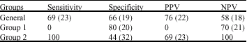 | |
| FEV1: Forced Expiratory Volume in one second; FVC: Forced Vital Capacity PPV: Positive Predictive Value, NPV: Negative Predictive Value, Values are presented as present (95% CI) | |
| Table 2: | Age, spirometry results and air trapping in different groups of the study |
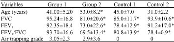 | |
| Values are expressed as mean±SD. FEV1: Forced Expiratory Volume in one second; FVC: Forced Vital Capacity. *p<0.05 compared with group 1 | |
| Table 3: | Prevalence (%) of different respiratory symptoms among groups |
 | |
There were no significant difference in age between exposed group (mean±SD, 42.33±6) and non exposed group (42.23±12.8, p = 0.96). The median of cigarette smoking was 1.5 (pack/years) in group 1 and 17.5 (pack/years) in group 2 (p<0.05). No emphysema was found in non smokers either in control 1 or in control 2.
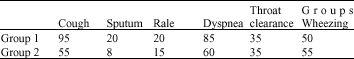
Cough and sputum were more frequent in cigarette smokers with history of SM exposure while other symptoms did not differ between groups (Table 3).
According to chest HRCT findings, different subtypes of emphysema had similar prevalence in group 1 (p = 0.19). Where as, central (7 of 40, 35%) and para-septal emphysema (7 of 40, 35%) were the most common subtypes in group 2 (p<0.05). No alpha1-antitrypsin deficiency was found in all 70 individuals.
PFT (FEV1/FVC) could not discriminate emphysematous patients in smokers with history of SM exposure. However it has a good diagnostic value in non exposed smokers (PPV = 60%, sensitivity = 100%). Smokers with history of SM exposure experienced respiratory symptoms earlier than other smokers. In average they were about 12 years younger than others. Thus other factors, except alpha-1 antitrypsin deficiency, may cause symptomatic emphysema in younger ages than expected. Comparing with non exposed smokers, smokers with SM exposure are suffering from similar symptoms, with lower grades of emphysema in younger ages. As smokers in Exposed group had more risk factors for respiratory problems, they showed more cough and sputum than others even in younger ages along with higher grades of air trapping.
We assume that when symptomatic emphysema occurs in younger ages than expected, a previous exposure to a respiratory toxin (i.e., SM) should be suspected. Concerning that our patients had sub clinical exposures to SM, we suggest that even mild exposure to toxic fumes may accelerate emphysema and related respiratory symptoms. Thus even patients do not remember any acute symptoms of exposure to respiratory toxins; previous exposure to toxic fumes should be taken into account in early onset emphysema. On the other hand in such conditions routine spirometry is not accurate enough and chest HRCT will be the modality of choice to diagnose emphysema.
Previous studies on patients with symptoms of emphysema found a good correlation between chest HRCT and lung function tests (Remy-Jardin et al., 1993a, b). But recent studies reported that chest HRCT revealed emphysematous change in smokers, despite normal PFT (Remy-Jardin et al., 1993b). Sanders et al. (1988) found CT evidences of emphysema in 69% (24/35) of patients who did not have functional findings of emphysema. Similar findings have been recently reported by Lee et al. (2000) who observed that PFT results were within normal ranges in all participants, regardless of the presence of air trapping. FEV1 and FEV1/FVC% could be also close to normal and almost identical among smokers with and without emphysematous lesions (Tylen et al., 2000).
This apparent discrepancy may be due to the possibility of identifying focal abnormalities on CT scans, whereas the spirometric measurements provide a more global measure of lung function (Tylen et al., 2000). Accordingly, cigarette smokers with previous exposure to respiratory toxins are susceptible to develop emphysema at younger ages while spirometry is not fully capable of diagnosing emphysema in them. Thus routine screening tests in patients with known history of exposure to toxic fumes should include chest HRCT beside PFT especially in symptomatic individuals.
Chest HRCT is the modality of choice for diagnosing early emphysema in symptomatic smokers with an additional risk factor of respiratory disease (as SM exposure). Although measurements of residual volume, total lung capacity and inspiratory capacity might be predictive of pulmonary hyperinflation, FEV1/FVC which is usually performed in routine spirometry would not help. It was previously shown that chest HRCT may be more useful in the early diagnosis of smoking-related lung disease (Tylen et al., 2000). It allows detection of emphysema in symptomatic smokers even when pulmonary function appears to be normal and mild emphysema is assumed (Sanders et al., 1988; Srinakarin et al., 2003). Chest HRCT scans provide sensitivity of 100% and a specificity of 91% in comparison with pathology (Quanjer et al., 1993). Thus chest HRCT should be performed for symptomatic current smokers with even mild exposure to respiratory toxins in order to detect emphysema early on.
SM exposure does not cause emphysema. Similar to normal population, chest HRCT of exposed patients showed less areas of emphysema or air trapping in non smokers than smokers (Lee et al., 2000; Remy-Jardin et al., 1993a, b; Ghanei et al., 2006). Primarily, emphysema was reported in association with SM exposure, but concerning confounding effect of smoking, evidences did not support the correlation between emphysema and exposure to SM (Ghanei et al., 2004a, b). No emphysema was found in nonsmokers. That`s why cigarette smoking is the main cause of emphysema in SM exposure as well as general population (Snider et al., 1985; Soejima et al., 2000).
Emphysema increases significantly with age. As age increases, lung undergoes a predictable set of morphologic changes, which include increased alveolar duct air; decreased complexity of the alveolar surface or surface to volume ratio; loss of alveolar wall tissue, elastic tissue and bronchiolar muscle and increased frequency of emphysema (Lee et al., 2000). Considering that group 2 were older than group 1, we can say that chest HRCT was more helpful in group 1 because of earlier stage of disease. Group 2 developed emphysema (11 of 20, 55%) more frequently than group 1 (5 of 20, 20%). Alternatively, results showed a higher grade of emphysema in non exposed smokers than the other group but it was not statistically significant. Emphysema and chronic bronchitis are a significant cost burdens to society, which together accounted for $14.5 billion in direct costs in 1996 in United States. Per patient costs demonstrate the higher disease severity of emphysema patients (Wilson et al., 2000). This study may lead us to diagnose emphysema in younger ages and also early stages, in symptomatic smokers with exposure to hazardous materials so that we can reduce hospitalization and medication burden which account for most of the costs (Wilson et al., 2000). Previous studies showed that knowledge of risk factor (i.e., alpha-1 antitrypsin deficiency) status may lead to positive health changes such as attempts to quit smoking (Wewers, 1989). Thus we should inform smokers with history of exposure to toxic fumes about the risk of emphysema and this may make them quit smoking.
The present study had some limitations. Although we are not aware of any sampling bias, the selection of patients from the outpatient clinic was not performed randomly. The difference in cigarette smoking between groups was due to the fact that exposed patients are not able to smoke as much as others due to their underlying respiratory problems. PFT results in this article were analyzed according to GOLD criteria. We should take into account that the cut off point of 70% for FEV1/FVC may be inappropriate for special populations such as our study samples. Thus inappropriate cut off point may affect the diagnostic value in patients with special risk factors.
This study showed the value of chest HRCT in current smokers with symptoms of emphysema. Even in asymptomatic subjects, significant air trapping is probably pathological and once bronchial asthma has been excluded, it may be related to cigarette smoking and indicates early inflammatory bronchiolar damage (early smokers). Future studies on asymptomatic patients will help us to diagnose emphysema in earlier phases. We should know more about indications of performing chest HRCT in suspected patients prior to developing symptoms or pulmonary dysfunctions.
In summary, smokers with additional risk factor, such as exposure to respiratory toxins, develop emphysema at younger ages while they have normal PFT. Chest HRCT should be regarded as a useful tool in the early diagnosis of emphysema in smokers with history of exposure to toxic fumes. This additional respiratory risk can accelerate the symptoms in early stages. Diagnosis of emphysema may be possible prior to occurrence of the symptoms; but future studies should focus on diagnostic values of routine tests in asymptomatic patients.
REFERENCES
- Austin, J.H., N.L. Muller, P.J. Friedman, D.M. Hansell and D.P. Naidich et al., 1996. Glossary of terms for CT of the lungs: Recommendations of the nomenclature committee of the fleischner society. Radiology, 200: 327-331.
PubMed - Bagheri, M.H., S.K. Hosseini, S.H. Mostafavi and S.A. Alavi, 2003. High-resolution CT in chronic pulmonary changes after mustard gas exposure. Acta Radiol., 44: 241-245.
CrossRefPubMedDirect Link - Dompeling, E., Q. Jobsis, N.M. Vandevijver, G. Wesseling and H. Hendriks, 2004. Chronic bronchiolitis in a 5-year-old child after exposure to sulphur mustard gas. Eur. Respir. J., 23: 343-346.
PubMed - Epler, G.R., 2001. Bronchiolitis obliterans organizing pneumonia. Arch. Intern Med., 161: 158-164.
Direct Link - Foster, J., P.C. Pratt, V.L. Roggli, J.D. Godwin, R.A. Halvorsen and C.E. Putman, 1986. Centrilobular emphysema: CT-pathologic correlation. Radiology, 159: 27-32.
Direct Link - Ghanei, M., H. Fathi, M.M. Mohammad, J. Aslani and F. Nematizadeh, 2004. Long-term respiratory disorders of claimers with subclinical exposure to chemical warfare agents. Inhal. Toxicol., 16: 491-495.
Direct Link - Ghanei, M., M. Mokhtari, M.M. Mohammad and J. Aslani, 2004. Bronchiolitis obliterans following exposure to sulfur mustard: Chest high resolution computed tomography. Eur. J. Radiol., 52: 164-169.
Direct Link - Ghanei, M., F.A. Moqadam, M. Mir-Mohammad and J. Aslani, 2006. Tracheobronchomalacia and air trapping after mustard gas exposure. Am. J. Respir. Crit. Care Med., 173: 304-309.
Direct Link - Gurney, J.W., K.K. Jones and R.A. Robbins et al., 1992. Regional distribution of emphysema: Correlation of high-resolution CT with pulmonary function tests in unselected smokers. Radiology, 183: 457-463.
Direct Link - Kubo, K., S. Eda, H. Yamamoto, K. Fujimoto, Y. Matsuzawa, Y. Maruyama, M. Hasegawa, S. Sone and F. Sakai, 1999. Expiratory and inspiratory chest computed tomography and pulmonary function tests in cigarette smokers. Eur. Respir. J., 13: 252-256.
PubMed - Lee, K.W., S.Y. Chung, I. Yang, Y. Lee, E.Y. Ko and M.J. Park, 2000. Correlation of aging and smoking with air trapping at thin-section CT of the lung in asymptomatic subjects. Radiology, 214: 831-836.
Direct Link - Mastora, I., M. Remy-Jardin, A. Sobaszek, C. Boulenguez, J. Remy and J.L. Edme, 2001. Thin-section CT finding in 250 volunteers: Assessment of the relationship of CT findings with smoking history and pulmonary function test results. Radiology, 218: 695-702.
Direct Link - Morrison, N.J., R.T. Abboud and F. Ramadan et al., 1989. Comparison of single breath carbon monoxide diffusing capacity and pressure±volume curve in detecting emphysema. Am. Rev. Respir. Dis., 139: 1179-1187.
PubMed - Quanjer, P.H., G.J. Tammeling, J.E. Cotes, O.F. Pedersen, R. Peslin and J.C. Yerault, 1993. Lung volumes and forced ventilatory fellows. Official statement of the European respiratory society. Eur. Respir. J., 16: 5-40.
PubMed - Remy-Jardin, M., J. Remy, C. Boulenguez, A. Sobaszek, J.L. Edme and D. Furon, 1993. Morphologic effects cigarette smoking on airways and pulmonary parenchyma in healthy adult volunteers: CT evaluation and correlation with pulmonary function tests. Radiology, 186: 107-115.
Direct Link - Remy-Jardin, M., J. Remy, B. Gosselin, V. Becette and J.L. Edme, 1993. Lung parenchymal changes secondary to cigarette smoking: Pathologic CT correlations. Radiology, 186: 643-651.
PubMed - Sanders, C., P.H. Nath and W.C. Bailey, 1988. Detection of emphysema with computed tomography. Correlation with pulmonary function tests and chest radiography. Invest. Radiol., 23: 262-266.
PubMed - Sanders, C., 1991. The radiographic diagnosis of emphysema. Radiol. Clin. North Am., 29: 1019-1030.
PubMed - Sartin, J.S., 2000. Gulf war illnesses: Cases and controversies. Mayo Clin. Proc., 75: 811-819.
Direct Link - Snider, G.L., J. Kleinerman, W.M. Thurlbeck and Z.H. Bengali, 1985. The definition of emphysema: Report of a national heart, lung and blood institute, division of lung diseases workshop. Am. Rev. Respir. Dis., 132: 182-185.
PubMed - Soejima, K., K. Yamaguchi, E. Kohda, K. Takeshita and Y. Ito et al., 2000. Longitudinal follow-up study of smoking-induced lung density changes by high-resolution computed tomography. Am. J. Respir. Crit. Care Med., 161: 1264-1273.
Direct Link - Srinakarin, J., J. Thammaroj and W. Boonsawat, 2003. Comparison of high-resolution computed tomography with pulmonary function testing in symptomatic smokers. J. Med. Assoc. Thai., 86: 522-528.
Direct Link - Thurlbeck, W.M. and N.L. Muller, 1994. Emphysema: Definition, imaging and quantification. Am. J. Roentgenol., 163: 1017-1025.
Direct Link - Tylen, U., M. Boijsen, A. Ekberg-Jansson, B. Bake and C.G. Lofdahl, 2000. Emphysematous lesions and lung function in healthy smokers 60 years of age. Respir. Med., 94: 38-43.
Direct Link - Wewers, M., 1989. Pathogenesis of emphysema. Assessment of basic science concepts through clinical investigation. Chest, 95: 190-195.
CrossRef - Wilson, L., E.B. Devine and K. So, 2000. Medical costs of chronic obstructive pulmonary disease: Chronic bronchitis and emphysema. Respir. Med., 94: 204-213.
Direct Link - Zhang, J., I. Hasegawa, H. Hatabu, D. Feller-Kopman and P.M. Boiselle, 2004. Frequency and severity of air trapping at dynamic expiratory CT in patients with tracheobronchomalacia. Am. J. Roentgenol., 182: 81-85.
Direct Link