
S.H. Garba
Department of Human Anatomy:
College of Medical Sciences,
P.M.B. 1069,
University of Maiduguri, Nigeria
M.M. Shehu
Department of Human Anatomy:
College of Medical Sciences,
P.M.B. 1069,
University of Maiduguri, Nigeria
A.B. Adelaiye
Department of Human Physiology, Faculw of Medical Sciences,
Ahmadu Bello University, Zaria, Nigeria
Journal of Medical Sciences
Year: 2007 | Volume: 7 | Issue: 1 | Page No.: 94-99


ABSTRACT
The effect of inhaling mosquito coil smoke on the haematology and histology of the rats spleen was studied. A total of 30 albino rats of the Wister strain were used in this study, they were divided into six groups of five rats each. Rats in Group I served as control (no exposure to mosquito coil smoke). While Groups II-VI were exposed to mosquito coil smoke for 12 h, 7, 14, 21 and 28 days, respectively. At the end of each experimental period, blood was collected from each rat for the analysis of Red Blood Cell (RBC) count, White Blood Cell (WBC) count, Haemoglobin (Hb) concentration, Packed Cell Volume (PCV) and the percentages of Neutrophils, Monocytes, Eosinophils, Basophils and Lymphocytes. The rats were then sacrificed and the spleen obtained, was processed for routine histological analysis. Haematological analysis of the blood obtained revealed a significant (p<0.01, 0.05) increase in WBC count in all exposure periods, while analysis of differential leucocyte count revealed a significant (p<0.05) increase in basophil and lymphocyte percentages. Histological analysis of the spleen tissue revealed severe congestion of venous sinusoids, hyperplasia and regression of both the red and white pulps. Results from this study demonstrates that mosquito coil smoke inhalation challenges the immune system in experimental rats, however, the precise mechanisms remain to be clarified in more detailed studies.
PDF Abstract XML References Citation
How to cite this article
S.H. Garba, M.M. Shehu and A.B. Adelaiye, 2007. Toxicological Effects of Inhaled Mosquito Coil Smoke on the Rat Spleen: A Haematological and Histological Study. Journal of Medical Sciences, 7: 94-99.
DOI: 10.3923/jms.2007.94.99
URL: https://scialert.net/abstract/?doi=jms.2007.94.99
DOI: 10.3923/jms.2007.94.99
URL: https://scialert.net/abstract/?doi=jms.2007.94.99
INTRODUCTION
Mosquitoes have long been identified as the main vectors of human and animal diseases like West Nile virus, malaria and dengue. This had made many families to adopt several methods to control mosquito populations around residential areas. The annual worldwide consumption of the four major types of residential insecticides products-aerosols, mosquito coils, liquid vaporisers and vaporising units are in the billions of units (Krieger et al., 2003).
Mosquito coils are the preferred antimosquito products in low income communities because they are cheap and readily available (Mulla et al., 2001) and this coils are burned indoors as a common practice in many households of Asia, South America and African countries including Nigeria.
Mosquito coils consist of an insecticide/repellent, organic fillers capable of burning with smouldering ,binder and additives such as synergists, dyes and fungicides (Krieger et al., 2003). The most common active ingredients in mosquito coils are various pyrethroids that are effective against many genera of mosquitoes including Aedes, Anopheles and Mansonia (Krieger et al., 2003).
Mosquito coils are often used overnight in sleeping quarters where elevated exposure may occur, children and their parents are often exposed to this chemically complex mosquito coil smoke containing small particles (1 μm), metal fumes and vapours (Liu and Sun, 1988) that may reach the alveolar region of the lung (Cheng et al., 1992). Researchers have also found that the gas phase of mosquito coil smoke contain carbonyl compounds (formaldehyde and acetaldehyde) with properties that can produce strong irritating effects on the upper respiratory tract (Chang and Lin, 1998).
Epidemiologic studies have also shown that long term exposure to mosquito coil smoke can induce asthma and persistent wheeze in children (Azizi and Henry, 1991; Fagbule and Ekanem, 1994; Koo and Ho, 1994). Toxicological studies using mosquito coils in rats showed focal deciliation of the tracheal epithelium, metaplasia of epithelial cells and morphologic alteration of the alveolar macrophages (Liu and Sun, 1988; Liu and Wong, 1987).
Despite the fact that inhaling mosquito coil smoke may have potential adverse health effects, large populations in developing countries still use the coils in their daily lives. This study was therefore designed to examine the effect of inhaling mosquito coil smoke on the haematology and histology of the spleen in Wister albino rats.
MATERIALS AND METHODS
Animal and husbandry: This study was carried out in the Departments of Human Anatomy and Human Physiology, University of Maiduguri, Nigeria between March and October, 2005.
A total of thirty (30) adult albino rats of the Wister strain (210 g±20) of both sexes were used. They were purchased from the animal facility centre of the National Veterinary Research Institute Vom, Plateau State, Nigeria. Following an acclimation period of 2 weeks, the rats were individually identified by color tattoo and weighed. The rats were kept in plastic cages at room temperature of 32±4°C and <30% relative humidity with a 12 h light/dark cycle. They had access to drinking water and standard laboratory diet (Sanders SEEPC feed PLC, Jos, Nigeria) ad libitum.
Test article: Mosquito coils were purchased from various retail outlets located within Maiduguri, Nigeria. The brand commercially purchased for the experiment contained pyrethroids (d-trans-allethrin) 0.2%w/w and inert ingredient 99.8% w/w. The mosquito coil used measured 12 cm diameter, 85 cm length and 14.0 g weight
Experimental design: The experiments were conducted in two (A and B) undisturbed rooms of size 26.2 m3 (3.0x3.5x 2.5) with cross ventilation. The rats were randomly divided into six groups of five rats per group. Group I served as control and were not subjected to mosquito coil smoke inhalation ; Group II were used for the 12 h acute mosquito coil smoke inhalation study while Groups III-VI were used for the subchronic mosquito coil smoke inhalation study.
Acute mosquito coil smoke inhalation study: The rats in group II were exposed via whole body inhalation to the commercially available mosquito coil for 12 h (8 pm-8 am). At the end of the experimental period of 12 h blood and spleen were obtained from each rat for haematological and histological analysis, respectively.
Subchronic mosquito coil smoke inhalation study: The rats in group III, IV, V and VI were also exposed to the commercially available mosquito coil via whole body inhalation. The experiment was carried out by igniting one mosquito coil every day for 8 h (10 pm to 6 am) for 7, 14, 21 and 28 days, respectively. At the end of each experimental period, blood and spleen were also obtained from each rat for haematological and histological analysis, respectively.
Blood sample collection: Blood samples were collected from sterilised tails of the rats and haematological analysis was performed according to the standard methods of Dacie and Lewis (1984), i.e., haemoglobin estimation by the cyanmethaemoglobulin method, PCV by the microhaematocrit method and all cell counts were manually executed using the improved Neubauer haemocytometer. Differential counts were made on 200 cells counting in longitudinal strips. All counts were made by the same observer.
Histological analysis: The rats were anaesthetised and the spleen was carefully dissected out, fixed in Bouins fluid, embedded in paraffin and sections cut at 5 μm. Sections were stained with Haematoxylin and Eosin and mounted in Canada balsam. Light microscopic examination of the sections was then carried out.
Statistical analysis: Numerical data obtained from this study were expressed as the mean value±standard error of mean. Differences among means of control and exposed groups were determined using Statistical Package for Social Scientist (SPSS 11.0). A probability level of less than 5% (p<0.05) was considered significant
RESULTS
Effect of mosquito coil smoke inhalation on mean body weight: Mean body weight was slightly increased in the control rats and slightly reduced in all the groups exposed to the mosquito coil smoke though the slight increase and reduction in body weight were not statistically significant (p>0.05) (Table 1).
Effect of mosquito coil smoke inhalation on haematological indices: The effects of mosquito coil smoke inhalation on hematological indices are presented in Table 2.
Red cell counts and packed cell volume were increased in all exposed groups but was only significant (p<0.01) in the group of rats exposed to the mosquito coil smoke for 28 days (group VI) when compared with the control rats. Haemoglobin concentrations showed no significant change (p>0.05) when exposed rats were compared to the non-exposed rats.
White cell counts were significantly (p<0.01, 0.05) increased in all the groups exposed to mosquito coil smoke. Differential leucocyte count analysis showed a decrease in neutrophils percentage which was only statistically significant (p<0.05) in group IV rats that were exposed to mosquito coil smoke for 28 days (Table 2). Basophil counts were significantly (p<0.05) increased in rats of groups III and VI that were exposed for 7 and 14 days, respectively. There was a reduction in lymphocyte counts in all groups, but statistical significance (p<0.05) was only established in group VI rats that were exposed to for mosquito coil smoke 28 days.
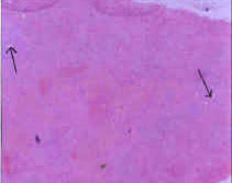 | |
| Fig. 1: | Section in the spleen of a control rat showing normal distribution of white pulp with normal central arterioles (arrows). H and E Stain x 100 |
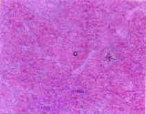 | |
| Fig. 2: | Section in the spleen of a rat exposed to mosquito coil smoke for 12 h showing hyperplasia of red pulp (o) and congestion of venous sinusoids (arrow). H and E Stain x 100 |
| Table 1: | Effect of mosquito coil smoke inhalation on mean body weights of rats |
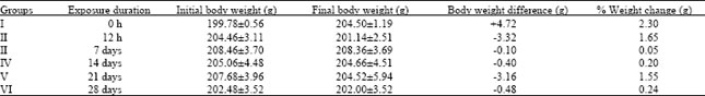 | |
| Results are presented as Means±SEM N = 5 | |
| Table 2: | Effect of mosquito coil smoke inhalation on haematological indices |
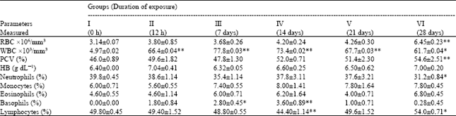 | |
| Significance relative to control (Group I)** p<0.01, * p<0.05, N = 5, Results are presented as Means±SEM. RBC = Red Blood Cell, WBC = White Blood Cell, PCV = Packed Cell Volume and Hb = Hemoglobin | |
 | |
| Fig. 3: | Section in the spleen of a rat exposed to mosquito coil smoke for 7 days showing hyperplasia of white pulp (N) and regressed red pulp. H and E Stain x 100 |
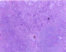 | |
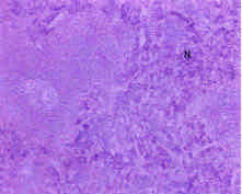
| Fig. 4: | Section in the spleen of a rat exposed to mosquito coil smoke for 14 days showing normal white pulp activity (D) and prominent trabeculae (E). H and E Stain x 100 |
Histopathologic findings: No histological or macroscopic alterations were observed in the splenic tissues of the control rats (Fig. 1).
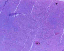 | |
| Fig. 5: | Section in the spleen of a rat exposed to mosquito coil smoke for 21 days showing congestion of the splenic veins (a) with the red pulp mostly confined to the cortical areas (T). H and E Stain x 100 |
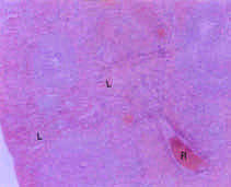 | |
| Fig. 6: | Section in the spleen of a rat exposed to mosquito coil smoke for 28 days showing normal predominance of red pulp (L) and normal distribution of splenic trabeculae (R). H and E Stain x 100 |
Histopathological analysis of rats in group II that were exposed to the mosquito coil smoke for 12 h showed hyperplasia of red pulp, normal white pulp and congestion of venous sinusoids (Fig. 2) while the rats in group III that were exposed to the mosquito coil smoke for 7 days showed hyperplasia of white pulp and regression the red pulp (Fig. 3).
Exposure of the rats in group IV to mosquito coil smoke for 14 days showed further regression of white pulp, prominent splenic trabeculae and a less active germinal centre (Fig. 4). Histological study of the splenic trabeculae of rats in group V that were exposed to mosquito coil smoke for 21 days was characterized by prominent congestion of splenic veins, normal white pulp activity and confinement of the red pulp to the cortical areas (Fig. 5).
The rats in group VI exposed to the mosquito coil smoke for 21 days had a normal dominance and extension of the red pulp into the medullary areas (Fig. 6).
DISCUSSION
The present study which was designed to mimic the local and everyday use of this insecticide in residential areas using rats as models demonstrates the potential health implications of mosquito coil smoke exposure. The body weight loss observed in this study though not significant agrees with other works of the same nature (Parker et al., 1984a; EPA , 1986a; Ishmael and Lithchfield, 1988; Schoenig, 1995). The basophilic leucocytosis observed in this study could be ascribed to an acute inflammatory response to pulmonary irritation; because signs of respiratory irritation have been reported in humans (Lessenger, 1992) and in laboratory animals (Flucke and Thyssen, 1980; Hext, 1987) that were exposed to pyrethrins so also is the immunological and lymphoreteticular effects that have also been reported in a human subject in which hypersensitivity pneumonitis with elevated levels of IgG, IgM and IgE were observed following repeated indoor use of a pyrethrum based insecticide (Carlson and Villaveces, 1977). Subsequent skin tests resulted in skin reactions and allergic pulmonary response to pyrethroids. This also agrees with a similar result from a pyrethroid poisoning in a human subject (He et al., 1989). Eosinophilic Chemotactic Factor (ECF) is released from basophils as well as IL-5, which causes eosinophilic leucocytosis by increasing the differentiation of eosinophilic precursor cells. Basophils express receptors for IgE complexed antigens which are phagocytosed in a non-specific manner by neutrophils and macrophages. Eosinophils modulate the sometimes hazardous effect of basophils and the eosinophilic leucocytosis observed in this study could be attributed to this fact.
Hypersensitivity pneumonitis is associated with shortness of breath and over a period of time could cause a significant decrease of O2 delivery to the tissues. This hypoxic situation could lead to the stimulation of erythropoietin synthesizing cells to express erythropoietin and hence stimulate red blood cell production. This could be responsible for the increased number of red blood cells observed in the course of this study, with a significant (p<0.01) increase observed in group VI animals exposed for the longest period (28 days). Packed Cell Volume (PCV) and Hb concentrations were also observed in this order. Though other authors have reported different results using other pyrethroids based substances; anaemia in rats repeatedly exposed to pyrethrins at mean analytical airborne concentrations ≥ 30 mg m-3 for 90 days (Schoenig, 1995) and significant increase in RBC count, Hb content and hematocrit (Parker et al., 1984b; EPA, 1991a; Shakoori et al., 1992).
Total WBC counts increased significantly (p<0.01) in the 7, 14 and 21 days exposed rats corresponding to the period of marked Eosinophilic and basophilic leucocytosis.
The spleen is to the circulatory system as the lymph nodes are to the lymphatic system. Most anatomic disorders of this organ are secondary to some systemic disorder, being a major secondary organ in the immune system (Aster and Kumar, 1999). The hyperplasia of the splenic red pulp and congestion of venous sinusoids (Fig. 2) suggests an inflammatory response in animals exposed for 12 h. In those exposed for seven days regression of the splenic red pulp and concurrent hyperplasia of the white pulp with germinal centres could be attributed to a stimulation of an immunological response, also observed in Fig. 4. Haematological findings also show in this period significantly increased basophilic leucocytosis. These effects are however, attenuated in the group VI rats suggesting an immunological recovery in the presence of a persisting challenges.
Macrophages, endothelial cells, lymphocytes and natural killer cells express a variety of cytokines, which are able to act in an autocrine fashion as well as affect other immune system (endocrine) components. The observations in this study suggest that mosquito coil inhalation challenges the immune system in experimental rats, however, the precise mechanisms remain to be clarified in more detailed studies.
ACKNOWLEDGMENTS
We wish to acknowledge the technical assistance of Ibrahim Wiam and Justus Jibrin of the Departments of Veterinary Anatomy and Human Pharmacology, University of Maiduguri, Nigeria.
REFERENCES
- Azizi, B.H.O. and R.L. Henry, 1991. The effects of indoor environmental factors on respiratory illness in primary school children in Kuala Lumpur. Int. J. Epidemiol., 20: 144-150.
Direct Link - Chang, J.Y. and J.M. Lin, 1998. Aliphatic aldehydes and allethrin in mosquito-coil smoke. Chemosphere, 36: 617-624.
Direct Link - Cheng, V., H.R. Lee and C.S. Chen, 1992. Morphological changes in the respiratory system of mice after inhalation of mosquito-coil smoke. Toxicol. Lett., 62: 163-177.
Direct Link - Fagbule, D. and E.E. Ekanem, 1994. Some environmental risk factors for childhood asthma: A case-control study. Ann. Trop. Paediatr., 14: 15-19.
Direct Link - Ishmael, J.M. and M.H. Lithfield, 1988. Chronic toxicity and carcinogenic evaluation of permethrin in rats and mice. Fundam. Applied Toxicol., 11: 308-322.
PubMed - Koo, L.C.L. and J.H.C. Ho, 1994. Mosquito coil smoke and respiratory health among Hong Kong Chinese epidemiological studies. Indoor Environ., 3: 304-310.
Direct Link - Krieger, R.L., T.M. Dinoff and X. Zhang, 2003. Octachlorodipropyl ether (S-2) mosquito coils are inadequately studied for residential use in Asia and illegal in the United States. Environ. Health Perspect., 111: 1439-1442.
Direct Link - Lessenger, J.E., 1992. Five office workers inadvertently exposed to cypermethrin. J. Toxicol. Environ. Health, 35: 261-267.
Direct Link - Mulla, M.S., U. Thavara, A. Tawatsin, W. Kong-Ngamsuk and J. Champoosri, 2001. Mosquito burden and impact on the poor; measures and costs for personal protection in some, communities in Thailand. J. Am. Mosquito Control Assoc., 17: 153-159.
Direct Link - Shakoori, A.R., F. Aslam and M. Sabir, 1992. Effect of prolonged administration of insecticide (cyhalothrin/karate) on the blood and liver of rabbits. Folia. Biol., 40: 91-99.
Direct Link