
Panagiotis Kafas
Department of Oral Surgery, School of Dentistry, Aristotle University of Thessalonika, Greece
Nikomacheia Chiotaki
Private Practice, School of Dentistry, Aristotle University of Thessalonika, Greece
George Kafas
University Hospitals of Morecambe Bay, NHS Trust, United Kingdom
Journal of Medical Sciences
Year: 2007 | Volume: 7 | Issue: 1 | Page No.: 158-160


ABSTRACT
Internal derangement is the medical condition, which may affect the joint and indirectly the masticatory muscles. A newly designed dental and physiotherapeutic approach of the head and neck area is presented and assessed with pain relieving results in a patient suffered by internal derangement of temporomandibular joint (TMJ) with reduction. The combined use of soft mandibular splint, phonophoresis of glucosaminoglycan at 1MHz and massage used to relief mandibular function and relieve acute disability. The pre-treatment inter-incisal distance measured at 25 mm, while the post-treatment mouth opening measured at 38 mm. In the follow up, three months later the patient was pain free and discharged. The current treatment plan was not reported before in the literature and composed of non- surgical methods. The physiotherapy may be helpful in symptomatic relief of internal derangement and muscular tension of the craniofacial system and considered to be a non-invasive, topical and physically supportive to the first line dental treatment modalities.
PDF Abstract XML References Citation
How to cite this article
Panagiotis Kafas, Nikomacheia Chiotaki and George Kafas, 2007. Glucosaminoglycan Phonophoresis of the TMJ in the Symptomatic Treatment of Internal Derangement. Journal of Medical Sciences, 7: 158-160.
DOI: 10.3923/jms.2007.158.160
URL: https://scialert.net/abstract/?doi=jms.2007.158.160
DOI: 10.3923/jms.2007.158.160
URL: https://scialert.net/abstract/?doi=jms.2007.158.160
INTRODUCTION
Internal derangement of temporomandibular joint (TMJ) with reduction is a clinical condition specified by the presence of temporomandibular joint(s) pain which may be associated with degenerative adaptive or osseocartilaginous processes and pathological involvement of the associated muscles (Defabianis, 2003). Informed reassurance should be the initial management step. This technique has been found to be effective in about 80% of the cases when used in conjunction with simple analgesics, physiotherapy and occlusal splints (Mitchell and Mitchell, 1999). Various thermal techniques, such as ultrasounds may be used to relieve musculoskeletal pain (Veitiene and Tamulaitiene, 2005).
We report a case of internal derangement with reduction, managed with physical treatment techniques. The combined dental and physiotherapeutic approach described on this article was found effective in the symptomatic treatment. As far as we know, this article is the second in the literature using phonophoresis for the management of TMJ’s internal derangement.
CASE REPORT
A forty years old female was referred to the clinic because of bilateral pain in the stomatognathic system lasting for two months. Additionally, the patient complaint for diminished mouth opening and clicking of the joints. Using the ten centimeters Visual Analogue Scale (VAS) the patient marked the intensity of pain as eight. Inspection of the facial area considered being normal due to absence of swellings and other signs of inflammation. Head examination was diagnostic due to tenderness on palpation of the bilateral masseter, lateral pterygoids muscles and both TMJ areas. The sensation on the TMJ’s was characterized as stabbing feeling.
On neck examination, muscular trigger points were found to refer pain to face. In more details, palpation of right and left sternocleidomastoid muscles referred pain to the right and left preauricular areas, respectively. Auscultation of the TMJ joints using a pediatric stethoscope was useful for evaluation of the bilateral TMJ clicking during mouth opening.
Intra orally, mucosa features inspected normal. Palpation of the lateral pterygoids directly in the pterygoid plates revealed the pathological involvement of these muscles. Medial pterygoids were normal functioning due to absence of pathological signs. The maximum inter-incisal distance measured at 25 mm. The diagnosis of internal derangement with reduction was established by the history and clinical examination. The dental panoramic tomography did not reveal any pathology.
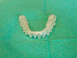 | |
| Fig. 1: | Semi-hard mandibular flat splint |
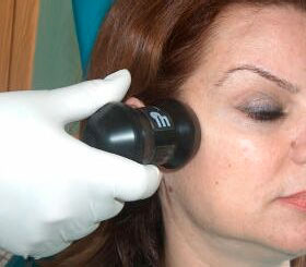 | |
| Fig. 2: | Phonophoresis of TMJ |
The management composed of flat mandibular splint (Fig. 1), ultrasound phonophoresis (Fig. 2) of glucosamine at 1 MHZ, 0.3 Watt cm-2 programmed in continuous output for 10 min of both TMJs and massage of the tender areas including the trigger points’ release for 20 min per session. No analgesics prescribed per os. This treatment plan was found effective in releasing stabbing sensation of the TMJs and trigger points of the sternocleidomastoid muscles in two weeks (VAS = 2). The above-mentioned protocol was performed every second day. The mouth opening measured at 37 mm at the end of the second week. At the follow up, three months later the patient reported lack of signs and symptoms of the TMJ condition.
DISCUSSION
Internal derangement is a difficult to cure condition that often requires a multi disciplinary therapeutic approach. The role of physiotherapy is not well defined regarding the cranio-facial muscular system disorders. The combined therapeutic approach of this condition using dental and physiotherapeutic methods is under consideration. The diadermic phonophoresis of active substances contained in gel forms is regularly used in various body sites. As far as we know, there is only one article in the literature, a randomized controlled trial, regarding the use of phonophoresis on temporomandibular joint with 1% indomethacin showing symptomatic relief (Shin and Choi, 1997). Furthermore, in the facial region the medical use of phonophoresis was limited in the treatment of facial neuritis using hydrocortisone (Antropova, 1974; Grinshtein and Shevchenko, 1973; Grinshstein et al., 1971).
It was our clinical interest to investigate the non-invasive technique of glucosamine phonophoresis in the TMJ derangement condition. This repairing substrate is inexpensive and non-toxic (Salvatore et al., 2000). Additionally, this technique enables the clinicians to avoid prescribing per os glucosamine limiting the systemic loading. Since the systemic use of glucosamine might be used for the treatment of osteoarthritis (Talent and Gracy, 1996), the question for the topical ultrasound assisted application on temporomandibular joint has been raised.
Although the sounds of the joint were not diminished fully at the end of the course (14 days) the pain free condition considered being normal since the head and neck muscular disability was clinically absent. The patient has been recommended to continue wearing the lower jaw splint for two months period during the night after the main course of treatment.
Our study showed that acute internal derangements with reduction might be initially multi disciplinary approached. The role of physiotherapy in the TMJ conditions is not well established in the field of evidenced based rehabilitation. The use of the glucosamine phonophoresis may be found useful for the symptomatic relief if combined with lower flat splint and massage of the tender muscular system if involved. The need for future clinical research using that protocol is encouraged for the establishment of the therapeutic precision.
REFERENCES
- Defabianis, P., 2003. Post-traumatic TMJ internal derangement: Impact on facial growth (findings in a pediatric age group). J. Clin. Pediatr. Dent., 27: 297-303.
Direct Link - Salvatore, S., R. Heuschkel, S. Tomlin, S.E. Davies and S. Edwards et al., 2000. A pilot study of N-acetyl glucosamine, a nutritional substrate for glycosaminoglycan synthesis, in paediatric chronic inflammatory bowel disease. Aliment Pharmacol. Ther., 14: 1567-1579.
CrossRefPubMedDirect Link - Shin, S.M. and J.K. Choi, 1997. Effect of indomethacin phonophoresis on the relief of temporomandibular joint pain. Cranio, 15: 345-348.
Direct Link - Talent, J.M. and R.W. Gracy, 1996. Pilot study of oral polymeric N-acetyl-D-glucosamine as a potential treatment for patients with osteoarthritis. Clin. Ther., 18: 1184-1190.
Direct Link - Veitiene, D. and M. Tamulaitiene, 2005. Comparison of self-management methods for osteoarthritis and rheumatoid arthritis. J. Rehabil. Med., 37: 58-60.
Direct Link