
Susi Endrini
Department of Biochemistry, School ofMedicine, YARSI University,
Cempaka Putih, 10510 Jakarta, Indonesia
Asmah Rahmat
Department of Nutrition and Health Sciences, Universiti Putra Malaysia, 43400, Serdang, Selangor D.E., Malaysia
Patimah Ismail
Department of Biomedicine, Faculty of Medicine and Health Sciences, Universiti Putra Malaysia, 43400, Serdang, Selangor D.E., Malaysia
Y.H. Taufiq-Yap
Department of Chemistry, Faculty of Science and Environmental Study,
Laboratory of Enzyme and Microbial Technology, Institute of Biosciences,
Universiti Putra Malaysia, 43400, Serdang, Selangor D.E., Malaysia
Journal of Medical Sciences
Year: 2007 | Volume: 7 | Issue: 7 | Page No.: 1098-1102


ABSTRACT
This research was conducted to study and compare the cytotoxic effect of the extracts of two plants, henna (Lawsonia inermis) and kejibeling (Strobilanthes crispus) on several kinds of cancer cell lines. The mechanism of the effect was also studied through the expression of cancer-caused gene, c-myc. This research was done in vitro using several kinds of cancer cell lines such as human colon cancer cell lines (Caco-2), liver cancer cell lines (HepG2), hormone-dependent breast cancer cell lines (MCF-7) and hormone-independent breast cancer cell lines (MDA-MB-231) and Chang Liver cell lines. The cytotoxic effect was measured through MTT assay and the potential cytotoxic value was calculated by determining the toxic concentration which may kill up to 50% of the total cell used (IC50). Meanwhile, the cytotoxic mechanism was studied by determining the effect of adding both extracts to the c-myc gene expression. The methods for determination were RT-PCR and sequencing process. The results showed that chloroform extract from henna can be used against human colon cancer cell lines (Caco-2) and liver cancer cell lines (HepG2) with an IC50-value of 25.1 and 28 μg mL–1, respectively. Kejibeling was also found to be cytotoxic against human liver cancer cell lines (HepG2) and hormone-dependent breast cancer cell lines (MCF-7) with an IC50-value of 0.3 and 24.8 μg mL–1, respectively. However, the extract was not cytotoxic against human colon cancer cell lines (Caco-2). The smallest value of IC50 was seen when the kejibeling extract was compared with henna. The cytotoxic effect of both plant extracts may be mediated by the down-regulation of c-myc expression.
PDF Abstract XML References Citation
How to cite this article
Susi Endrini, Asmah Rahmat, Patimah Ismail and Y.H. Taufiq-Yap, 2007. Comparing of the Cytotoxicity Properties and Mechanism of Lawsonia inermis and Strobilanthes crispus Extract Against Several Cancer Cell Lines. Journal of Medical Sciences, 7: 1098-1102.
DOI: 10.3923/jms.2007.1098.1102
URL: https://scialert.net/abstract/?doi=jms.2007.1098.1102
DOI: 10.3923/jms.2007.1098.1102
URL: https://scialert.net/abstract/?doi=jms.2007.1098.1102
INTRODUCTION
Henna (Lawsonia inermis Linn) is a plant, which grow wild in abandoned areas (Muhammad and Mustafa, 1994) and commonly known as ‘inai’ in Sumatra or ‘Pachar kuku’ in Java. This plant is a worldwide known cosmestic agent used to color hair, skin and nails (Hanna et al., 1998). However, it is not only relevant to cosmetics. Henna was also reported to have tuberculostatic activity (Sharma, 1990). The leaves are also used as a prophylactic against skin diseases. They are used externally in the form of paste or decoction against boils, burns, bruises and skin inflammations. A decoction is used as a gurgle against sore throat (Rout et al., 2001). The roots of this plant are useful in burning sensation, leprosy, strangury and premature greying of hair (Vaidyaratnam, 1995).
The major phytochemical constituents of henna, lawsone, is found to possess significant antiinflammatory, analgesic and antipyretic activities (Ali et al., 1995). Recently, this compound has been reported to have a growth inhibitory effect against human colon carcinoma, HCT-15 cells (Kamei et al., 1998).
Strobilanthes crispus ZII 109 (L) Bremek or Saricocalix crispus ZII 109 (L) Bremek (Acanthaceae) plant is a native to countries from Madagascar to Indonesia (Sunarto, 1977) and was first quoted by Anderson, Thomas who classified the plant under Spermatophyta (Flowering plants and Gymnosperma) (Brummit and Powell, 1992).
A study in Indonesia found that an infusion of the dried leaves of S.crispus has been used as antidiabetic, diuretic, antilytic and laxative agents. A recent study indicated that the water extract of S. crispus contained compounds with very high binding affinity to protein molecules that bind the active part of reverse transcriptase. It inhibits the proliferation of retrovirus; an agent in viral disease such as acquired immune deficiency syndrome (AIDS) and Adult T-cell Leukemia (Kusumoto et al., 1992).
The present study aimed at comparing two different plants, Lawsonia inermis and Strobilanthes crispus with regard to their cytotoxic and mechanism activities in several kinds of human cancer cell lines.
MATERIALS AND METHODS
Cytotoxicity studies
Plant materials and extractions: The research work was done at Department of Nutrition and Health Sciences, Universiti Putra Malaysia from June 2002 until June 2003. The leaves of L. inermis and S. crispus were harvested at the Faculty of Medicine and Health Sciences, UPM, Serdang, Selangor. The herbarium voucher specimen were identified and deposited by Mr. Ahmed Zainuddin from the Department of Botany, Faculty of Science and Technology, Universiti Kebangsaan Malaysia. The voucher number of L. inermis and S. crispus were AZ-6804 and AZ-6803, respectively.
The extraction methods were obtained from Ali et al. (1996) with slight modification.
Culturing of cells: HepG-2, Caco-2, MDA-MB-231, MCF-7 and Chang Liver cell lines were obtained from American Type Culture Collection (ATCC, USA). The medium for HepG-2 and Chang liver were Minimum Essential Medium with Earle’s salt (Gibco, USA). While Caco-2, MDA-MB-231 and MCF-7 were grown by using Dulbecco’s Modified Eagle medium (Gibco, USA). The cells were cultured in their own medium supplemented with 10% of fetal calf serum, 100 IU mL–1 penicillin and 100 μg mL–1 of streptomycin (Gibco, USA) using 25 cm2 flasks (Nunc, Denmark) in a CO2 incubator (Sanyo, Japan) at 37°C.
MTT assay: Cytotoxicity effect was determined by the tetrazolium salt method (MTT method), according to the manufacturer’s instructions (Roche Diagnostic, USA).
Study of the mechanism
Reverse Transcriptase-Polymerase Chain Reaction (RT-PCR): The isolation of mRNA was performed by using the Micro-FastTrack™ 2.0 kit (Invitrogen, USA). The RT-PCR process was carried out by using cDNA Cycle kit (Invitrogen, USA). The polymerase Chain Reactions were performed by 30 cycles amplification for 1 min at 94°C, 2 min at 55°C and for 3 min at 72°C. The PCR products were analyzed by electrophoresis on a 1.5% agarose gel.
The sequences of primers were as follows :
c-myc sense : 5’-CAAGAGGCGAAGACACAACGTCT-3’
c-myc antisense : 5’-AACTGTTCTCGTCGTTTCCGCAA-3’
Sequencing: The sequencing technique was done by using an Automatic Sequencer (USA) and the chromatograms were analysed by using the Chromatos software. The similarities in the sequences of samples were compared with the sequence in the database of genes using the BLAST program.
RESULTS
The chloroform extract of S. crispus has shown to be cytotoxic against Caco-2 (IC50=25.1 μg mL–1) and human liver cancer cell lines, HepG2 (IC50 = 28 μg mL–1) (Fig. 1).
 | |
| Fig. 1: | The effect of S. crispus on different human cell lines. Cells were plated with 1x10⁵ cells per well in 96-well culture plates. After 72 h incubation at 37°C, MTT assay was applied to assess cytotoxic effect. IC50 of 25.1 and 28 μg mL–1 were obtained against Caco-2 and HepG2, respectively |
 | |
| Fig. 2: | The effect of chloroform extract of L. inermis on different human cell lines. The protocol applied was similar to the previous one. IC50 of 0.3 and 24.8 μg mL–1 were obtained against HepG2 and MCF-7, respectively (Susi Endrini et al., 2002) |
On the other hand, the chloroform extract of L. inermis has shown to be very cytotoxic to human liver cancer cell line (HepG2) and also toward hormone-dependent breast cancer cell line (MCF-7) with IC50 value of 0.3 and 24.8 μg mL–1, respectively. No cytotoxic effect was detected in Caco-2 (Fig. 2).
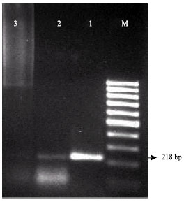
The results showed that the c-myc genes (218 bp) were expressed in untreated HepG2 and Caco-2 cell lines (Fig. 3 and 4).
In contrast, Fig. 3 showed that the c-myc genes were not expressed in HepG2 cells treated with 20 and 30 μg mL–1 L. inermis crude extract. The c-myc gene was also not expressed in HepG2 cell treated with 30 μg mL–1 S. crispus crude extract.
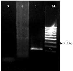 | |
| Fig. 3: | Effect of L. inermis crude extract on the expression of c-myc gene in HepG2 cell line. PCR products were analysed on a 1.5% agarose gel. M, 100 bp DNA ladder marker; lane 1, HepG2 control (untreated); lane 2, HepG2 treated with 20 μg mL–1 L. inermis extract; lane 3, HepG2 treated with 30 μg mL–1 L. inermis extract. C-myc gene at lane 1 was 218 bp in length. The gene expression was not observed in lane 2 and 3 |
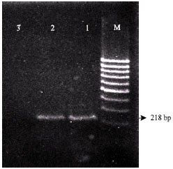 | |
| Fig. 4: | Effect of S. crispus crude extract on the expression of c-myc gene in HepG2 cell line. PCR products were analysed on a 1.5% agarose gel. M, 100 bp DNA ladder marker; lane 1, HepG2 control (untreated); lane 2, HepG2 treated with 20 μg mL–1 S. crispus extract; lane 3, HepG2 treated with 30 μg mL–1 S. crispus extract. C-myc genes at lane 1 and 2 was 218 bp in length. The gene expression was not observed in lane 3. |
However, the c-myc expression was still observed in HepG2 cells treated with a low dose (20 μg mL–1) of this extract (Fig. 4). The similar pattern were also observed in Caco-2 cell treated with this extract (Fig. 5).
 | |
| Fig. 5: | Effect of S. crispus crude extract on the expression of c-myc gene in Caco-2 cell line. PCR products were analysed on a 1.5% agarose gel. M, 100 bp DNA ladder marker; lane 1, Caco-2 control (untreated); lane 2, Caco-2 treated with 20 μg mL–1 S. crispus crude; lane 3, Caco-2 treated with 30 μg mL–1 S. crispus crude. C-myc gene at lane 1 and 2 was 218 bp in length. The gene expression was not observed in lane 3 |
The confirmation of c-myc gene was performed by using sequencing technique and the percent similarity was achieved by using BLAST software from gene bank database. In this study, the 218 bp DNA ladder was confirmed as human c-myc gene with 91% similarity.
DISCUSSION
The choloroform extract of henna displayed the strongest cytotoxic effect on human liver cancer cell line (HepG2) with an IC50- value of 0.3 μg mL–1. On the other hand, the chloroform extract of S. crispus has shown to be cytotoxic against Caco-2 and HepG2 with an IC50- value of 25.1 and 28 μg mL–1, respectively. According to Wall et al. (1987) any plant extracts with an IC50-value below 20 μg mL–1 can be accepted as a potent cytotoxic extract.
To observe the effectiveness of henna and S. crispus crude extracts in suppressing oncogenes, mRNAs were extracted from the treated cells. Due to instability of RNA and for the PCR purpose, mRNA was converted to cDNA before proceeding to the PCR process. The PCR has been selected as the most suitable technique to amplify the quantity of oncogenes, so that the suppression of oncogenes can be visualized clearly after a gel electrophoresis analysis. Besides, different oncogenes have their own temperature for denaturation, annealing, elongation in a number of cycles to get the best PCR products.
Expression of the nuclear proto-oncogenes, c-myc is indicative of early response during cell proliferation and it has been found to be frequently overexpressed in a variety of tissues and cultured cancer cell lines (Saito et al., 1991). Many investigators have found that down-regulation of c-myc expression may be mandatory for the induction of apoptosis in leukemia cells (Alnemri et al., 1992), macrophages cells (Oritani et al., 1992), prostate cancer (Balaji et al., 1997) and lung cancer (Van Waardenburg et al., 1997).
Many plant extracts have been reported to inhibit cell proliferation through the down regulation of c-myc expression (Jiang et al., 2006; Woo and Choi, 2005). In this study, the c-myc expression was suppressed by crude extract from both plants. The effect depended on the doses given and it seemed to be correlated with the IC50- value of each treatment.
CONCLUSION
This present study verifies that L. inermis was most cytotoxic against human liver carcinoma cell lines, HepG-2. On the other hand, S. crispus was shown to be more cytotoxic against colon carcinoma cell lines, Caco-2 than L. inermis. The cytotoxic effect of both plant extracts may be mediated by the down-regulation of c-myc expression.
ACKNOWLEDGMENT
The authors thank IRPA grant 06-02-04-0050.
REFERENCES
- Alia, B.H., A.K. Bashir and M.O.M. Tanira, 1995. Anti-inflammatory, antipyretic and analgesic effect of Lawsonia inermis L. (henna) in rats. Pharmacology, 51: 356-363.
Direct Link - Ali, A.M., M.M. Mackeen, I. Intan-Safinar, M. Hamid, N.H. Lajis, S.H. El-Sharkawy and M. Murakoshi, 1996. Antitumour-promoting and antitumour activities of the crude extract from the leaves of Juniperus chinensis. J. Ethnopharmacol., 53: 165-169.
CrossRefDirect Link - Alnemri, E.S., T.F. Fernandes, S. Haldar, C.M. Croce and G. Litwack, 1992. Involvement of BCL-2 in glucocorticoid-induced apoptosis of human pre-B-leukemias. Cancer Res., 52: 491-495.
PubMedDirect Link - Balaji, K.C., H. Koul, S. Mitra, C. Maramag and P. Reddy et al., 1997. Antiproliferative effects of c-myc antisense oligonucleotide in prostate cancer cells: A novel therapy in prostate cancer. Urology, 50: 1007-1015.
CrossRefDirect Link - Rostkowska, H., M.J. Nowak, L. Lapinski and L. Adamowicz, 1998. Molecular structure and infrared spectra of 2-hydroxy-1,4-naphthoquinone; Experimental matrix isolation and theoretical Hartree-Fock and post Hartree-Fock study. Spectrochimica Acta Part A: Mol. Biomol. Spectrosc., 54: 1091-1103.
CrossRefDirect Link - Jiang, J.J., V. Slivova and D. Sliva, 2006. Ganoderma lucidum inhibits proliferation of human breast cancer cells by down-regulation of estrogen receptor and NF-kB signaling. Int. J. Oncol., 29: 695-703.
Direct Link - Kamei, H., T. Koide, T. Kojima, Y. Hashimoto and M. Hasegawa, 1998. Inhibition of cell growth in culture by quinones. Cancer Biother. Radiopharm., 13: 185-188.
CrossRefPubMedDirect Link - Kusumoto, I.T., I. Shimada, N. Kakiuchi, M. Hattori, T. Namba and S. Supriyatna, 1992. Inhibitory effects of Indonesian plant extracts on reverse transcriptase of an RNA tumour virus (I). Phytother. Res., 6: 241-244.
CrossRefDirect Link - Oritani, K., T. Kaisho, K. Nakajima and T. Hirano, 1992. Retinoic acid inhibits interleukin-6-induced macrophage differentiation and apoptosis in a murine hematopoietic cell line, Y6. Blood, 80: 2298-2305.
PubMedDirect Link - Rout, G.R., G. Das, S. Samantaray and P. Das, 2001. In vitro micropropagation of Lawsonia inermis (Lythraceae). Rev. Biol. Trop., 49: 957-963.
PubMedDirect Link - Saito, H., T. Morizane, T. Watanabe, T. Kagawa, S. Miyaguchi, Kumagai and M. Tsuchiya, 1991. Differentiating effect of sodium butyrate on human hepatoma cell lines PLC/PRF/5, HCC-M and HCC-T. Int. J. Cancer, 48: 291-296.
CrossRefPubMedDirect Link - Endrini, S., A. Rahmat, P. Ismail and T.Y. Yun Hin, 2002. Anticarcinogenic properties and antioxidant activity of henna (Lawsonia inermis). J. Med. Sci., 2: 194-197.
CrossRefDirect Link - Woo, H.J. and Y.H. Choi, 2005. Growth inhibition of A549 human lung carcinoma cells by B-lapachone through induction of apoptosis and inhibition of telomerase activity. Int. J. Oncol., 26: 1017-1023.
Direct Link