
Medhat Haroun
Department ofBiotechnology
Institute of Graduate Studies and Research
Alexandria University Alexandria, Egypt
Journal of Medical Sciences
Year: 2004 | Volume: 4 | Issue: 4 | Page No.: 294-299


ABSTRACT
The aim of this study was to investigate the presence of anti-immunoglobulin antibodies in patients with rheumatoid arthritis, using as a target goat immunoglobulins, in an attempt to elucidate further complex immuno-pathogenetic interactions of the disease. Serum immunoglobulin A concentrations were measured by ELISA technique in twenty patients with rheumatoid arthritis and twenty matched normal adult individuals. The effect of anti-goat immunoglobulin antibodies leads to an overestimation of serum IgA level in patients; this increase is different for each sample estimated by ELISA. Visualization of IgA by immunoblotting technique confirms that goat immunoglobulins were co-precipitated with the antibody-antigen complex. Initial results before purification from the interfering anti-goat immunoglobulin antibodies suggested that patients had increment levels of IgA in their sera. It was found that normal individuals had a mean IgA level of 2.77 mg mL-1 and patients had a mean IgA level of 5.13 mg mL-1 (p<0.00004). While, the mean level of IgA in patients after purification from anti-goat immunoglobulins was 2.92 mg mL-1. Therefore, there was no significant difference in IgA level in patients purified from anti-goat immunoglobulin antibodies compared to normal individuals (p<0.33). A circulating immunoglobulin reacting with other immunoglobulins is thus present in patients with rheumatoid arthritis and may well play a part in the complex immuno pathogenetic interactions.
PDF Abstract XML References
How to cite this article
Medhat Haroun, 2004. Avoiding Antibodies Interferences on Serum IgA Detection in Rheumatoid Arthritis. Journal of Medical Sciences, 4: 294-299.
DOI: 10.3923/jms.2004.294.299
URL: https://scialert.net/abstract/?doi=jms.2004.294.299
DOI: 10.3923/jms.2004.294.299
URL: https://scialert.net/abstract/?doi=jms.2004.294.299
INTRODUCTION
Autoimmune diseases affect between 3 and 5% of the population, often causing chronic illnesses. Autoimmune diseases are mediated by the immune response to self-antigens and characterized by the site of tissue destruction[1]. Rheumatoid Arthritis (RA) is an autoimmune disease with unknown etiology. Polyclonal B cell activation is one of immunological abnormalities commonly found in RA patients[2,3]. B-lymphocytes play a major pathogenetic role by the generation of autoantibodies, such as Rheumatoid Factors (RF) and antinuclear antibodies[4]. An increase in circulating IgA concentrations is a generalized phenomenon among RA patients and RA complications are associated with a significant increase in serum IgA concentration[5]. RA patients with serum IgA level beyond the normal range was more likely to be positive for the autoantibodies[6].
A number of humoral immune factors are present in the circulation in the early stages of RA patients and are thought to play an important part in the complex immunopathological interactions occurring during the establishment of the severe course of RA with systemic damages[7]. Immunoglobulins, which bind other immunoglobulins, add another facet to the abnormal immune response of RA.
The present study was therefore designed to investigate the presence of these anti-immunoglobulin antibodies, using as a target goat immunoglobulins, in RA patients. Immunoglobulins, which bind goat immunoglobulins, have been shown to be present in RA patients studied when compared with matched normal control group. This binding was found in human serum IgA, which obscure the analysis of specific one type of immunoglobulin found in human RA sera. The human antibodies, which reacted in this way, were termed anti-ruminant antibodies[8].
Previous studies indicated that ELISA was more sensitive and specific than nephelometry or latex agglutination for the detection of IgM RF and in particular to autoantibodies that may have a high diagnostic value for RA[9,10]. In support of this report, ELISA technique was used as a sensitive method for detecting antibodies. Antibodies specific for bovidae proteins {sheep, goat and cow} can also produce double precipitation circles during quantification of any immunoglobulin by radial immunodiffusion assay[11,12]. This problem can be avoided by using antiserum prepared in other animals, such as rabbit[9]. Despite this complexity, data in this study provides more reliable results than have been obtained without purification from anti-goat immunoglobulin antibodies.
MATERIALS AND METHODS
Anti-human IgA antiserum (raised in rabbit), human IgA, rabbit anti-human IgA conjugated to horseradish peroxidase (HRP), donkey anti-rabbit IgA conjugated to HRP, diaminobenzidine and tetramethyl-benzidine were purchased from Sigma (Sigma-Aldrich Company Ltd, UK) and all other chemicals were supplied from BDH (VWR International Ltd, UK).
Subjects: The study population consisted of 20 normal adult male individuals (group 1) as a control group, 20 adult male patients with rheumatoid arthritis with recent onset of disease symptoms (group 2) and the same RA individual serum samples purified from anti-goat immunoglobulins antibodies (group 3) were used for analysis of IgA levels by enzyme-linked immunosorbent assay (ELISA). Both group 1 and 2 were matched for age (range 40-52 years). Patients, had no clinical or laboratory findings indicating infection or other disease. No patients had been treated with immuno-suppressant drugs at the time of IgA determination. All sera were collected within three months and stored in small aliquots at -20°C until tested under code.
Sample preparation for immunoblotting: Rheumatoid arthritis serum samples [4 μL] were mixed with 20 μL of 2x Laemmli sample buffer (0.125 M Tris-HCl pH (6.8), 4% (w/v) SDS, 20% (w/v) glycerol, 10% (v/v) β-mercaptoethanol, 0.005% (w/v) bromophenol blue), water [20 μL], 1 M iodoacetamide [8 μL] and then incubated at 95°C for 3 min. A fraction of this mixture [20 μL] was electrophoresed overnight on a discontinuous 10% polyacrylamide gel containing 0.1% (w/v) SDS at a constant voltage of 45 V at room temperature[13].
Immunoblotting of gel: After electrophoresis, the protein was electroblotted onto a sheet of nitrocellulose [Millipore HAHY 00010] at 500 mA for 1 h[14,15]. The nitrocellulose was blocked by incubation with 5% (w/v) Marvel (dried skimmed milk) in PBS (phosphate buffered saline; 0.25 M NaCl, 0.0268 M KCl, 0.081 M Na2HPO4 and 0.0146 M KH2PO4) for 1 h, washed three times with PBST (PBS containing 0.1% (w/v) Tween 80; 10 min per wash). The filter was then incubated with a 1:200 dilution of rabbit anti-human IgA serum in PBSM (PBS containing 0.1% (w/v) Marvel) for 1 h, followed by washing three times in PBST. The rabbit immunoglobulin was detected by incubation in a 1:1000 dilution of donkey anti-rabbit immunoglobulin conjugated to horseradish peroxidase in PBSM. The peroxidase was visualized by staining with 100 mL of a solution containing 0.5 mg mL-1 of diaminobenzidine in 25 mM phosphate buffer pH 7.4, 0.03% (w/v) CoCl2, 0.03% (w/v) ammonium phosphate to which 5 μL of 100 Vol. H2O2 was added immediately prior to staining[16].
Immunoglobulin A measurement by ELISA: Coating antibody [anti-human IgA antiserum] was diluted 1 in 1000 in 1x coating buffer (0.02 M Tris-HCl, 1.5 M NaCl pH 9.0) and 100 μL was added to each of the wells of a microtitre plate[17,18]. After overnight incubation at 4°C the plate was washed 4 times with PBST20 (0.1% (w/v) [Tween 20 in 1x PBS (phosphate buffered saline; 0.25 M NaCl, 0.0268 M KCl, 0.081 M Na2HPO4 and 0.0146 M KH2PO4)]. Sites unoccupied by antibody were blocked by addition of 5% (w/v) Marvel (dried skimmed milk) in PBS for 1 h at room temperature followed by washing 6 times with PBST20. The human serum samples were initially diluted 1 in 2000 in 1x PBS and 2 fold serial dilutions subsequently performed on the plate. Diluted samples were allowed to bind to the first antibody and the plate was then washed 6 times in PBST20.
Rabbit anti-human IgA conjugated to HRP [second antibody] was diluted 1 in 1000 in 1x PBS, 100 μL was added to each well of the microtitre plate, incubated at room temperature for 1 h and then washed 6 times in PBST20. The amount of bound second antibody was determined by adding 200 μL of the substrate solution [tetramethylbenzidine 6 mg mL-1 in 0.1 M sodium acetate buffer pH 6.0] to each well. After incubation in the dark at room temperature for 20 min, the reaction was stopped by adding 50 μL of 10% (w/v) H2SO4 to each well after that the absorbance was measured at 450 nm. The concentration of IgA (mg mL-1) was calculated from the standard dilution series. A standard curve was constructed by plotting absorbance against concentration for the standard solutions and the concentration of IgA in the samples was determined.
Purification of RA sera from the effect of anti-goat immunoglobulins: Goat immunoglobulins were isolated previously from goat serum by affinity chromatography using the appropriate sepharose-bound antibody. The final purified antibody preparation contains only antigen- specific active antibody plus a small amount of denatured antibody resulting from elution procedure (Tago, 4100 series Ab). Goat immunoglobulins, 200 μL, at a concentration of 10 mg L-1 in PBS, pH 7.2 were mixed with 200 μL of human serum samples from each of 20 RA individuals (diluted 1 in 10) to minimize further cross-reactivity to human sera. The absorption was carried out for 1 h at 37°C, followed overnight at 4°C. The rheumatoid arthritis sera were clarified by centrifugation at 10000xg for 15 min at 4°C before testing[19]. The absorption of RA sera with goat immunoglobulins completely removed the positive reaction of these sera and then the amount of IgA present in each of these samples was determined by ELISA as described above.
Statistical analysis: After tabulating the data, the arithmetic mean for each group was calculated. The variation or variability in each group was represented by the standard deviation (SD). The means of the groups were compared to see if the differences were significant. Student's t-test was used to assess the significance of the difference between groups.
RESULTS
Examination of human rheumatoid arthritis immunoglobulin heavy chains using immunoblotting technique with anti-human IgA reveals differences in band intensities. Results showed (Fig. 1) that the level of IgA in RA sera purified from anti-goat immunoglobulin antibodies was lower than that seen in the same RA sera without pre-treatment. Furthermore, ELISA measurements showed (Fig. 2) that group 2 has a high level of serum IgA compared to group 3. The absorption of RA sera with goat immunoglobulins was carried out to eliminate the positive reaction of these sera; the dramatic increase determined by ELISA was varied between different RA samples.
Results showed (Fig. 3) that group 2 has a higher level of serum IgA compared to group 1 and 3. The quantitative analysis of serum IgA level (mean±SD) found that group 1 had a mean level of IgA (2.77±0.45 mg mL-1) lower than group 2 (5.13±0.71 mg mL-1). These represented significant increases in IgA level in the sera of group 2 compared to group 1 (p<0.00004). On the contrary, the mean level of IgA in the sera of group 3 was (2.92±0.48 mg mL-1) within the normal level.
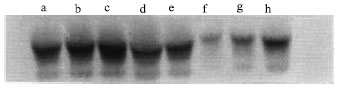 | |
| Fig. 1: | Visualization of IgA by immunoblotting after denaturing polyacrylamide gel electrophoresis. IgA heavy chain was detected by immunoblotting. Lanes a, b, c and d contained purified rheumatoid arthritis serum samples and lanes e, f, g and h contained non-purified same rheumatoid arthritis serum samples |
 | |
| Fig. 2: | Scatter diagram showing the effect of anti-goat immunoglobulin antibodies on ELISA IgA determinations |
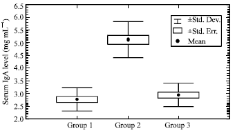 | |
| Fig. 3: | Serum IgA levels in groups of rheumatoid arthritis and unaffected control. Comparison of average serum IgA (mean±S.D) |
Therefore, there was no significant difference in IgA level in the sera of group 3 compared to group 1 (p<0.33).
DISCUSSION
An understanding of the immunologic defects that contribute to the development of autoimmunity will provide an insight into the pathogenesis of the autoimmune process. Many autoimmune disorders have been strongly associated with a high number of immune abnormalities; both humoral and cellular, occur either transiently or permanently in rheumatoid arthritis[4,20,21]. Despite this abundance of reports, most researchers consider that whether the role of immunological factors is primarily pathogenetic, co-causative, secondary or simply chronologically associated to pathogenetic events has not been definitively answered[22,23]. This problem has directed research towards other possible immunological factors likely to be present in RA, in the attempt to elucidate further the complex immuno-pathogenetic interactions of the disease: our finding of anti-immunoglobulin antibody in RA sera, leading to inaccuracies in immunoglobulin estimation by immunoassay, represents a move in this direction.
In order to overcome this problem of interfering due to the effect of anti-immunoglobulin antibodies on immunoassay, the RA human sera were pre-treated with goat immunoglobulins to eliminate cross-reaction or interfering of other irrelevant antibodies found in RA sera. Interestingly, the levels of these antibodies decline after purification, hence this proves that it is not reliable to use the human RA sera directly without previous purification. Thus, this sort of interaction led to over-estimation of immunoglobulin levels in RA sera, which in turn produces unreliable results.
Anti-ruminant antibodies effect noticed by a target goat immunoglobulins showed that the major dramatic effect was accompanied the IgA molecule using ELISA measurement. Therefore, it was necessary to examine with other non-immunochemical assay, such as immunoblotting technique, to find out what sort of complex can be seen due to this interaction. Results suggested that other immunoglobulins interact and co-precipitate with the antibody-antigen complex heavy chains. The fact that different antibodies recognize different epitopes together with the fact that the antibody molecules are dimeric means that anti-goat immunoglobulin antibodies found in RA sera is able to form a cross-linked structure when mixed with antiserum. Hence, an antibody in antiserum binds to antibodies in RA sera is not indicative that the bound antibody is the antigen. These concluded that there was an anti-ruminant antibody interaction arises during the analysis of immunoglobulin A.
It was found that immunoglobulin determinations by radial immunodiffusion are influenced by the chosen reference antigen and by subclass ratios in the samples tested[11,24]. In order to avoid the influence of using various reference antigens or standards as well as the source of antibody in the immunochemical quantitation of immunoglobulin A, standard curves from analyses were compared with different sources of antibodies such as antisera from rabbit, goat and sheep. The results indicate that rabbit antisera were reliable for quantitation of serum IgA in rheumatoid arthritis patients based on ELISA absorbance. Anti-goat immunoglobulins found in RA sera greatly overestimate serum IgA concentration measurements in RA patients, regardless of the source of antibody used. The possibility of interference with the antigen-detection immunoassay for RA patients by anti-sera developed in goat was investigated and concluded that antigen detection ELISA for captures and detection might misdiagnose RA serum immunoglobulin concentration. Results indicate that this bias will be avoided if reagents for capture and detection are derived from different species such as rabbit. This is in agreement with previous reports[12,25,26]. Defects within one component of the immune system may alter the way a pathogen induces an immune response and lead to an inflammatory response directed at self-antigens. Where, the increased IgA levels in the sera of RA patients suggest a possible role for IgA in the pathogenesis of the vascular complications of rheumatoid arthritis.
In conclusion, the presence of these anti-goat immunoglobulins in rheumatoid arthritis may reflect the increase production of autoantibodies and then, lead to humoral immune abnormalities. This is best explained by suggesting that there is an interaction producing spurious immuno-precipitation as well as a circulating immunoglobulin which is capable of binding other autologous immunoglobulins may well interact with other immune factors, thus participating in vivo in the complex immunopathological events which occur in rheumatoid arthritis. Moreover, this study indicates the risk factor of antibodies reacting with human immunoglobulins in sera from RA patients. Such false positive results may obscure the diagnosis of immunoglobulins in rheumatoid arthritis patients. However, the time course for the development of antibodies before onset of clinical RA is unknown, which might be most sensitive or specific for predicting future development of the disease. A finding of an elevated serum level of IgA in a healthy individual implies a high risk for the development of RA. Therefore, IgA testing with appropriately high specificity may assist in the early detection of RA in high-risk populations.
REFERENCES
- Harris, E.D., 1990. Rheumatoid arthritis. Pathophysiology and implications for therapy. N. Engl. J. Med., 322: 1277-1289.
PubMed - Laemmli, U.K., 1970. Cleavage of structural proteins during the assembly of the head of bacteriophage T4. Nature, 227: 680-685.
CrossRefDirect Link - Engvall, E. and P. Perlmann, 1971. Enzyme-Linked Immunosorbent Assay (ELISA) quantitative assay of immunoglobulin G. Immunochemistry, 8: 871-874.
CrossRefPubMedDirect Link