
Amit Kumar
Department of Veterinary Microbiology, UP Pt. Deen Dayal Uphadhyaya Veterinary University and Gau Anusandhan Sanathan (DUVASU), Mathura, 281001 (UP), India
N.C. Srivastava
Division of Bacteriology and Mycology, Indian Veterinary Research Institute (IVRI), Izatnagar (UP), India
V.P. Singh
Division of Bacteriology and Mycology, Indian Veterinary Research Institute (IVRI), Izatnagar (UP), India
Research Journal of Microbiology
Year: 2014 | Volume: 9 | Issue: 1 | Page No.: 59-65


ABSTRACT
Contagious agalactia is an endemic disease in most of the parts of the world with the classical etiology of M. agalactiae which accounts for 90% outbreaks of contagious agalactia syndrome in goats. The disease is responsible for high economic losses due to loss in milk yield and kids/lambs because of abortions, neonatal deaths and loss of animals. It has been reported from various parts of the country including India. However, till date no specific diagnostic is available in India due to cross reactivity of mycoplasma antigens. The conventional methods of diagnosis are cumbersome and lack specificity and sensitivity. Keeping this in mind the present study was undertaken with the objective to conduct the antigenic analysis of Indian isolates of M. agalactiae by separation of proteins by SDS-PAGE and estimating their immunogenicity and diagnostic potential. SDS-PAGE revealed 24 polypeptides in Whole Cell Antigens (WCA) and Sonicated Supernatant Antigens (SSA) of both the isolates, respectively in the range of 20.89-181.97 kDa with the seven major proteins of 63.10, 60.25, 58.88, 47.86, 44.66, 33.88 and 28.84 kDa molecular weights. On immunoblotting with polyclonal rabbit serum raised against M. agalactiae RPNS 216. All the major proteins appeared immunogenic with 12-14 immunogenic polypeptides. Out of them proteins of 47.86, 44.66, 33.88 and 28.84 kDa might be of protective and diagnostic importance for M. agalactiae and could be the source for future diagnostics in India.
PDF Abstract XML References Citation
Received: August 27, 2013;
Accepted: October 29, 2013;
Published: March 01, 2014
How to cite this article
Amit Kumar, N.C. Srivastava and V.P. Singh, 2014. Antigenic Characterization of Mycoplasma agalactiae by SDS-PAGE and
Immunoblotting. Research Journal of Microbiology, 9: 59-65.
URL: https://scialert.net/abstract/?doi=jm.2014.59.65
URL: https://scialert.net/abstract/?doi=jm.2014.59.65
INTRODUCTION
Contagious agalactia is an endemic disease in most of the parts of the world which severely affects the sheep and goats (Lambert, 1987; Sarris, 1996; Kumar et al., 2011, 2012). The disease originally included mastitis, arthritis and kerato conjunctivitis with the classical etiology of M. agalactiae which accounts for 90% outbreaks of contagious agalactia syndrome in goats (Garrido et al., 1987; Kumar et al., 2012) and almost 100% in sheep (Lambert, 1987; Nicholas et al., 2000). The disease is responsible for high economic losses due to loss in milk yield and kids/lambs because of abortions, neonatal deaths and loss of animals (MacOwan et al., 1984; Lagakis, 1996; Kumar et al., 2002). In India, Bawa (1944) first time reported the disease. Later on it has been reported from various parts of the country (Dhanda et al., 1959; Banerjee et al., 1979; Sikdar, 1979; Gupta and Verma, 1984; Kumar and Chandiramani, 1987; Srivastava et al., 1996). As India has large sheep and goat population, contributing significantly in Indian economy (FAO, 2008). Therefore it is important to have better understanding of mechanisms implicated in the pathogenesis as well as knowledge about the nature of the major antigenic substances of M. agalactiae for controlling contagious agalactia and to develop a diagnostic add for the early diagnostic of the infected and carrier animals (Kumar et al., 2010b). It can be developed based on the immunogenic protein of M. agalactiae (Kumar et al., 2010a; Kumar and Singh, 2011). Keeping this in mind the present study was undertaken with the objective to conduct the antigenic analysis of Indian isolates of M. agalactiae by separation of proteins by SDS PAGE and estimating their immunogenicity which can be used, in future, for the development of diagnostic as well as immuno prophylactic agents for the control of contagious agalactia in India.
MATERIALS AND METHODS
Mycoplasma strains: Two strains of M. agalactiae RPNS 216 and RPNS 200 isolated from pneumonic goats were obtained from National Referral Laboratory on Mycoplasma, Division of Bacteriology and Mycology, IVRI.
Culture media: The Modified Beef Horse Serum liquid (MBHS-L) medium was prepared according to the method of Srivastava (1982) for the propagation of M. agalactiae strains. The MBHS-solid medium was prepared by the addition of 1.2% bacto agar (Difco) in MBHS-L medium.
Whole cell antigens (WCA): WCA were prepared as per the method of Solsana et al. (1996) with slight modifications. Actively growing 2-5 mL of M. agalactiae culture was inoculated in 10 mL MBHS-liquid medium and incubated at 37°C for 48 h. The growth was confirmed by the change in pH (change of color red to yellowish orange). This growth was subsequently transferred to larger volume of media and incubated for 4-5 days to obtain sufficient growth. Simultaneously the growth was checked for purity on Robertson cooked meat, Sabouraud’s dextrose agar and Blood agar media. The growth was centrifuged at 10,000 rpm for 25 min using a refrigerated centrifuge (Sorvell, RC-5C). The pellets washed thrice with PBS (pH 7.2) and finally resuspended in 10 mL of PBS. The protein concentrations of WCA were estimated by the method of Lowry et al. (1951).
Sonicated supernatant antigen (SSA): Sonicated antigens of both the isolates were prepared from the whole cell antigen by the method of Bhanuprakash and Srivastava (1996) and the protein concentration of SSA was estimated by the method of Lowry et al. (1951).
Raising of hyperimmune sera: The antiserum against M. agalactiae (RPNS 216) was raised in white New Zealand rabbits (obtained fron LAR, IVRI, Izatnagar), according to the method of Jones (1989). These antisera were tested by slide agglutination test (Srivastava et al., 1986) and titer was estimated by Indirect Haemagglutination (IHA) by the method of Krogsgaard-Jensen (1971) as modified by Srivastava et al. (1992). Hyperimmune sera raised in rabbits were finally filtered through 0.20 μm filter (Sartorius) and then stored at -20°C for further use.
Sodium dodecyl sulphate polyacrylamide gel electrophoresis (SDS-PAGE): The SDS-PAGE of both WCA and SSA under denaturating, reducing conditions was performed by the method of Laemmli (1970) as modified by Solsana et al. (1996). The determination of molecular weights was based on the distance migrated by the polypeptides in the gels in comparison to the distance migrated by polypeptide markers of known molecular weights (Biored) (Neville, 1971).
Immunoblotting: The WCA and SSA were separated on 12.5% (w/v) SDS-PAGE slabs (Laemmli, 1970) and transferred electrophoretically on nitrocellulose membrane papers (NCP) (Sartorius) by the method of Towbin et al. (1979).
RESULTS AND DISCUSSION
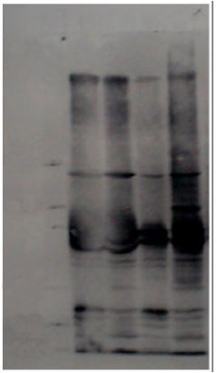
The sonicated supernatant and whole cell antigens of the isolates of M. agalactiae were separated under reducing and denaturating conditions by SDS-PAGE (Fig. 1). SDS-PAGE revealed 24 polypeptides in WCA and SSA of the isolates, respectively in the range of 20.89 to 181.97 kDa. The protein profiles of WCA and SSA of respective isolates were similar and identical. The polypeptide of 120.22 kDa was present only in M. agalactiae RPNS 216 whereas the polypeptide of 62.85 kDa was present only in the profile of M. agalactiae RPNS 200. These are of diagnostic importance and can be used for the differentiation of isolates. The major proteins appeared in the range of 20 to 80 kDa.
 | |
| Fig. 1: | SDS-PAGE profile of M. agalactiae, Lane A: WCA of M. agalactiae RPNS 216, Lane B: WCA of M. agalactiae RPNS 200, Lane C: SSA of M. agalactiae RPNS 216, Lane D: SSA of M. agalactiae RPNS 200 |
 | |
| Fig. 2: | Immunoblot of M. agalactiae, Lane A: WCA of M. agalactiae RPNS 216, Lane B: WCA of M. agalactiae RPNS 200, Lane C: SSA of M. agalactiae RPNS 216, Lane D: SSA of M. agalactiae RPNS 200 |
As per the intensity of proteins the seven major proteins were of 63.10, 60.25, 58.88, 47.86, 44.66, 33.88 and 28.84 kDa molecular weight. The protein profile of the Indian isolate resemble to that of M. agalactiae isolates from other parts of the world (Boothby et al., 1983; Bergonier et al.,1996; Gummelt et al., 1996) with the difference of number of polypeptides and minor differences in major proteins. Similar to present study, Bergonier et al. (1996) observed almost identical protein profile of 52 isolates of M. agalactiae, isolated from different geographical regions with 5-7 major polypeptides in the range of 20-90 kDa. Boothby et al. (1983) and Gummelt et al. (1996) also reported 5-8 major polypeptides and almost similar protein profiles of different isolates with minor differences in whole cell proteins. Moreover the naïve PAGE also confirmed the presence of 5 major proteins in M. agalactiae (Kumar et al., 2010a) which appear to remain intact even after fractionation (Kumar and Singh, 2011). These proteins which persisted in fractions and are persistent in presence can be the antigens of choice for diagnostics and vaccine candidature (Kumar et al., 2011). There are reports of using whole cell of M. agalactiae in vaccine preparation in different animal models (Sunder et al., 2001; Kumar et al., 2009). However, the use of whole organism is a burden upon the host immune system if a suitable protein is capable enough to protect against pathogen. Thus immunoblotting, a more sensitive and more specific confirmatory method to find out the immunogenic proteins when the results of common serological tests are ambiguous (Hung et al., 1991) was applied to provide the information of immunogenic proteins responsible for protective immunity (Solsana et al., 1996; Kumar et al., 2011). The polypeptides bands were electrophoretically transferred on nitrocellulose membranes and blotted with polyclonal rabbit serum produced against M. agalactiae RPNS 216 (Fig. 2). All the major proteins appeared immunogenic with 12-14 immunogenic polypeptides. Both the polypeptides (120.22 and 62.85 kDa) which were present in one isolates (M. agalactiae RPNS 216 and M. agalactiae RPNS 200, respectively) were found non immunogenic. The findings are in resemblance to the findings of Boothby et al. (1983) who also reported almost similar immunogenic protein profile with the major immunogenic proteins in the range of 20-80 kDa. In contrast to the findings of present study (Gummelt et al., 1996) reported high variability in 19 strains of M. agalactiae with M. agalactiae serum however, the range and major proteins coincide to present study. Solsana et al. (1996) recommended 29 kDa proteins as the mean of diagnosis on the basis of immunoblot analysis of 31 strains of M. agalactiae. Similarly the present study also suggests that the major polypeptides may be of protective and diagnostic importance for M. agalactiae. Thus the presence of major immunogenic polypeptides suggested the candidate for diagnostics as well as vaccine development.
CONCLUSION
The presence of major immunogenic proteins particularly the proteins of 28.84 kDa suggested the potential diagnostic as well vaccine candidates for the prevention and control of the disease conditions produced by M. agalactiae. However, further challenge as well as exhaustive studies to assess the selectivity is required.
ACKNOWLEDGMENTS
The authors are highly thankful to the Director, Indian Veterinary Research Institute, Izatnagar for all the necessary facilities.
REFERENCES
- Banerjee, M., N. Singh and P.P. Gupta, 1979. Isolation of mycoplasmas and acholeplasmas from pneumonic lesions in sheep and goats in India. Zentralblatt fur Veterinarmedizin Reihe B, 26: 6689-6695.
CrossRefDirect Link - Bergonier, D., M. Solsana, J. Frey, R. Miserez, J. Nicolet and F. Poumarat, 1996. PCR on16SrRNA Gene, REA and SDS-PAGE Patterns of M. agalactiae Isolates. In: Cost 826: Mycoplasmas of Ruminants: Pathogenicity, Diagnostics, Epidemiology and Molecular Genetics, Frey, J. and K. Sarris (Eds.). European Commission, Brussels, Belgium, ISBN-13: 9789282777633, pp: 91-92.
- Bhanuprakash, V. and N.C. Srivastava, 1996. Partial purification and electrophoretic profile of Mycoplasma arginini antigenic preparations. Indian J. Anim. Sci., 66: 542-544.
Direct Link - Boothby, J.T., D.E. Jasper and M.H. Rollins, 1983. Characterization of antigens from mycoplasmas of animal origin. Am. J. Vet. Res., 44: 433-439.
PubMed - Hung, A.L., A. Alvarado, T. Lopez, R. Pearles, O. Li and E. Garcia, 1991. Detection of antibodies to mycoplasmas in South American camelids. Res. Vet. Sci., 51: 250-253.
CrossRefDirect Link - Krogsgaard-Jensen, A., 1971. Indirect hemagglutination with Mycoplasma antigens: Effects of pH on antigen sensitization of tanned fresh and formalinized sheep erythrocytes. Applied Microbiol., 22: 756-759.
Direct Link - Kumar, A., A.K. Verma and A. Rahal, 2011. Mycoplasma bovis, a multi disease producing pathogen: An overview. Asian J. Anim. Vet. Adv., 6: 537-546.
CrossRefDirect Link - Kumar, A., N.C. Srivastava and V.P. Singh, 2009. Immunogenicity of Mycoplasma agalactiae saponin vaccine in mice. Indian J. Comp. Microbiol. Immunol. Infect. Dis., 30: 61-62.
Direct Link - Kumar, A., N.C. Srivastava and V.P. Singh, 2010. Analysis of Mycoplasma agalactiae and Mycoplasma bovis antigens by Polyacrylamide Gel Electrophoresis (PAGE). Indian J. Small Rumin., 16: 271-273.
Direct Link - Kumar, A., N.C. Srivastava and V.P. Singh, 2010. Pathogenicity of Mycoplasma agalactiae in mice. Indian J. Small Rumin., 16: 269-270.
Direct Link - Kumar, A., A.K. Verma, N.K. Gangwar and A. Rahal, 2012. Isolation, characterization and antibiogram of Mycoplasma bovis in sheep pneumonia. Asian J. Anim. Vet. Adv., 7: 149-157.
CrossRefDirect Link - Lagakis, K., 1996. An Economic Appraisal of the Acute form of Contagious Agalactia in the Transhumant Sheep and Goats in Greece. In: Cost 826: Mycoplasmas of Ruminants: Pathogenicity, Diagnostics, Epidemiology and Molecular Genetics, Frey, J. and K. Sarris (Eds.). European Commission, Brussels, Belgium, ISBN-13: 9789282777633, pp: 100-103.
- Lambert, M., 1987. Contagious agalactia of sheep and goats. Rev. Sci. Tech. Off. Int. Epiz, 6: 699-711.
Direct Link - Laemmli, U.K., 1970. Cleavage of structural proteins during the assembly of the head of bacteriophage T4. Nature, 227: 680-685.
CrossRefDirect Link - Lowry, O.H., N.J. Rosebrough, A.L. Farr and R.J. Randall, 1951. Protein measurement with the folin phenol reagent. J. Biol. Chem., 193: 265-275.
CrossRefPubMedDirect Link - MacOwan, K.J., T.F. Brand, N. McGillveray and A.R. Hunter, 1984. Experimental infection of castrated lambs with Mycoplasma agalactiae. J. Hygiene, 93: 455-463.
CrossRefDirect Link - Neville, D.M., 1971. Molecular weight determination of protein-dodecyl sulfate complexes by gel electrophoresis in a discontinuous buffer system. J. Biol. Chem., 246: 6328-6334.
Direct Link - Solsana, M., M. Lambert and F. Poumarat, 1996. Genomic, protein homogeneity and antigenic variability of Mycoplasma agalactiae. Vet. Microbiol., 50: 45-58.
CrossRefDirect Link - Sunder, J., N.C. Srivastava, V.P. Singh, M. Kumar and A. Kumar, 2001. Humoral immune response in rabbits against killed mycoplasma vaccine. Indian J. Anim. Sci., 71: 231-232.
Direct Link - Towbin, H., T. Staehelin and J. Gordon, 1979. Electrophoretic transfer of proteins from polyacrylamide gels to nitrocellulose sheets: Procedure and some applications. Proc. Natl. Acad. Sci. USA., 76: 4350-4354.
PubMedDirect Link