
I. Zalina
Department of Aquaculture, Faculty of Agriculture, Universiti Putra Malaysia, 43400 Serdang, Selangor, Malaysia
C.R. Saad
Department of Aquaculture, Faculty of Agriculture, Universiti Putra Malaysia, 43400 Serdang, Selangor, Malaysia
A. Christianus
Department of Aquaculture, Faculty of Agriculture, Universiti Putra Malaysia, 43400 Serdang, Selangor, Malaysia
S.A. Harmin
Center for Land and Aquatic Technology (CeLAT), Faculty of Science and Biotechnology, Universiti Industri Selangor (UNISEL), 45600 Bestari Jaya, Selangor, Malaysia
Journal of Fisheries and Aquatic Science
Year: 2012 | Volume: 7 | Issue: 5 | Page No.: 291-306


ABSTRACT
This study described the induced breeding and embryonic stage of Anabas testudineus using commercial hormone LHRHa with the intensity level of 2, 20, 200 μg kg-1 of body weight, respectively. It was found that all intensity of LHRHa hormone level could enhance the fish to breed with the exception of the control group. Fertilization rate, hatching rate, latency period, fecundity, oocytes diameter and GSI were quantified in each set of experiment. It was observed that the fertilised eggs of A. testudineus were almost spherical in shape, clear pearl likes in appearance and free floating on water surface. Fecundity and GSI were significantly higher in fish treated with 200 μg kg-1 as compared to fish treated with 2 and 20 μg kg-1 of body weight of LHRHa hormone. There was no significant (p>0.05) effect between hormone level on fertilization rate, hatching rate and eggs diameter. The diameters of fertilised eggs ranged from 800-850 μm. The first cleavage occurred at 1:30 h, epiboly began at 5 h, the embryonic body was formed at 12 h and hatching occurred at 20 h after fertilization at water temperature of 26°C. Newly hatched larvae were approximately 0.6-1 mm (total length) and pigment spots were present over the yolk and head. The embryonic development of the fish is described. The present study indicated that the administration of LHRHa hormone is effective for ovulation and hormone level at 2 μg kg-1 is recommended.
PDF Abstract XML References Citation
Received: November 04, 2011;
Accepted: February 13, 2012;
Published: March 26, 2012
How to cite this article
I. Zalina, C.R. Saad, A. Christianus and S.A. Harmin, 2012. Induced Breeding and Embryonic Development of Climbing Perch (Anabas testudineus, Bloch). Journal of Fisheries and Aquatic Science, 7: 291-306.
DOI: 10.3923/jfas.2012.291.306
URL: https://scialert.net/abstract/?doi=jfas.2012.291.306
DOI: 10.3923/jfas.2012.291.306
URL: https://scialert.net/abstract/?doi=jfas.2012.291.306
INTRODUCTION
Anabas testudineus (Bloch) is an important fish for farming in freshwater and brackish water. It is also locally known as ‘puyu’ and can be found in most tropical or subtropical Asia including Malaysia, Southern China, Thailand, Vietnam, Filipina, India, Taiwan and Indonesia (Rainboth, 1996; Wang et al., 1999; Kottelat, 2001; Tan and Lim, 2004). It is an omnivorous species and often found in swamps, lakes, canal, rice paddies and streams (Sarkar et al., 2005; Iwata et al., 2003; Rainboth, 1996). According to Yakupitiyage et al. (1998), this species is also described as carnivorous or an insectivore.
A. testudineus contains high iron and copper which is essential for haemoglobin synthesis (Sarma et al., 2010), thus it is one of the important local fish species because it is a healthy diet especially for sick and convalescent people since this fish in addition, it also contains an easily digestible poly-unsaturated fats and many essential amino acids (Kohinoor et al., 1991). A. testudineus also has a potential used in mosquito control in rice fields and temporary pools. The habitat similarity with the mosquito immature and the ability of these fish to tolerate low level of oxygen in aquatic systems favour their augmentative release as a part of a biological control program (Bhattacharjee et al., 2009). At present due to high price and increasing market demand, climbing perch culture has been expanding especially in certain parts of India including North Eastern state (Sarkar et al., 2005). However, larval rearing of climbing perch is still a problem for fish farming because of its low survival.
Various hormone preparations has been applied on A. testudineus, to stimulate maturation and ovulation such as Ovaprim (Bhattacharyya and Homechaudhuri, 2009), Wova-FH (Sarkar et al., 2005), LHRHa (Superfact) combination with a dopamine inhibitor (Motillium) (Morioka et al., 2009) and heteroplastic pituitary gland extract (Moitra et al., 1987) had shown successful attempts. For the present study, LHRHa was used in order to stimulate maturation and ovulation in A. testudineus.
Luteinizing hormone releasing hormone (LHRHa) is a peptide that is similar in structure to native LHRH that has been employed successfully to induce the reproductive hormonal cascade (D-Ala6-des-Gly10 LHRH ethylamide) (Mortezavizadeh et al., 2010). This hormone is produced by a part of the brain called the hypothalamus. It stimulates the pituitary gland to release Lutenising Hormone (LH) and Follicle Stimulating Hormone (FSH) into the bloodstream, via which they act on the ovaries or testes. LHRHa has been successfully used for maturation and spawning of various fish including Atlantic salmon (Crim and Glebe, 1984) and Seabass and rabbit fish (Harvey et al., 1985).
Embryonic development in fishes is dependent on many factors in addition to temperature, pH and dissolved oxygen (Das et al., 2006). The embryonic stages are considered to be very sensitive indicators of environmental disturbances (Puvaneswari et al., 2009). Embryonic development is a complex process in which cellular differentiation and proliferation occur simultaneously though their rate is different (Gould, 1977; Hall, 1922). The embryonic stage occurs inside the chorion and ends at hatching.
There is only scarcely literature of work on early embryonic development stage especially in Malaysia for A. testudineus. Studies done by Jalilah et al. (2011), Singh and Mishra (1980) and Hughes et al. (1986) did not give a sufficient details on growth and morphology development of larvae. However, Morioka et al. (2009) had done a good study on larvae development.
Information on early embryonic, larvae develop and organogeny is of critical important in understanding the basis biology of a particular species and their dietary needs as well as environmental preferences (Koumoundouros et al., 2001; Borcato et al., 2004). Studies on embryonic and early larvae development are imperative and consequential to the successful rearing of larvae for large scale seed production and aquaculture (Khan and Mollah, 1998; Rahman et al., 2004). Furthermore, early life stage information is also necessary for any consideration of evolutionary ecology and phylogeny in the Anabantoidei (Morioka et al., 2009).
The present study was designed to determine and compare the efficiency of LHRHa on breeding and to standardize the doses for aquaculture control condition. In addition, the purpose of this present study also was to observe and record systematically the detailed embryonic development at different stage of eggs until hatched.
MATERIALS AND METHODS
Selected broodstocks of A. testudineus were acquired (28 female and 28 male) from a commercial farmer located in Beranang, Selangor, Malaysia and transported back to Aquaculture Research Centre in Puchong, Selangor, Malaysia. They were stocked and acclimatised in round fiber tanks (300 L) at water temperature of 26-27°C. The fish were fed with pellet (40% protein) at 2.0% of body weight twice a day. The water quality parameter of pH (6.6-7.5) and DO (6-8 mg L-1) were observed in the breeding and broodstocks holding tanks. No mortality was noticed during the acclimatization period. The brooders were gravid and were easily distinguishable between male and female. Adult fish showed sexual dimorphism. Males were darker in colour and the genital papilla rather pointed and narrow with free oozing milt while applying slight pressure on the abdomen. In case of female, the genital papilla was found swollen and slight pink in color, the abdomen was bulging and soft in appearance.
Hormone administration: LHRHa hormone (Syndel Laboratories Ltd., Canada) was dissolved in a solution of saline NaCl (0.7%) and stored in glass vials and frozen at -20°C until use.
Ovulatory response: Fish were examined for Germinal Vesicle (GV) position by applying the eggs to clearing solution (ethanol: formalin: acetic acid, 6:3:1 v/v) for about 5 min prior to observation under microscope. All females fish had central GV were selected and marked by placing it in partition aquarium tanks. About 10 oocytes from the ovaries were cleared of yolk to identify the GV stage. The diameters of oocytes also were measured by taking 10 samples of oocytes from each of the selected females using digital microscope, LEICA DM750.
Experimental design: Two experiments were carried out. In the first experiment, 28 females and 28 males were separated into four groups consisting of seven sexually matured females and males of A. testudineus. For females, their body weight varies from 44.4±4.1 to 56.1±4.4 g and 14.1±0.4 to 14.6±0.2 cm in total length. As for males, their body weight range from 27.85±2.3 to 35.42±3.5 g and 12.38±0.39 to 13.05±0.3 cm in total lengths (Mean±SE). Morphologically, sex identification for A. testudineus can easily be confirmed by biopsy of the gonad. At the point of capture, an initial selection was made for male with pointed genital and sperm and female with swollen genital pore. Three treatment; (T1; 2 μg kg-1, T2; 20 μg kg-1 and T3; 200 μg kg-1) in separate 10 L aquaria were set up. A control group was also maintained whereby the brood fishes been injected with 0.6% saline only. Free oozing male and female were taken in the ratio of 1:1, respectively for breeding. All females and males in all treatment were injected with ‘LHRHa’ intramuscularly. Immediately after administering the hormone, the brooders were released into specially prepared aquaria that have been made a partition to separate the brooders. Approximately after 9-11 h, the fish reached final oocytes maturity stage by checking the female at least every 1 h to determine when the eggs flow freely from the vent. Before stripping, water and slime from the brood vent and tail area were dried with a towel to ensure no water dripping into the eggs as it could activate the sperm and also would cause the opening (micropyle) through which sperm enter to close. The fertilised eggs from each pair of brood stock were mixed together gently and divided into 100 eggs per petri dish and were triplicate for fertilization rate (after 1 h) and hatching rate (after 20 h) and recorded, respectively. Along this, latency period, eggs fecundity was also measured. Effective fecundity was determined by taking a representative sample of eggs (1 g) from the total eggs released by the female. The total number of eggs in 1 g were counted and multiplied with total weight off eggs released. Gonadosomatic index was estimated by calculating:
corrected for stripping. The 1 day hatchlings were maintained in aquaria.
In the second experiment, one male (27.9 g) and one female (47.5 g) fish were obtained and selected based on the external morphological features described by Morioka et al. (2009). Matured male fish were identified by a slightly pointed genital papilla and females by a swollen abdomen and a reddish swollen vent. In addition, the maturity of the female was confirmed by a slightly pressing the ventral side of the fish for oozing of eggs. Both the female and male fish were artificially induced by intra-muscular injection with a commercial hormone (LHRHa) at 2 μg kg-1 b.wt. Hormone injected male and female fish were then released separately into individual aquarium (10 L) containing freshwater. Approximately 11 h after the administration of hormone, the female was checked for its ovulatory response. The release of eggs through the genital pore following gentle pressure on the abdomen was considered as commencement of ovulation. Eggs from ovulated female were stripped into a bowl. Following ovulation, the testes were removed from the male fish and sperm was pressed into a sterile dry Petri dish. Stripped eggs were then fertilized with the diluted sperm suspension. After 2 min of gentle stirring, the fertilized eggs were washed several times with fresh water to remove excess milt. The fertilized eggs were immediately transferred to three glass aquaria (10 L) for incubation. The eggs were then examined under a microscope 10 to 15 min after gamete-mixing to see whether the blastodisc had formed as an indication of successful fertilization. One hour after insemination, the unfertilized eggs turned whitish while the fertilized eggs remained translucent. The unfertilized eggs were removed carefully from the incubation tank.
Observation on embryonic development: Twenty developing eggs were sampled at 30 min to 1 h intervals until hatched. In the present study, the developmental stages were divided into embryonic and newly hatched larvae. The eggs were monitored until hatched (20 h) and the morphological characteristic regarding the development of eggs through to the late yolk sac larval stage was followed (Mousa, 2010). The measurement and observation were carried out and photographs were taken using a LEICA microscope (DM 750). The observed eggs were then preserved in 10% concentration of formalin.
Statistical analysis: Data collected in this study were determined by one way analysis of variance (ANOVA), p<0.05, followed by Duncan’s multiple comparison tests using statistical software package SPSS version 16.0.
RESULTS
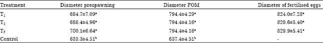
Experiment 1: In this study, out of 28 female selected for induced breeding in the experiment, all 21 from the treated fish responded positively and produced viable eggs. No spawning was observed in the control group. The female for this group did not reach the ovulation stage. Table 1 showed there was a significant different between treatment and control in eggs diameter during prespawning where the control had an eggs diameter of 633.3±4.51 μm. Whereas among the three treatments there was no significant different in eggs diameter of prespawning with T1; 684.7±7.09 μm, T2; 688.4±4.96 μm and T3; 700.1±6.64 μm. Similarly, for the diameter of Final Oocytes Maturation (FOM), there was a significant different (p<0.05) between treatments and control group where GV in control did not show any changes throughout the experiment and only moved from 633.3±4.51 to 637.42±4.51 μm. This showed that the eggs in the control group did not ovulate.
| Table 1: | Oocytes diameter of female Anabas testudineus at different stage of ovulation in treatment (2, 20, 200 μg kg-1) |
 | |
| Different superscript in the same column indicate significant different amongst different stage of oocytes (p<0.05), One-way ANOVA followed by Duncan’s multiple range test). Values are expressed as Mean±SE (n = 10) | |
| Table 2: | Result of captive breeding experiments of Anabas testudineus treated with LHRH hormone |
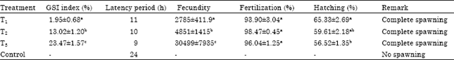 | |
| Different superscript in the same column indicate significant different among different dosage of LHRH hormone (p<0.05); One-way ANOVA followed by Duncan’s multiple range test). Values are expressed as Mean±SE (n = 28) | |
As for the treatment group, there was no significant different (p>0.05) in the diameter of eggs where T1: 794.4±4.29 μm, T2: 794.4±4.16 μm and T3: 794.4±4.16 μm. The diameter of fertilised eggs showed no significant different (p>0.05) among treatments where by the diameter of eggs from T1 are smaller with a value of 824.0±7.28 μm as compared to T2 with a value of 829.6±5.40 μm and T3 with a value of 829.9±5.41 μm. For the control group, there was no spawning occurred and the eggs did not ovulate and fertilised. For the control group eggs were bright golden in colour whereas in the treatment group, the eggs were fertilised and clear pearl like in appearance, epipelagic, spherical in shape and free floating in nature, non adhesive and rise to the water surface.
Table 2 showed that, in a Gonadosomatic index (GSI) parameter, there was significant different (p<0.05) among treatment where fish in T3 had the highest GSI value of 23.47±1.57%, followed by fish in T2 with the value of 13.02±1.20% and the lowest in fish T3 with the value of 1.95±0.68%. All females (n = 7) in T1 had spawned at 11 h after injection. In T2, female spawned after 10 h and for T3 all females spawned at a time of 9 h after injection. For the control group, after 24 h the females still did not show any ovulation. There was a significant different (p<0.05) observed for fecundity. Fish in T3 had the highest fecundity with a value of 30499±7935, followed by fish in T2 with the value of 4851.3±1415 and lastly fish in T1 with the value of 2785.0±411.9. Fertilisation rate among treatments showed no significant different (p>0.05) where fish in T1 had the lowest value of 93.90±3.04%, followed by fish in T3 with the value of 96.04±0.45% and the highest by fish in T2 with the value of 98.47±1.25%. There was a significant different (p<0.05) for hatching rate. The highest hatching rate was found in fish T1 with the value of 65.33±2.69%, followed by fish in T2 with 59.61±2.18% and lastly by fish in T3 with the value of 56.52±1.35%.
Induced breeding: Prespawning female contain vitellogenic oocytes varying in diameter from 600-650 μm. Vitellogenic oocytes had a centrally located Germinal Vesicle (GV) (Mousa, 2010). High rate of final oocytes maturation (100%), ovulation and spawning present from female injected with commercial hormone 2 μg kg-1 b.wt. All injected female spawned at 11 h after injection. The rate of fecundity, fertilization and hatching ranged between 3061-5536, 91 and 62%, respectively.
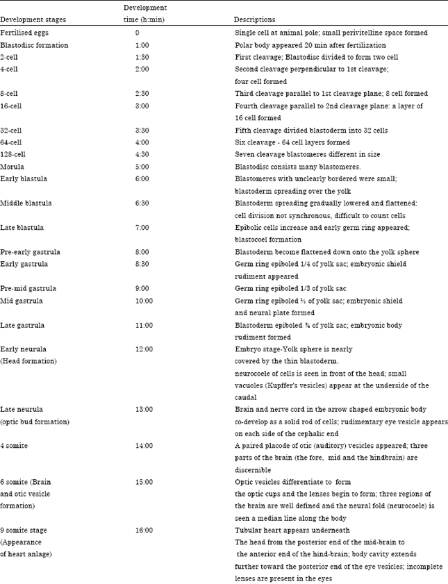
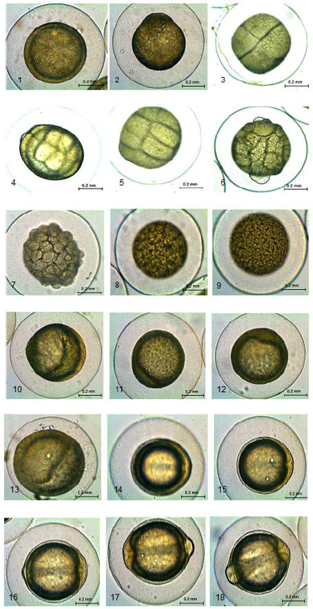
Eggs development: The fertilised eggs have a diameter ranging from 800-850 μm. The eggs were spherical in shape and clear pearl like in appearance. The eggs have narrow perivetelline space (Fig. a1). The embryonic development characteristic stages are shown in Table 3.
| Table 3: | Embryonic Characteristic and duration of development in A. testudineus |
 | |
 | |
| A total 540 eggs were examined | |
Cleavage: First cleavage dividing into two blastomeres occurred 1:30 h after fertilization. The second cleavage, perpendicular to the first, follows within 2 h. In another 2:30 min the eight-cell stage is reached (Fig. a5). The fourth cleavage which is parallel to the second one, is effected 3 h later and the 16 cell stage is obtained. The 32 cell stage follows in the next 3:30 h and is followed by the sixth cleavage in another 4 h. As successive cleavages occur, the blastomeres decrease in size. The eggs continuous further divisions to 128 cell stages at 4:30 h after fertilization (Fig. a9). Morula: Morula stage was observed at 5:00 h after injection (Fig. a10). Blastodisc consists of numerous blastomeres.
Blastula: Blastula describes the period when the blastodisc become multilayered (Mousa, 2010). At this stage, cell division becomes less synchronous and difficult to count cell (Fig. a11-13).
Gastrula: The eggs enter the gastrula stage at 8:00 h after fertilization. Blastoderm began proliferates the surface of the yolk sac. At this stage differentiation of the embryonic tissue began (Fig. a14-18).
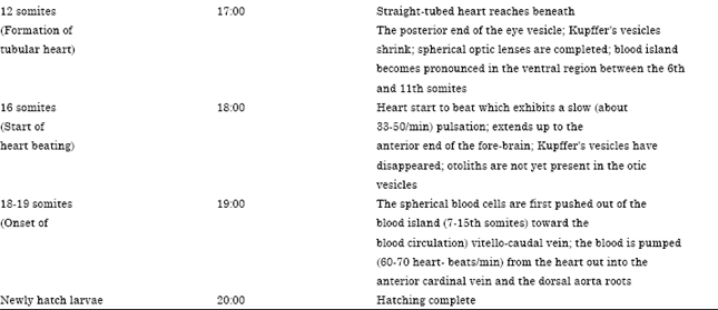
Embryonic body formation: An embryonic body was formed 12:00 h after fertilization (Fig. a19). Here head start to formed where neurocoel cell is seen in front of the head. At 13:00 h after fertilization, rudiment eyes vesicle appears on each of side of the cephalic end (Fig. a20). Kupffer’s vesicles enlarge. Optic vesicle (14:00 h) was observed appeared at the posterior region of the head were 4 somite can be counted. Then the brain and otic vesicles were observed at 15:00 h (Fig. a22) where optic cups and lenses begin to formed. Three divisions of brain forebrain (narrow prosencephalon), midbrain (slightly wider mesencephalon) and hindbrain (short rhombencephalon) are well defined. After 16:00 h of fertilization, formation of heart appeared underneath from posterior end of midbrain to hindbrain and this stage 9 somite can be counted.
 | |
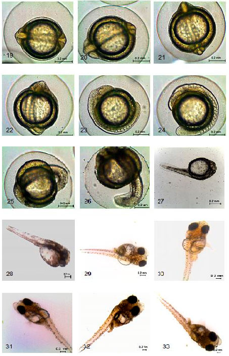 | |
| Fig. a(1-33): | Embryonic development in Anabas testudineus, (1) Fertilised eggs, (2) Blastodisc formation, (3) 2 cell, (4) 4 cell, (5) 8 cell, (6) 16 cell, (7) 32 cell, (8) 64 cell, (9) 128 cell, (10) Morula, (11) Early blastula, (12) Middle blastula, (13) Late blastula, (14) Pre-early gastrula, (15) Early gastrula, (16) Pre-mid gastrula, (17) Mid gastrula, (18) Late gastrula, (19) Early neurula (Head formation), (20) Late neurula (optic bud formation), (21) 4 somite, (22) 6 somite (Brain and otic vesicle formation), (23) 9 somite (Appearance of heart anlage), (24) 12 somite (Heart formation), (25) 16 somite (atart of heart beaf), (26) 18-19 somite (onset of blood circulation), (27) Newly hatched larvae, (28) 1st day larvae; 1.8 mm, (29) 2 day old larvae; 2.8 mm, (30) 3 day old larvae; 3.8 mm, (31) 4 day old larvae; 4.3 mm, (32) 5 day old larvae; 4.8 mm and (33) 6 day old larvae; 5 mm |
The formation of heart start at 17:00 h (Fig. a24) followed by beating at 1 h (Fig. a25) later with slow pulsation (33-50 min-1). Blood start to circulate at 19:00 h after fertilization where bloods were pumped every 60-70 heart beats/min) (Fig. a26).
Hatching (Newly hatch larvae): Twenty hour after fertilization (Fig. a27), the eggs started to hatch at water temperature of 26-27°C. The newly hatched larva has unpigmented eyes, the mouth not formed but anus situated posterior to the yolk mass. Total length of newly hatched larvae was 0.6 -1 mm. There were 22 pairs of somites. The fish was transparent microscopically with some star or branch-shaped melanophores distributed on both side of the body and the back of the head and somites. Star shaped, dark black-brown melanophores were present on both sides of the yolk sac. The digestive duct were thin and undistinguished, as it was attached tightly to the yolk sac. The mouth and anus was not open and the branchial skeleton still undeveloped. The optic vesicles are not unpigmented. The pigmentation extended uniformly from the head along the trunk, except for the final section of the tail. The larvae body was straight, floating on the water surface.
First day old larvae: Total length of first day old larvae (Fig. a28) ranged from 1-1.8 mm. Mouth and anus started to open. Lateral line was visible. Swim bladder and nostril also formed. Majority of larvae still remained motionless in the surface layer of water. The pigmentation extended to the yolk sac both dorsally and ventrally. Typical star-shaped melanophores clusters appear around the final tract of the intestine.
Two day old larvae: Total length of two day old larvae (Fig. a29) ranged from 2.8 mm. Upper and lower jaws were formed. Mouth and anus opened and nostril were also formed. At this point, larvae started to eat rotifer and they were passively carried by water current in the aquarium.
Three day old larvae: Total length of three day old larvae (Fig. a30) ranged from 3.8 mm. Eyes became pigmented. Larvae began settling to bottom of the aquarium. They were still carried by water current. Myomeres were observable.
Four day old larvae: Total length of four-day-old larvae (Fig. a31) ranged from 4.3 mm. Caudal fin ray started to appeared. Several stellate or punctate melanophores were appeared on upper jaws. Larvae had a transparent pectoral fin fold.
Fifth day old larvae: Total length of fifth-day-old larvae (Fig. a32) ranged from 4.8 mm Myomeres began to develop. Caudal fin development proceeded. Dorsal, anal and pectoral fin rays appeared. The mouth was opened and good development of upper jaws and lower jaws, able to take food.
Six day old larvae: Total length of six-day-old larvae (Fig. a33) ranged from 5.0 mm. Dorsal and anal fin ray anlagen appearing. Soft ray caudal also developed. Melanophores appearing in lower jaw and caudal fin. At this point, more larvae were settling to bottom of the aquarium.
DISCUSSION
The climbing perch (A. testudineus) is a highly demanded freshwater fish species and hence has an excellent culture potential. To expand climbing perch culture, knowledge of early larval development and feeding is imperative. But the embryonic and larval development of this fish is poorly understood and only few studies have ever been made (Morioka et al., 2009; Moitra et al., 1987). Therefore the present study was conducted to investigate and also to provide detailed information about the breeding and embryonic development of this important fish species.
Reproduction in fishes can be categories in two types, iteroparous and semelparous (Sivashanthini et al., 2010). Anabas testudineus most reproductive season is considered being during the pre-rainy, the rainy and post-rainy season, March to October. This species also known as extended breeding period, although the peak breeding season is April to August (Morioka et al., 2009). In the present study, LHRHa were used in order to stimulate maturation and ovulation in A. testudineus. The observation indicates that the use of LHRHa hormone at the doses of 2, 20, 200 μg kg-1 b.wt. of female is effective to induce breed A. testudineus similar to that reported by Morioka et al. (2009). It was observed that the hormone dosage affected the percentage of eggs production and gonadosomatic index, respectively. However, the control group (saline injected) did not ovulate in captivity. Similar result was noted by Sarkar et al. (2005) and Azuadi et al. (2011) when A. testudineus and T. tambroides reared in captivity did not ovulate when induced with saline solution. The use of LHRHa for induced breeding A. testudineus is not yet been fully applied and only with a combination of other solution such as Motilium (Morioka et al., 2009). Report on other hormone like Ovaprim (Bhattacharyya and Homechaudhuri, 2009) Wova-FH (Sarkar et al., 2005) and heteroplastic pituitary gland extract (Moitra et al., 1987) has been experimented by several researcher and shown a successfully attempts.
In the present study, different latency period was observed. Shorter latency period at a dose of 200 μg kg-1 of body weight indicates differences in the mode of action of the hormone (Sarkar et al., 2005). Longer latency period on low dose of LHRHa was noticed where similar observation was reported by Pandey et al. (2002) in low dose of synthetic hormone Ovatide. The latency period of Ovaprim induced air breathing fishes were 18 h for Channa punctatus and H. fossilis (Haniffa et al., 2000). Latency period of 11-12 h was recorded with water temperature of 26-27°C in Claria gariepinus (Adebayo and Papoola, 2008). Pandey et al. (2002) reported varied interspawning period between 8 and 15 h in H. fossilis injected in the doses of 0.3-1.0 mL kg-1 of body weight of synthetic hormone Ovatide (M/S Hemmo Pharma, Mumbai, India). According to Billard et al. (1984) and Peter et al. (1986), differences in dose requirement may be attributed to varied level of dopamine activity in different species of fish.
Fecundity is the number of ripening eggs in the female and varied from species depends on age, length, weight and environment (Sivashanthini et al., 2008). Fecundity increases with the increased of length, weight and eggs (Ghafari and Jamili, 2010; Lawson, 2011). Early report showed that, A. testudineus a freshwater fish had high fecundity (Amornsakun et al., 2005). In this study, eggs production of female treated with 2 and 20 μg kg-1 were low (2785±411.9 and 4851±1415 eggs/fish) compared to group treated with 200 μg kg-1, where the production of eggs were 30499±7935 eggs/fish in size range of 56.1±4.4 g, the number of eggs was found to increase linearly with the increase in gonad weight. Zalina et al. (2011) reported high eggs production (5126 eggs/fish) of female treated with 2 μg kg-1 of LHRH hormone of the same fish. This eggs production was higher than Trichogaster pectoralis, Siamese gourami where the fecundity was 26,261 ova/fish (Amornsakun et al., 2004). Sarkar et al. (2005) reported the number of eggs released by the female ranged from 52,000 to 130,000 which indicate high fecundity. The present data of fecundity seems compatible as compared with other reports available in India. Khan and Mukhopadhya, 1972 observed fecundity ranging from 10,002 to 36,477 in fish size range of 99 to 169 mm. However, Banerjee and Prasad (1974) reported the fecundity of 4588-34993 in Bihar region in the fish size range 84-100.2 g. Chanchal et al. (1978) reported a minimum of 3481 to maximum of 42564 in the fish weight range of 9.0-53.1 g. Banerjee and Thakur (1981) reported a number of 2000-13000 eggs in seven sets of induced bred A. testudineus (24.8-40.1 g) in glass aquaria. The size of fertilised eggs of A. testudineus measured in the present study was close to climbing perch (830 μm) when injected with Suprefact and Motilium (Amornsakun et al., 2005). However they were larger (720±0.05 μm) than A. testudineus when injected with Wova-FH (Sarkar et al., 2005). Variation of eggs size might be related to the existence of different strains, conditions and size of the female in the wild conditions (Thakur, 1980). Alternatively, it may also depend on the individual parental investment, moderated by the food availability experienced by the female fish to induce and spawn eggs (Puvaneswari et al., 2009). Hatching rate in the present study showed no significant different between the three groups of treatment with 65.33±2.69, 59.61±2.18 and 56.52±35% (2, 20, 200 μg kg-1). This hatching rate seems low compared to other studies of same fish in captive breeding. Sarkar et al. (2005) reported a high hatching rate of 90.5±3.65%, Bhattacharjee et al. (2009), 68.57 to 73.11% and Morioka et al. (2009), 100% hatching. This result might relate to the lower temperature (26-27°C) during the period of the experiment which temperature is consider one of the main environmental factors (Gadomski and Caddell, 1991) influencing hatching percentage and survival of embryo and larvae (Das et al., 2006).
The embryonic stage occurs inside the chorion and ends at hatching. The egg membrane is fully separated from the egg and has a small perivitelline space which is filled with fluids and this fluid cushion may protect the eggs and the embryo from any external injury (Khan, 1972).
In the second experiment, the first cleavage occurred within 1 h and 16 cells within 3 h of post-fertilization. Klimogianni et al. (2011) reported first meroblastic cleavage occurred 1 h after fertilization. Thakur et al. (1974) observed that the first cleavage, 16 cell and morula stages in H. fossilis were attained within 30, 70-80 and 100 min, respectively after fertilization. Jalilah et al. (2011) reported two blastomeres formed after 30 min of fertilization. In A. testudineus, the invasion of the yolk by the blastoderm was completed at 10 h after spawning (Munshi and Hughes, 1991). In the present study, this invasion was completed after 7 h. Just 1-2 h before hatching, the embryo of A. testudineus showed twisting movements inside the egg envelops. The similar hatching behaviour is reported in the catfish by Thakur et al. (1974) and also commonly observed in other catfish species, C. batrachus (Thakur, 1980) and P. sutchi (Islam, 2005). The observation of early development and pre hatching behaviour of embryo in A. testudineus agrees well with the results obtained by Moitra et al. (1987). In the present study, the incubation period lasted for 20 h at a water temperature of 26°C. Munshi and Hughes (1991) reported the incubation period of 10.5 h of the same fish. Zaki and Abdula (1983) and Herath (1988) reported a shorter incubation period at higher temperatures. The development and incubation periods of embryo in most fishes are fully temperature-dependent and varied from species to species (De Graaf and Janssen, 1996). The observation of early development and pre hatching behaviour of embryo in A. testudineus agrees well with the results obtained by Moitra et al. (1987).
In the present study, the incubation period lasted for 20 h at a water temperature of 28°C. Moitra et al. (1987) reported the incubation period of 16 h at a temperature of 29°C, Munshi and Hughes (1991) reported the incubation period to be around 10.5 h after fertilization of the same fish. Kohli and Vidyarthi (1990) reported the incubation period of 16-18 h in H. fossilis at a temperature of 26°C. Ramanathan et al. (1985) reported the incubation period in Mystus punctatus (Jerdon) to be varied from 18-24 h at a temperature of 28.5±1.8°C. Adebayo et al. (2007) noted that hatching started at 22.0±1.0 at the temperature of 25.50°C in African catfish. Zaki and Abdula (1983) and Herath (1988) reported shorter incubation periods at higher temperatures. The development and incubation periods of embryo in most fishes are fully temperature-dependent and varied from species to species (De Graaf and Janssen, 1996).
In the present observation, the newly hatched larva of this species was 1-1.8 mm in length. According to Parameshwaran and Kamal (1988), the length of newly hatched and yolk absorbed snakehead fish larvae were 3.88-4.47 and 6.8-7.1 mm in C. marulius. These variations can be related to the size of the eggs. According to Bagarinao and Chua (1986), egg diameter is positively correlated with larval length and weight at hatching.
The larvae development of A. testudineus in this studies from day one until day six is not much different with development for this species that been observed by Morioka et al. (2009) where the morphological development is similar.
CONCLUSIONS
This study provided some basic information on the breeding and embryonic development of A. testudineus. Furthermore, the comparative developmental studies of climbing perch species could provide a better understanding on its embryonic and larval stages and also their behaviour. The objective of this study was fulfilled and based on the present experiments, LHRH hormone at 2 μg kg-1 dose is recommended in order to stimulate spawning. In conclusion, it is recommended that the seed of A. testudineus could be produced in captivity through proper management of eggs, larvae and hatchlings.
ACKNOWLEDGMENTS
We express our sincere gratitude to Head of the Department of Aquaculture, staff of Research Aquaculture centre, Puchong and staff of Aquaculture laboratory, University Putra Malaysia for providing infrastructural facilities. The valuable assistance of everyone who contributed to the research is greatly appreciated.
REFERENCES
- Adebayo, O.T., K.A. Ayinde and O.M. Popoola, 2007. Effect of cassava effluent on the hatching and survival of African catfish, Clarias gariepinus larvae. J. Fish. Aquatic Sci., 2: 371-374.
CrossRefDirect Link - Adebayo, O.T. and O.M. Popoola, 2008. Comparative evaluation of efficacy and cost of synthetic and non-synthetic hormones for artificial breeding of African catfish (Clarias gariepinus Burchell, 1822). J. Fish. Aquat. Sci., 3: 66-71.
CrossRefDirect Link - Amornsakun, T., W. Sriwatana and P. Promkaew, 2005. Some aspect in early life of climbing perch, Anabas testudineus larvae. Songklanakarin J. Sci. Technol., 27: 403-418.
Direct Link - Amornsakun, T., W. Sriwatana and P. Promkaew, 2004. Some aspects in early life stage of Siamese gourami, Trichogaster pectoralis (Regan) larvae. Songklanakarin J. Sci. Technol., 26: 347-356.
Direct Link - Bagarinao, T. and T.E. Chua, 1986. Egg size and larval size among teleosts implications to survival potential. Proceedings of the 1st Asian Fisheries Forum, May 26-31, 1986, Manila, Philippines, pp: 651-656.
Direct Link - Banerjee, S.R. and N.K. Thakur, 1981. Observation on the spawning behaviour of Anabas testudineus (Bloch). Indian J. Anim. Sci., 51: 651-654.
Direct Link - Bhattacharyya, M. and S. Homechaudhuri, 2009. Assessment of captive breeding of Anabas testudineus with the synthetic hormone, Ovaprim. Proc. Zool. Soc., 62: 23-27.
CrossRef - Bhattacharjee, I., G. Aditya and G. Chandra, 2009. Laboratory and field assessment of the potential of larvivorous, air-breathing fishes as predators of culicine mosquitoes. Biol. Control, 49: 126-133.
CrossRef - Billard, R., K. Bieniarz, R.E. Peter, M. Sokolowka, C. Weil and L.W. Crim, 1984. Effects of LHRH and LHRH-A on plasma GtH levels and maturation/ovulation in the common carp, Cyprinus carpio, kept under various environmental conditions. Aquaculture, 41: 245-254.
CrossRef - Crim, L.W. and B.D. Glebe, 1984. Advancement and synchrony of ovulation in Atlantic salmon with pelleted LHRH analog. Aquaculture, 43: 47-56.
CrossRef - Das, T., A.K. Pal, S.K. Chakraborty, S.M. Manush, R.S. Dalvi, K. Sarma and S.C. Mukherjee, 2006. Thermal dependence of embryonic development and hatching rate in Labeo rohita (Hamilton, 1822). Aquaculture, 255: 536-541.
CrossRef - Gadomski, D.M. and S.M. Caddell, 1991. Effects of temperature on early life history stages of California halibut, Paralichthys californicus. Fish. Bull., 89: 567-576.
Direct Link - Ghafari, S.M. and S. Jamili, 2010. Certain aspects of the reproductive biology of berzem (Barbus pectoralis) in karoon river. J. Fish. Aquat. Sci., 5: 33-41.
CrossRefDirect Link - Harvey, B., J. Nacario, L.W. Crim, J.V. Juario and C.L. Marte, 1985. Induced spawning of sea bass, Lates calcarifer and rabbitfish, Siganus guttatus, after implantation of pelleted LHRH analogue. Aquaculture, 47: 53-59.
CrossRef - Hughes, G.M., J.S.D. Munshi and J. Ojha, 1986. Post embryonic development of water and air breathing organs of Anabas testudineus (Bloch). J. Fish Biol., 29: 443-450.
CrossRefDirect Link - Islam, A., 2005. Embryonic and larval development of Thai Pangas (Pangasius sutchi Fowler, 1937). Dev. Growth Differ., 47: 1-6.
CrossRef - Klimogianni, A., M. Kalanji, G. Pyrenis, A. Zoulioti and G. Trakos, 2011. Ontogeny of embryonic and yolk-sac larval stage of the sparid sharpsnout sea bream (Diplodus puntazzo Cetti, 1777). J. Fish. Aquat. Sci., 6: 62-73.
CrossRef - Koumoundouros, G., P. Divanach and M. Kentouri, 2001. Osteological development of Dentex dentexK (Osteichthyes: Sparidae): Dorsal anal, paired fins and squamation. Mar. Biol., 138: 399-406.
Direct Link - Lawson, E.O., 2011. Length-weight relationships and fecundity estimates in mudskipper, Periophthalmus papilio (Bloch and Schneider 1801) caught from the mangrove swamps of Lagos Lagoon, Nigeria. J. Fish. Aquat. Sci., 6: 264-271.
CrossRefDirect Link - Moitra, A., K.T. Ghosh, A. Pandey and J.S.D. Munshi, 1987. Scanning electron microscopy of the post-embryonic stages of the climbing perch, Anabas testudineus. Jpn. J. Ichthyol., 34: 53-58.
Direct Link - Morioka, S., S. Ito, S. Kitamura and B. Vongvichith, 2009. Growth and morphological development of laboratory-reared larval and juvenile climbing perch Anabas testudineus. Ichthyol. Res., 56: 162-171.
CrossRef - Mortezavizadeh, S.A., M.Y. Feshalami and F.B. Kahkesh, 2010. Effect of GnRHa(D-Ala6,des-Gly10 mGnRHa), LHRH-a-(des-Gly10,[D-Ala6] LH-RH Ethylamid) and carp pituitary in artificial propagation of Gattan, Barbus xanthopetrus (heckle, 1843). World J. Fish Mar. Sic., 2: 280-284.
Direct Link - Mousa, M.A., 2010. Induced spawning and embryonic development of Liza ramada reared in freshwater ponds. Anim. Reprod. Sci., 119: 115-122.
CrossRef - Puvaneswari, S., K. Marimuthu, R. Karuppasamy and M.A. Haniffa, 2009. Early embryonic and larval development of Indian catfish, Heteropneustes fossilis. EurAsian J. Biol. Sci., 3: 84-96.
Direct Link - Rahman, M.R., M.A. Rahman, M.N. Khan and M.G. Hussain, 2004. Observation on the embryonic and larval development of silurid catfish, gulsha (Mystus cavasius Ham.). Pak. J. Biol. Sci., 7: 1070-1075.
CrossRefDirect Link - Ramanathan, N., P. Natarajan and N. Sukumaran, 1985. Studies on the induced spawning and larval rearing of a fresh water catfish Mystus punctatus (Jerdon). Proc. Anim. Sci., 94: 389-398.
CrossRef - Sarkar, U.K., P.K. Deepak, D. Kapoor, R.S. Negi, S.K. Paul and S. Singh, 2005. Captive breeding of climbing perch Anabas testudineus (Bloch, 1792) with Wova-FH for conservation and aquaculture. Aquacult. Res., 36: 941-945.
CrossRef - Sarma, K., A.K. Pal, S. Ayyappan, T. Das, S.M. Manush, D. Debnath and K. Baruah, 2010. Acclimation of Anabas testudineus (Bloch) to three test temperatures influences thermal tolerance and oxygen consumption. Fish. Physiol. Biochem., 36: 85-90.
CrossRef - Sivashanthini, K., W.S. Thulasitha and G.A. Charles, 2010. Reproductive characteristics of squid Sepioteuthis lessoniana (Lesson, 1830) from the northern coast of Sri Lanka. J. Fish. Aquat. Sci., 5: 12-22.
CrossRefDirect Link - Sivashanthini, K., G.A. Charles and S. Shutharshan, 2008. Fecundity studies of Gerres abbreviatus (BLEEKER, 1850) from the Jaffna lagoon, Sri Lanka. J. Fish. Aquatic Sci., 3: 320-327.
CrossRefDirect Link - Wang, T.Y., C.S. Tzeng and S.C. Shen, 1999. Conservation and phylogeography of Taiwan paradise fish, Macropodus opercularis Linnaeus. Acta Zool. Taiwan., 10: 121-134.
Direct Link - Zalina, I., C.R. Saad, A.A. Rahim, A. Christianus and S.A. Harmin, 2011. Breeding performance and the effect of stocking density on the growth and survival of climbing perch, Anabas testudineus. J. Fish. Aquatic Sci., 6: 834-839.
CrossRefDirect Link