
Ahmed Elkhtam
Department of Parasitology, Faculty of Veterinary Medicine, University of Sadat City, 32897 Menofia, Egypt
Walid Mousa
Department of Animal Medicine and Infectious Diseases, Faculty of Veterinary Medicine, University of Sadat City, 32897 Menofia, Egypt
Journal of Entomology
Year: 2016 | Volume: 13 | Issue: 5 | Page No.: 203-209


ABSTRACT
Background and Objective: Psoroptic mange is a serious contagious skin disease causes economic losses in buffaloes. The present study was carried out in naturally mange infected buffaloes (308) from different localities of Menofia province, Egypt to determine the prevalence, hematobiochemical parameters, risk factors associated with mange and describe the morphology of causative mite. Materials and Methods: Three hundred and eight buffaloes were physically inspected for mange lesions and signs. Microscopic examination of 33 clinically infested buffaloes was performed. Different hematological and biochemical parameters were evaluated in infested and non infested buffaloes. The morphological description and measurements of recovered Psoroptes were recorded. Results: Clinical examinations revealed that 53.90% (166/308) of examined buffaloes were infected with mange. The highest infestation rate was recorded in spring season in all ages with highly significance in buffaloes of more than 5 years of age. The infestation rate of mange was higher in females than males. Back was the most affected part with an infestation rate of 58.43 while the ear was the lowest affected part (1.81%). Microscopic examinations indicated that 100% were Psoroptes spp. There were significant decrease in packed cell volume, hemoglobin, total protein, albumin and globulin in highly infested buffaloes. Conclusion: The age, sex and season had significant effect on the prevalence of psoroptic mange in Egyptian buffaloes at Menofia Governorate, Egypt with significant decrease in hematological and biochemical parameters in highly infested buffaloes.
PDF Abstract XML References Citation
Received: August 14, 2016;
Accepted: September 12, 2016;
Published: October 15, 2016
How to cite this article
Ahmed Elkhtam and Walid Mousa, 2016. Epidemiology and Hematobiochemical Changes in Egyptian Buffaloes Infested by Psoroptic Mange. Journal of Entomology, 13: 203-209.
DOI: 10.3923/je.2016.203.209
URL: https://scialert.net/abstract/?doi=je.2016.203.209
DOI: 10.3923/je.2016.203.209
URL: https://scialert.net/abstract/?doi=je.2016.203.209
INTRODUCTION
Psoroptic mites are important cause of mange in buffaloes and other domesticated animals, transmitted by contact with infected animals and indirectly with contaminated objects1, causing important economic losses2,3. Psoroptic mange in buffaloes is important skin disease in several countries4, caused by Psoroptes natalensis causing intense pruritus, irritation and itching that is manifested by biting and rubbing5. Non burrowing mites as Psoroptes spp. feed on skin cells, lymph and exudates by making abrasions in epidermis6. Moreover, actively feeding psoroptes mites cause inflammation, lymph exudation and formation of crusts on the skin7. Coproantigen from Psoroptes spp. may lead to hosts hypersensitivities8. These hypersensitivities cause loss of hairs, decreased body weight gain9 and in sever serious infected cases, death may occur due to pneumonia and dehydration10,11. Also, the Infection with psoroptic mite associated with loss of appetite and subsequently weight loss12,13. Loss of appetite may develop due to changes in hepatic function and structure and decrease in nutrients digestion14,15. Psoroptes mites morphologically have compressed dorsoventrally oval body. Morphological characteristics form the identification basis and classification of the genus Psoroptes at the level of the species16. The genus Psoroptes is differentiated from others by the presence of pretarsus containing sucker like ambulacral disk on a long segmented ambulacral stalk17. Mixed infestations by Psoroptes and Sarcoptes in buffaloes cause loss of weight, decreased lymphocytes, low albumin globulin ratio and decreased serum zinc level18,19. Psoroptic mange in buffaloes was early recorded in Egypt by Carpano20. The present study aimed to determine the prevalence, morphology of Psoroptes spp., risk factors and the haematobiochemical changes in infested buffaloes in Menofia Governorate, Egypt.
MATERIALS AND METHODS
Animals and localities: A total of 308 buffaloes were examined for presence of mites from different localities of Menofia province (Ashmoun and Menoof) during the period extended from April, 2015 to March, 2016.
Sample collection: Thirty three skin scrapings were examined; each skin scraping was collected separately in plastic bags labeled with all data about the examined buffaloes (sex, age, locality and date of collection). Skin scrapings in petri dishes were examined microscopically for the presence of live mites.
Skin-scraping samples were placed in test tubes containing 10% potassium hydroxide solution. After gentle heating, the tubes were centrifuged, the supernatant was discarded and the sediment examined for presence of mites according to Soulsby5, Pritchard and Kruse21 and Aujla et al.22.
Clinical examination: All buffaloes were examined clinically for presence of mange signs and lesions. The animals had the signs of the mange (loss of hairs, crusts, dermatitis and attempt of the affected animals to rub the affected parts) considered infested animals.
Microscopical examination: Mite specimens were cleared in Nesbitt’s fluid (40 g chloral hydrate, 25 mL distilled water and 2.5 mL of conc. HCl) as mentioned by Sanders et al.17 for 3 days and mounted in Hoyer’s medium (50 mL of distilled water, 30 g of gum arabic, 200 g of chloral hydrate and 20 mL of glycerin). Mounted specimens were examined under a light microscope provided with ocular micrometer.
Collection of blood and serum samples: Blood and serum samples from 10 buffaloes (5 non infested and 5 infested with Psoroptes) were collected. These buffaloes were examined parasitological and were free from any parasites (internal and external parasites).
Hematological parameters evaluation: Hemoglobin concentration (Hb) and Packed Cell Volume (PCV) of 10 samples were evaluated according to the routine hematological procedures Jelalu23.
Estimation of serum biochemical parameters: Biochemical parameters of 10 samples including (total protein, albumin, globulin and albumin: globulin ratio) were determined according to procedure of Jelalu23.
Statistical analysis: The results were analyzed by Chi-square and t-test using SPSS version 20.
RESULTS
Prevalence of psoroptic mange: Clinical examinations recorded that 53.90% (166 out of 308) of examined buffaloes were infested with mange. The infestation rate in group 1 (the examined buffaloes were divided into three groups according to their ages; group 1 includes buffaloes less than 1 year, group 2 includes buffaloes of 1-5 years old age and group 3 includes buffaloes more than 5 years) less than 1 year old aged buffaloes was (34.84%). While, 33 of 79 and 110 of 163 with an infestation rates of 41.77 and 67.48% in group 2 and 3 had mange infestation, respectively. Mange lesions were in the form of crusts, dermatitis, loss of hairs and pruritus. The overall prevalence of mange was 27.71, 50 and 65.97% in summer, winter and spring season, respectively (Table 1). The highest infestation rate was recorded in spring season in all ages with highly significance in buffaloes of more than 5 years of age.
Overall, the most affected part was back with an infestation rate of 58.43% followed by 45.78% in wither but the lowest infestation rate of 1.81% was in ear (Table 2). In group 1 the highest infestation was 39.13% in back followed by neck 30.43% without any infestation in perianal skin. Also, the most infested part was back with an infestation rate of 54.54% in group 2 followed by 33.33% in neck without any infestations recorded in ear and leg. Regarding to group 3, the infestation rate of mange in back and wither was 63.63 and 57.27%, respectively without any infestations reported in neck and scapular regions. The mange in neck was recorded in group 1 and group 2 with an infestation rate of 30.43 and 3.03%, respectively without any infestations in group 3. While, the mange in perianal skin was been recorded only in group 2 and group 3 with prevalence of 12.12 and 12.72%, respectively. The mange infestations in legs were 13.04 and 3.63% in group 1 and group 3 without any recorded lesions in group 2.
The total infestation rate of mange was higher (54.98%) in females than males (49.12%) (Table 3). The prevalence of mange in group 1 and group 2 in males was 39.28 and 58.62%, respectively. While, in females the prevalence was 31.57 and 32% in group 1 and group 2, respectively. There was no infestations recorded in group 3 in males but high infestation rate of 67.48% was recorded in females in this group.
There were significant different in PCV and Hb in infested and non infested groups. Moreover, there were marked decrease in total protein, albumin and globulin in infested group in comparing with non infested group. Total protein was 8.07±0.13 g dL–1 in non infested group but in infested group was 5.94±0.10 g dL–1. In addition, albumin was 3.92±0.04 and 3.10±0.03 g dL–1 in non infested and infested groups, respectively. Also, globulin was 4.15±0.12 and 2.84±0.12 g dL–1 in non infested and infested groups, respectively.
| Table 1: | Seasonal prevalence and infestation degree of psoroptic mange in different age groups of buffaloes |
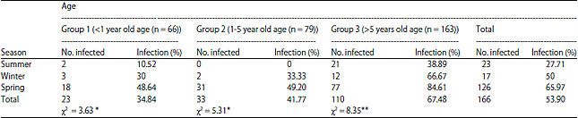 | |
| *Significant at 0.05 and **Highly significant at 0.01 | |
| Table 2: | Prevalence of psoroptic mange in different body parts of examined buffaloes |
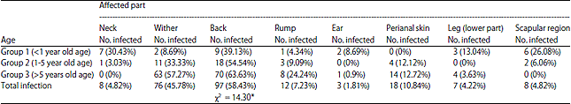 | |
| *Significant at 0.05 and **Highly significant at 0.01 | |
| Table 3: | Prevalence of psoroptic mange according to the sex of examined buffaloes |
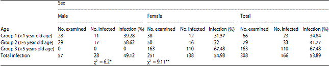 | |
| *Significant at 0.05 and **Highly significant at 0.01 | |
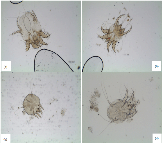 | |
| Fig. 1(a-d): | (a) Psoroptes female x4, (b) Psoroptes male x4, (c) Psoroptes nymph x4 and (d) Psoroptes larva x10 |
| Table 4: | Hematological and biochemical parameters in non infested and highly infested buffaloes by psoroptic mange |
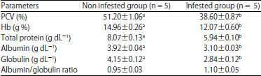 | |
Values are Means±SE, PCV: Packed cell volume, Hb: Hemoglobin and means within the same row having the different letters are significantly different at p<0.05 | |
Conclusively there were significant changes in different hematological and biochemical parameters in non infested and infested buffaloes by psoroptic mange in Table 4.
Morphological description and measurements of Psoroptes spp.: The recovered mites in all skin scraping were Psoroptes spp. having oval bodies, long legs, pointed capitulum and body without scales and spins. The males were oval in shape, measured 315 μm (280-350 μm) in length and 300 μm (260-340 μm) in width. The male had four pairs of long legs ended with suckers on 1st, 2nd and 3rd pairs. While the 4th pair end with bristle. Each sucker was found on segmented pretarsus. The posterior end of the abdomen in male was bilobed and had one pair of copulatory suckers. Females measured 605 μm (460-750 μm) in length and 405 μm (350-460 μm) in width had suckers on 1st, 2nd and 4th pairs of legs with bristle on the 3rd pair. The female body was oval in shape and larger in size than male as shown in Fig. 1.
Psoroptic mange signs and lesions: Lesions due to psoroptic mange were in the form of loss of hairs, redness, scales and dermatitis (Fig. 2). The infected animals showed the rubbing of the affected parts that lead to injuries and loss of hairs in the affected parts. Forty eight out of 166 (28.91) infected buffaloes were reported mange in more than one body parts.
DISCUSSION
The clinical examinations revealed the mange in 53.90% of the examined buffaloes, the highest infection rate in spring season in different ages. This infection rate was higher than that reported by El-Khodery et al.13 (16.6%) in middle Delta region. Lower infection rates of 28, 11 and 6.57% were also reported by Amer et al.24, Kazmi et al.25 and Mottelib and Mohamed26, respectively.
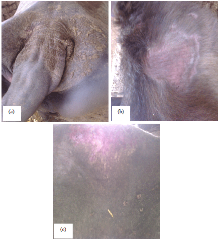 | |
| Fig. 2(a-c): | Signs and lesions of psoroptic mange in different body parts of buffaloes, (a) Psoroptic mange in perinal skin, (b) Psoroptic mange in scapular region and (c) Psoroptic mange in back region |
In contrast to previous studies, higher infection rate (83.2%) was recorded by Yassin27 in Giza Governorate, Egypt. This variation may be attributed to differences in geographic areas, animal breeds, hygienic measures, housing condition of surroundings animals and available veterinary care in examined areas.
The infestation rate of mange was higher in females (54.98%) than males (49.12%) with an infestation rate of and in females and males, respectively. Previously significant variation in mange infection between males and females has been reported at Assuit and Quena Governorates Mottelib and Mohamed26.
Results of highest infestation rate in spring season in all ages are disagreed with Vishe et al.28 who recorded that the mange infestation was the highest in winter season.
The influence the disease occurrences on age indicated that old age buffaloes over 5 years were the most affected with infection rate 67.48% compared with calves less than 1 year (34.38%) and those of 1-5 year old age (41.77%). These results are similar to Yassin27 who recorded old age buffaloes over 6 years was more susceptible to mite infestation with no recorded infestation in buffaloes of less 1 year. While other studies in Egypt26 in Assuit and Quena Governorates reported low infestation rate in calves below 6 months, this rate increased with age but gradual decrease in infestation with ageing was noticed in animals over 6 years of age.
This study compared the prevalence rate between two areas in Menofia province, (Ashmoun and Menoof) illustrated that Ashmoun area was the highly prevalence of infection (54.98%) than in Menoof (48.27%). This variation can be there due to difference in the awareness of farmers and veterinary and public health services offered between the two areas. Also, the socioeconomic level will have an effect on prevalence of that problem.
Morphologically, Psoroptes sp. had oval body, long legs, pointed capitulum and segmented pretarsus. Moreover, male Psoroptes had one pair of copulatory suckers and bilobed posterior end of the body5,17,24, 29.
There were significant decrease in PCV, Hb, total protein, albumin and globulin in highly infested buffaloes due to loss of appetite, changes in hepatic function, structure and decrease in nutrients digestion in infested buffaloes that agreed with Vishe et al.28 that reported that the infestation with Psoroptes was associated with anemia and decreasing in total protein. Infections with different species of mange lead to inflammation resulting into malnutrition and debility which are reflected by haematobiochemical responses30.
CONCLUSION
The infestation by psoroptic mange was significantly affected by sex, age and season. Also, the infested buffaloes showed decreased PCV, Hb and total protein especially in sever infestation.
SIGNIFICANT STATEMENT
Psoroptic mange is a serious contagious skin disease and causes economic losses in buffaloes. The age, sex and season had significant effect on the prevalence of psoroptic mange in Egyptian buffaloes at Menofia Governorate, Egypt with significant decrease in hematological and biochemical parameters in highly infested buffaloes.
ACKNOWLEDGMENT
Great thanks to staff members of Parasitology Department, Faculty of Veterinary Medicine, University of Sadat City, Egypt.
REFERENCES
- Jones, J., T. Jenkins, L. Webb, A. Davies and P. Bates, 2008. Psoroptic mange in cattle in South Wales. Vet. Rec., Vol. 162.
CrossRef - Osman, S.A., A. Hanafy and S.E. Amer, 2006. Clinical and therapeutic studies on mange in horses. Vet. Parasitol., 141: 191-195.
CrossRefDirect Link - Losson, B.J., 2012. Sheep psoroptic mange: An update. Vet. Parasitol., 189: 39-43.
CrossRefDirect Link - Jabeen, F., N. Ahmed, M.A. Chaudhy and I. Javed, 1998. Epidemiology and treatment of sarcoptic mange in buffalo calves around Lahore. Pak. Vet. J., 181: 39-42.
Direct Link - Rafferty, D.E. and J.S. Gray, 1987. The feeding behaviour of Psoroptes spp. mites on rabbits and sheep. J. Parasitol., 73: 901-906.
CrossRefDirect Link - Bisdorff, B. and R. Wall, 2008. Control and management of sheep mange and pediculosis in Great Britain. Vet. Parasitol., 155: 120-126.
CrossRefDirect Link - Jones, A., G. Caldow, N. Cameron and M. McGregor, 2014. Psoroptic mange in a Scottish beef herd. Vet. Rec., 174: 509-510.
CrossRefDirect Link - Losson, B. and J.F. Lonneux, 1996. Field efficacy of moxidectin 0.5% pour-on against Chorioptes bovis, Damalinia bovis, Linognathus vituli and Psoroptes ovis in naturally infected cattle. Vet. Parasitol., 63: 119-130.
CrossRefDirect Link - Stromberg, P.C., W.F. Fisher, F.S. Guillot, J.H. Pruett, R.E. Price and R.A. Green, 1986. Systemic pathologic responses in experimental Psoroptes ovis infestation of Hereford calves. Am. J. Vet. Res., 47: 1326-1331.
PubMedDirect Link - Hallal-Calleros, C., J. Morales-Montor, J.A. Vazquez-Montiel, K.L. Hoffman, A. Nieto-Rodriguez and F.I. Flores-Perez, 2013. Hormonal and behavioral changes induced by acute and chronic experimental infestation with Psoroptes cuniculi in the domestic rabbit Oryctolagus cuniculus. Parasites Vectors, Vol. 6.
CrossRef - Rahbari, S., S. Nabian and A.R. Bahonar, 2009. Some observations on sheep sarcoptic mange in Tehran province, Iran. Trop. Anim. Health Prod., 41: 397-401.
CrossRefDirect Link - El-Khodery, S.A., S.A. Osman, M. Ishii and M.H. Al-Gaabary, 2010. Risk factors of infestation by Psoroptes spp. mites in buffalo (Bubalus bubalis) at smallholder farms in the Nile Delta region, Egypt. Trop. Anim. Health Prod., 42: 275-281.
CrossRefPubMedDirect Link - Dimri, U. and M.C. Sharma, 2004. Effects of sarcoptic mange and its control with oil of Cedrus deodara, Pongamia glabra, Jatropha curcas and benzyl benzoate, both with and without ascorbic acid on growing sheep: Epidemiology; assessment of clinical, haematological, cell-mediated humoral immune responses and pathology. J. Vet. Med. Ser. A, 51: 71-78.
CrossRefDirect Link - Fisher, W.F. and H.R. Crookshank, 1982. Effects of Psoroptes ovis (Acarina: Psoroptidae) on certain biochemical constituents of cattle serum. Vet. Parasitol., 11: 241-251.
CrossRefDirect Link - Wall, R. and K. Kolbe, 2006. Taxonomic priority in Psoroptes mange mites: P. ovis or P. equi? Exp. Applied Acarol., 39: 159-162.
CrossRefDirect Link - Sanders, A., P. Froggatt, R. Wall and K.E. Smith, 2000. Life-cycle stage morphology of Psoroptes mange mites. Med. Vet. Entomol., 14: 131-141.
CrossRefPubMedDirect Link - Randhawa, C.S., R.S. Brar, D.R. Sharma and S.S. Randhawa, 1997. Biochemical responses in mixed chronic psoroptic and sarcoptic mange of buffaloes (Bubalus bubalis). Trop. Anim. Health Prod., 29: 253-254.
CrossRefPubMedDirect Link - Bala, A. and S.S. Rath, 2006. Studies on haemato-biochemical alterations due to Sarcoptic mange in buffalo calves. Indian Vet. J., 83: 230-231.
Direct Link - Aujla, R.S., L.D. Singla, P.D. Juyal and P.P. Gupta, 2000. Prevalence and pathology of mange-mite infestations in dogs. J. Vet. Parasitol., 14: 45-49.
Direct Link - Amer, S., T. Abd El Wahab, A.E.N. Metwaly, Y. Feng and L. Xiao, 2015. Morphologic and genotypic characterization of Psoroptes mites from water buffaloes in Egypt. PLoS One, Vol. 10.
CrossRefDirect Link - Kazmi, S.A.I., A. Maqbool, M.T. Tonio, A. Naureen, A. Ajmal and M.T. Anwar, 2009. Treatment of dairy buffaloes naturally infected with sarcoptic mange. J. Parasitic Dis., 33: 54-56.
CrossRefPubMedDirect Link - Yassin, M.K., 2011. Mange mites causing scabies in Egyptian buffaloes at Giza Governorate, Egypt. J. Egyptian Soc. Parasitol., 41: 55-64.
PubMedDirect Link - Vishe, H.P., K. Pawar, H.K. Gupta and G.S. Rao, 2012. Prevalence and hemato-biochemical studies in parasitic and non parasitic dermatological disorders in Surti buffalo and buffalo calves. Vet. World, 5: 230-235.
CrossRefDirect Link