
Safyzan Salim
British Malaysian Institute, Universiti Kuala Lumpur, Bt. 8, Jalan Sungai Pusu, 53100 Gombak, Selangor, Malaysia
Nur Adilah Abd Rahman
Biomedical Modelling and Simulation Research Group, Faculty of Electrical and Electronics Engineering,
University Tun Hussein Onn Malaysia, Batu Pahat, Johor, Malaysia
Muhammad Mahadi Abdul Jamil
Biomedical Modelling and Simulation Research Group, Faculty of Electrical and Electronics Engineering,
University Tun Hussein Onn Malaysia, Batu Pahat, Johor, Malaysia
Mansour Youseffi
School of Engineering, International College of Automotive Malaysia, Faculty of Engineering and Technology, Pekan, Pahang, Malaysia
Morgan C.T. Denyer
School of Life Sciences, Faculty of Engineering and Informatics, University of Bradford, Bradford BD7 1DP, United Kingdom
Journal of Biological Sciences
Year: 2016 | Volume: 16 | Issue: 7 | Page No.: 278-283


ABSTRACT
Electroporation (EP) is a method of controlling cell function by using pulses of electrical fields to create pore through a cell membrane and causes other substance around it to be absorbed into the cell. Where this method had been led to a variety of medical applications. While, micro contact printing (μCP) is a quite useful technique for patterning extracellular matrix as an adhesion molecule for cells that works for controlling the cell growth. This study focuses on a comparison of a cancer cell (HeLa cancer cell) cultured on two different type of substrates which is a protein surface and empty surface. In order to see the effect of cell proliferation of cancer cell with protein solution, which is in this experiment we used fibronectin for the protein solution and the preliminary result show a positive respond to the protein solution that act as self-assembled monolayer.
PDF Abstract XML References Citation
Received: May 01, 2016;
Accepted: August 13, 2016;
Published: September 15, 2016
How to cite this article
Safyzan Salim, Nur Adilah Abd Rahman, Muhammad Mahadi Abdul Jamil, Mansour Youseffi and Morgan C.T. Denyer, 2016. Investigation of Electroporation Technique on Cell Properties
Cultured on Self-assembled Monolayer. Journal of Biological Sciences, 16: 278-283.
DOI: 10.3923/jbs.2016.278.283
URL: https://scialert.net/abstract/?doi=jbs.2016.278.283
DOI: 10.3923/jbs.2016.278.283
URL: https://scialert.net/abstract/?doi=jbs.2016.278.283
INTRODUCTION
Microcontact printing has been developed about 17 years ago and it is an outstanding surface patterning technique in micron scale and also in nanoscale1. Surface science communities such as engineers and biologists have been promoting attention in μCP and therefore enriching in improvement to the μCP process itself2. The μCP is a soft lithography that used the release pattern on master polydimenthylsiloxane (PDMS) stamp to form a patterns of self-assembled monolayers (SAMs) of ink on the surface of a substrate through conformal contact3. In the original version of μCP, the micro-metre-scale patterned chemical modification of a large surface area is obtained by transferring different types of compounds using a soft polymer stamp4. Electroporation or electropermeabilization is a method to introduce molecules or a method for increasing cell membrane permeability to molecules. It had been used in various biotechnological and biomedical applications, such as the introduction of molecules into cells, cell fusion, tissue ablation and sterilization of water and liquid food. In molecular medicine and biotechnology, tissue electro-poration is performed with electrodes placed in the target area of the body5. Electroporation can be endorsed in 3 type of ways that is ex vivo, in vivo and in vitro. Cell electroporation in vitro is used mainly for transfection by DNA introduction but many other interventions are possible, including microbial killing. Ex vivo electroporation provides the manipulation of cells that are reintroduced into the body to provide therapy6. In vivo electroporation of tissues enhances molecular transport through the tissues and into their constitutive cells. By applying an electrical pulses across cells it can have a variety of outcomes from the result; from no effect to a reversible electroporation to irreversible electroporation7.
MATERIALS AND METHODS
Cell lines and culture conditions: In this study, HeLa cells were used. HeLa cells are cultured in standard culture flask in Rosewell Park Memorial Institute (RPMI) 1640 media (Sigma) with 10% fetal bovine serum until 90% confluence. The cell was maintained in the atmosphere of 5% CO2 at 37°C and were split once reaching confluence, usually every 5-6 days8 (Fig. 1).
Microcontact printing method: Figure 2 shows the concept of the fabricating high resolution patterns of protein using a μCP method with a PDMS stamp start9 at A and B.
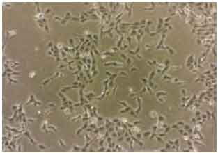 | |
| Fig. 1: | HeLa cell with 50% confluence |
 | |
| Fig. 2: | Process flow schematic of microcontact printing method |
Where, A show step of inking the PDMS stamp with protein solution and B shows the transferring the proteins to a substrate area or contact or it called step of stamping PDMS with protein at the new substrate by applying a small amount of force and C shows a patterned surface after remove the stamp from the stamping step, the ink is patterned on the glass substrate10.
Electroporation technique: For the electroporation technique, Fig. 3 and 4 shows the main components of the experimental setup.
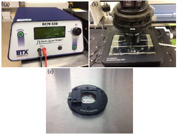 | |
| Fig. 3(a-c): | (a) High voltage pulsed system (ECM® 830), (b) Chamlide TC stage and (c) Microchamber slide (Chamlide EC) |
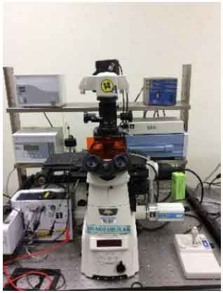 | |
| Fig. 4: | Nikon inverted microscope (Ti series) |
A nikon inverted research microscope (Ti series) offers improved speed, increased flexibility and efficient multi-mode microscopy as part of fully integrated microscope system for cell live imaging. In order to expose biological cells to high voltage pulse electric field, two main subsystems are needed: (1) Imaging chamber system (Chamlide EC) which is connected with the main components, dedicated to the applications of field stimulation, which supplied with a pair of platinum electrodes11 and (2) High voltage pulsed system (ECM® 830) which allow to deliver 10-600 μsec duration of pulses with the adjusted amplitude upto 3 kV12.
RESULTS AND DISCUSSION
Patterning test with Fetal Bovine Serum (FBS) proteins: Fetal Bovine Serum (FBS) is used initially to test μCP functionality as in protein patterning. Figure 5 shows that the protein patterning outcome by using microcontact printing method with variety size such as 100, 50, 25 and10 μm. The variety size of stamp used to identify which pattern size will be suitable in microcontact printing method to analyze the cell alignment activity close to 100%.
Electroporation effect on HeLa cell cultured on protein surface and empty surface: With the availability of the HeLa cancer cell, this test was conducted in order to see the effect of the electroporation technique on HeLa cancer cell with the protein solution, which is in this experiment we used fibronectin and the electroporation technique we used 2000 kV with pulse length of 30 µsec in order to achieve high electrical field intensity.
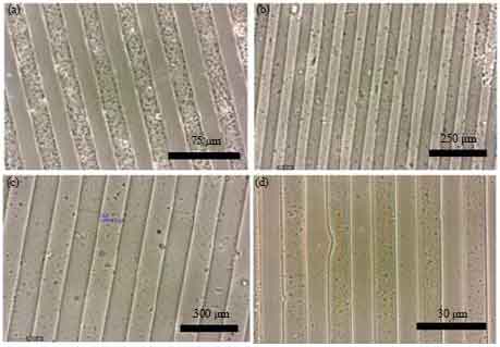 | |
| Fig. 5(a-d): | Patterns of proteins by microcontact printing method with variety of size, (a) 75 µm, (b) 250 µm, (c) 300 µm and (d) 30 µm |
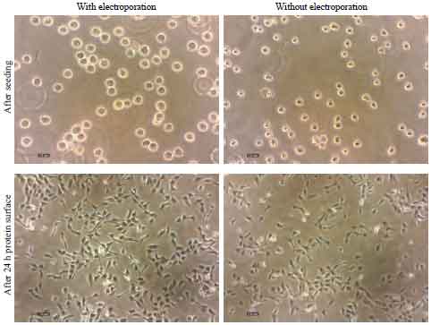  | |
| Fig. 6: | Electroporation effect on HeLa cell within 24 h |
 | |
| Fig. 7: | Graph shows a confluence rate of HeLa cell in this test |
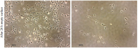
Where in this experiment, in one well, it divides into two sections of the substrate. Which is the left side is the empty surface while the right side is the protein surface (Fig. 6).
It had been left for 24 h to see the effect of the electroporation technique on HeLa with the protein solution. After 24 h, the result as Fig. 7 shows that the proliferation rate of HeLa cell with EP is increased dramatically at 24 h compared with HeLa cell without EP and also most of HeLa cell attracts with protein surface with the ratio confluence 3:7. This shows that HeLa cell can be used in the fabricating process on the next objective of this study.
CONCLUSION
In conclusion, the preliminary result shown in the previous section, confirm the cell adhesion and proliferation occurs mainly on the fibronectin coated area. By comparing the previous research on the microcontact printing and electroporation, there is minor study on the combination of these two method. The basic concepts and techniques of electroporation and microcontact printing were highlighted. Both EP and μCP were found to be related to wound healing process, depending on the level of their threshold and application (gene therapy, electrochemotherapy and wound healing or tissue ablation). Thus, investigations of EP and μCP showed that the two methods can be combined in in vitro to see the cell response to μCP with the PEF applied to it. Important parameters (pulse number and pulse duration) should be considered along with MCP method/technique, which include the cell type and system configuration. We strongly believe that the result of this study can lead to the development of wound healing and cancer treatment.
ACKNOWLEDGMENTS
The researchers wish to acknowledge the financial support by UTHM GIPS grant sponsor. We would also thank to individual who have helped in this study.
REFERENCES
- Qin, D., Y. Xia and G.M. Whitesides, 2010. Soft lithography for micro- and nanoscale patterning. Nat. Protocols, 5: 491-502.
CrossRefDirect Link - Mrksich, M., L.E. Dike, J. Tien, D.E. Ingber and G.M. Whitesides, 1997. Using microcontact printing to pattern the attachment of mammalian cells to self-assembled monolayers of alkanethiolates on transparent films of gold and silver. Exp. Cell Res., 235: 305-313.
CrossRefDirect Link - Jiang, C., R.V. Davalos and J.C. Bischof, 2015. A review of basic to clinical studies of irreversible electroporation therapy. IEEE Trans. Biomed. Eng., 62: 4-20.
CrossRefDirect Link - Maksud, M.I., M.S. Yusof and M.M. Abdul Jamil, 2013. An investigation of parameter effect on microcontact printing and feasibility study for application in microelectronic and biomedical. Proceedings of the 6th Biomedical Engineering International Conference, October 23-25, 2013, Amphur Muang, pp: 1-4.
CrossRef - Ito, Y., 1999. Surface micropatterning to regulate cell functions. Biomaterials, 20: 2333-2342.
CrossRefDirect Link - Tizatl, A.L.V., L.I.G. Jimenez, S.A.R. Cuevas, A.V. Hernandez and P.H. Rodriguez, 2013. 3D model and simulation of electroporation application on healthy and tumoral breast tissue. Proceedings of the 10th International Conference on Electrical Engineering, Computing Science and Automatic Control, September 30-October 4, 2013, Mexico, pp: 144-149.
CrossRef - Sundararajan, R., T. Salameh, I.G. Camarillo, R.R. Prabu, A. Natarajan and K. Sankaranarayanan, 2014. Irreversible Electroporation: A Drug-Free Cancer Treatment. In: Electroporation-Based Therapies for Cancer: From Basics to Clinical Applications, Sundararajan, R. (Ed.). Chapter 10, Elsevier Science, New York, USA., ISBN-13: 9781908818294, pp: 219-243.
- Rinaldi, R., P.P. Pompa, G. Maruccio, A. Biasco and P. Visconti et al., 2004. Self-assembling of proteins and enzymes at nanoscale for biodevice applications. IEE Proc.-Nanobiotechnol., 151: 101-108.
CrossRefDirect Link - Quist, A.P., E. Pavlovic and S. Oscarsson, 2005. Recent advances in microcontact printing. Anal. Bioanal. Chem., 381: 591-600.
CrossRefDirect Link - Azioune, A., N. Carpi, Q. Tseng, M. Thery and M. Piel, 2010. Protein micropatterns: A direct printing protocol using deep UVs. Meth. Cell Biol., 97: 133-146.
CrossRefDirect Link - Cheng, K., F. Lu and X. Liang, 2010. An effective electroporation protocol for Bacillus alcalophilus TCCC11004. Proceedings of the 4th International Conference on Bioinformatics and Biomedical Engineering, June 18-20, 2010, Chengdu, pp: 1-4.
CrossRef - Ivorra, A. and B. Rubinsky, 2006. Impedance analyzer for in vivo electroporation studies. Proceedings of the IEEE 28th Annual International Conference of Annual International Conference, August 30-September 3, 2006, New York, pp: 5056-5059.
CrossRefDirect Link