
Shashi Kumar
International Medical University, Kuala Lumpur, Malaysia
Srikumar Chakravarthi
International Medical University, Kuala Lumpur, Malaysia
Gan Seng Chiew
Department of Pre-clinical Sciences, Faculty of Medicine and Health Sciences, UTAR, Malaysia
Thavamanithevi Subramaniam
SIRIM Bhd, Shah Alam, Malaysia
Umadevi Palanisamy
School of Medicine and Health Sciences, Monash University Sunway Campus, Malaysia
Ammu Radhakrishnan
International Medical University, Kuala Lumpur, Malaysia
Nagaraja Haleagrahara
Discipline of Physiology and Pharmacology, School of Veterinary and Biomedical Sciences, James Cook University, Australia
Journal of Biological Sciences
Year: 2012 | Volume: 12 | Issue: 7 | Page No.: 385-392


ABSTRACT
Pathogenic mechanisms of arthritis are studied using Collagen-Induced Arthritis (CIA) animal models. Plant derived antioxidants are known to reduce the inflammatory response in CIA. The aim of the study was to assess the protective efficacy of Nephelium lappaceum ethanol extract against Collagen-Induced Arthritis (CIA) in dark agouti rats. Arthritis was induced with 4 mg kg-1 of collagen in complete Freund's adjuvant. CIA rats were orally treated with 100 and 200 mg kg-1 per oral of N. lappaceum from day 25-50. Changes in body weight, joint thickness, C-reactive protein were recorded and immunohistochemistry for matrix metalloproteinase-13 (MMP-13) and tissue inhibitor of matrix metalloproteinase-1 (TIMP-1) was done. N. lappaceum (100 and 200 mg kg-1) significantly reduced (p<0.05) the arthritis-induced changes in body weight and paw edema. There was a significant reduction (p<0.05) in the C-reactive protein in the treatment groups. A significant reduction (p<0.05) in the arthritis-induced histopathological changes was seen after treatment with N. lappaceum. Treatment with N. lappaceum showed dose dependent effects on MMP-13 and TIMP-1 levels. N. lappaceum rind extract significantly suppressed the physiological, biochemical and histopathological changes produced during collagen-induced arthritis in dark Agouti rats. N. lappaceum extract supplementation may be beneficial in preventing the tissue damage and inflammatory conditions in arthritis.
PDF Abstract XML References Citation
Received: July 11, 2012;
Accepted: October 18, 2012;
Published: December 27, 2012
How to cite this article
Shashi Kumar, Srikumar Chakravarthi, Gan Seng Chiew, Thavamanithevi Subramaniam, Umadevi Palanisamy, Ammu Radhakrishnan and Nagaraja Haleagrahara, 2012. Protective Effects of Nephelium lappaceum Rind Extract against Collagen-induced Arthritis in Dark Agouti Rats. Journal of Biological Sciences, 12: 385-392.
DOI: 10.3923/jbs.2012.385.392
URL: https://scialert.net/abstract/?doi=jbs.2012.385.392
DOI: 10.3923/jbs.2012.385.392
URL: https://scialert.net/abstract/?doi=jbs.2012.385.392
INTRODUCTION
Rheumatoid Arthritis (RA) is a chronic inflammatory disease that mainly targets the synovial tissue, cartilage and subchondral bone. Patients will suffer from joint inflammation, joint destruction and motor disability. Progressive and erosive destruction of the peripheral joints will lead to severe motor disability leading to decreased quality of life (Kumar et al., 2009). The hallmark of RA is a persistent inflammation of synovial membrane, pannus formation and cartilage thinning which ultimately leads to joint deformities (Choy and Panayi, 2001; Navarro-Cano et al., 2003).
Antioxidant micronutrients are presumed to play an important role in protecting against the tissue damage caused by Reactive Oxygen Species (ROS). Antioxidants’ provision in suppression of cytokines and collagenase expression induced by Tumor Necrosis Factor (TNF)-α has been demonstrated in RA patients (Halliwell et al., 1988; Mahajan and Tandon, 2004; Sato et al., 1996) which suggests additional mechanisms of protection against RA. The reports about the modulation of pro-inflammatory cytokine expressions by diet derived antioxidants have lead to the discussion that antioxidants have the potential to protect against rheumatoid arthritis. But there are very few researches available about the anti-inflammatory and antioxidant role of antioxidants from the plants in rheumatoid arthritis (Buchanan et al., 1991; Mangge et al., 1999; Panush, 1991; Shukla et al., 2008).
Rambutan (Nephelium lappaceum), a tropical fruit of the Sapindaceae family is native to Malaysia. This seasonal tropical fruit is an important commercial crop in Asia (Dembitsky et al., 2011). The rind of N. lappaceum, habitually discarded, is having extremely high anti-oxidant (Palanisamy et al., 2008; Thitilertdecha et al., 2010), antibacterial (Martinez-Castellanos et al., 2009; Thitilertdecha et al., 2008), anti-hyperglycemic and free radical scavenging activities (Palanisamy et al., 2011). Ethanolic N. lappaceum rind extract possess high free radical-scavenging activity and low pro-oxidant capability (Dembitsky et al., 2011; Palanisamy et al., 2011). The current study was designed to explore the anti-inflammatory effect of N. lappaceum rind extract on Collagen-Induced Arthritis (CIA) in rats. We hypothesize that the ethanol extract of N. lappaceum has significant anti-inflammatory effect and protects against arthritis-induced histopathological changes in Dark Agouti rats.
MATERIALS AND METHODS
Drugs and chemicals: Type II collagen, Complete Freund’s Adjuvant (CFA) and all other analytical chemicals used for the experiment were obtained from Sigma-Aldrich (St. Louis, MO, USA).
Plant collection, isolation and standardization: The extract was standardized using Geraniin as described previously (Palanisamy et al., 2011). N. lappaceum was obtained from Kuala Lumpur, Malaysia from 2009-2010 and plants were authenticated by the herbarium of the Forest Research Institute of Malaysia (FRIM), Malaysia. Plants (1 kg) were cleaned and dried at 40°C in the oven and then, powdered using the Fritsch dry miller. Ethanol extraction (1:15; w/v) was carried out at room temperature in an orbital shaker. The suspension obtained was filtered using a 114 Whatman filter paper and filtrate was collected. Ethanol filtrate was concentrated using a rotary evaporator to yield a dark brown ethanol extract of rambutan rind.
Determination of total phenolic content: Total phenolics were determined based on a colorimetric oxidation and reduction reaction using the Folin-Ciocalteu method (Palanisamy et al., 2008, 2011). Aliquot of the extracts (1 mL) was added to 5 mL of Folin-Ciocalteu reagent. After 3 min, 4 mL of 7.5% Na2CO3 solution in water was added to the mixture and the content was thoroughly mixed. The absorbance at 765 nm was read after 1 h. Blank consisted of Folin-Ciocalteu reagent (5 mL), ethanol/distilled water (1 mL) and 7.5% Na2CO3 solution (4 mL). A linear dose-response regression curve was generated using absorbance reading of gallic acid at the wavelength of 765 nm. The calibration curve using gallic acid was obtained in the same manner as above except that the absorbance was read after 30 min. Results were expressed as milligrams of gallic acid equivalent per gram of dry weight (mg GAE/g DW) of extracts (Palanisamy et al., 2008).
Experimental animals: The autoimmune arthritis is mediated by sex hormones and is associated with a female preponderance for development of arthritis (Holmdahl, 1995; Van den Berg, 2009). Inbred female Dark Agouti (DA) rats of 6-8 weeks of age (200-220 g) were maintained at room temperature (24±2°C), with a 12 h light-dark cycle, relative humidity 60-70% and allowed food and water ad libitum (Tudave et al., 2011).
Research protocol approval: All the experimental procedures were according to internationally approved ethical guidelines for the care of laboratory animals and the study got the approval from Institutional research and ethics committee.
Collagen-induced arthritis (CIA) and drug administration: Collagen-Induced Arthritis (CIA) in rats was induced as previously described, with minor modification (Brand et al., 2007). Approximately 4 mg kg-1 of type II collagen in complete Freund’s adjuvant was administered intradermally in the tail, 2 cm distal to the base of the tail for each rat (Tudave et al., 2011). On day 21 after the primary immunization, the rats were randomly divided into five groups (n = 6):
Group 1: Control
Group 2: Arthritis control
Group 3: 100 mg kg-1 N. lappaceum extract
Group 4: 200 mg kg-1 N. lappaceum extract
Group 5: 300 mg kg-1 glucosamine
These rats were orally administered with N. lappaceum extract and glucosamine once a day from day 25-50.
Body weight and paw thickness measurement: The severity of arthritis was assessed by measuring changes in paw edema and joint thickness using a digital caliper. The body weight changes of the rats were also measured during the study period.
Biochemical analysis: All the rats were sacrificed 24 h after the last day of treatment. Approximately 5 mL of blood samples were collected by cardiac puncture. Blood was immediately centrifuged and plasma was separated and stored (–80°C) until further analysis. From the plasma, alanine transaminase (AST), creatinine, Blood Urea Nitrogen (BUN) and total protein assay was done using commercially available kits. C-Reactive Protein (CRP) levels in plasma were determined using Millipore® Rat C-Reactive Protein ELISA kit (CYT294) in accordance with the manufacturer's instructions. The CRP concentration of each of the samples calculated based on the standard curve obtained.
Histopathological analysis: Paw joints were collected at the end of experiment and fixed in 10% (v/v) neutral formalin. The joints were trimmed into 5-6 mm thickness, decalcified and the tissue was processed and embedded into paraffin blocks. The slides, stained with haematoxylin and eosin (H and E) were examined under a Nikon Eclipse 80β (CF160) bright field microscope.
Immunohistochemistry for MMP-13 and TIMP-1: The paraffin embedded sections were deparaffinized with xylene and rehydrated in a gradient of ethanol. Endogenous peroxidase activity was quenched with 0.5% H2O2 in ethanol for 5 min. Immunohistochemistry was performed with the avidin-biotin peroxidase complex (ABC) technique. After being washed with Tris Buffer Saline (TBS), antigen retrieval was performed manually by using a hot water bath immersion of 75°C for an hour with Target retrieval solution (S1699, Dako) of pH 9.0 for 45 min. This lower-than-average temperature was used to prevent excessive destruction of the sample tissues (Lee et al., 2008). The sections were blocked with peroxidase-blocking solution (S2023, Dako) at room temperature for 30 min and then incubated overnight at 4°C with the anti-TIMP1 (ab1827) and anti-MMP13 (ab3208) antibodies (Abcam), respectively. After washing with TBS three times, they were incubated with HRP-conjugated secondary antibodies (K0609, Dako) at 37°C for 30 min. After washing with TBS three times, all sections were visualized with diaminobenzidine (DAB) (K3467, Dako). The slides were counterstained with Mayer’s hematoxylin and mounted with coverslip.
Statistical analysis: All the results were expressed as Mean±SEM. Results were analyzed by one-way analysis of variance (ANOVA) followed by Tukey’s test (all pair wise multiple comparison procedure). A value of p<0.05 was considered statistically significant.
RESULTS
Changes in paw thickness: The macroscopic sign of severe arthritis at 50th day included swelling, redness deformity and ankylosis in the hind paw and ankle joints. The symptoms of arthritic control rats showed significant difference (p<0.05) as compared to the hind paw of normal rats. The arthritic rats treated with N. lappaceum (100 mg kg-1), N. lappaceum (200 mg kg-1) and glucosamine (300 mg kg-1) showed no significant changes in the joints when compared to arthritis alone group (Fig. 1). A significant increase (p<0.05) in the hind paw thickness was observed in the arthritic control rats from 0 to 50th day during the development of arthritis (Fig. 2). N. lappaceum treatment depicted a significant reduction in paw thickness compared to arthritic control at 50th day. The arthritic rats treated with N. lappaceum (200 mg kg-1) also showed more significant reduction (p<0.05) in paw thickness during the development of arthritis as compared 100 mg kg-1 group (Fig. 2).
Changes in the body weight: Rats that developed arthritis showed reduction in body weight during first three weeks, found to be almost similar in all the groups of rats. However, after three weeks, the body weight of arthritic control rats was declined significantly (p<0.05) compared to its normal counterpart.
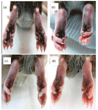 | |
| Fig. 1(a-d): | Hind-limbs distal inter-phalangeal joints of representative rat groups (a) Normal rat, (b) Arthritis (c) N. lappaceum treated 200 mg kg-1 and (d) N. lappaceum treated 100 mg kg-1 |
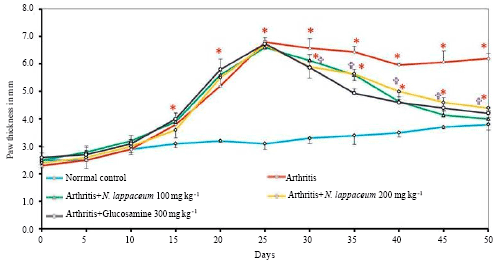 | |
| Fig. 2: | Changes in paw thickness in normal, arthritis, N. lappaceum (100 and 200 mg kg-1) and glucosamine (300 mg kg-1) treated rats. Data are expressed as Mean±SE of six rats per group. *p<0.05: Control vs. other groups, groups _p<0.05: Arthritis vs. other groups |
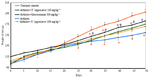 | |
| Fig. 3: | Changes in body weight in normal, arthritis and N. lappaceum (100 and 200 mg kg-1) and glucosamine (300 mg kg-1) treated rats, data are expressed as Mean±SE of six rats per group, *p<0.05: Control vs. other groups *p<0.05: Arthritis vs. other groups |
N. lappaceum (100 mg kg-1 and 200 mg kg-1) treated arthritic rats showed a significant increment (p<0.05) in their body weight as compared to arthritic control (Fig. 3).
Biochemical changes: There were no significant differences in the AST, BUN, Creatinine and total proteins analysed between the controls and treated groups. There was a significant increase (p<0.05) in the CRP levels in the arthritis rats than controls. The concentration of C-reactive protein was found to be significantly reduced (p<0.05) in N. lappaceum treated as well as the glucosamine treated groups (Fig. 4). Glucosamine treatment group had shown more significant decrease in the CRP levels (p<0.05).
Histopathological analysis of joints: There was extensive proliferation of synovial cells, resulting in pannus formation and infiltration of mononuclear cells and neutrophils to the sub synovial region, with damage to the articular surfaces and discontinuity in the cartilage in arthritis rats. All groups except the baseline normal control showed varying levels of pathological changes (Fig. 5a). Severe arthritic and degenerative changes were observed, which showed reduction in higher dosage group (N. lappaceum 200 mg kg-1) (Fig. 5b, d). The degree of reduction of morphological changes was more in glucosamine (300 mg kg-1) treated group (Fig. 5e). There was healthy regenerative synovium and areas of fibrosis, angiogenesis and fibroblasts proliferation in the treatment groups.
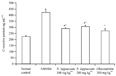 | |
| Fig. 4: | Plasma C-Reactive Protein (CRP) levels in arthritis and treated groups, *p<0.05: Control vs. other groups, †p<0.05: Arthritis vs. other groups |
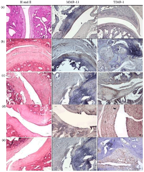 | |
| Fig. 5(a-e): | Histopathological analysis of joint morphology and immunohistochemistry for MMP-13 and TIMP-1 (200X) of (a) Normal control, (b) Arthritis, (c) N. lappaceum 100 mg kg-1, (d) N. lappaceum 200 mg kg-1 and (e) Glucosamine 300 mg kg-1 treated rats |
Immunohistochemistry of MMP-13 and TIMP-1: Immunohistochemical expression of MMP-13 and TIMP-1 were observed in the sections under light microscopy and graded for positivity. IHC analysis indicated that the joint specimens from arthritic control rats strongly stained for MMP-13, which is a key contributor to the destruction of cartilage, however treatment with N. lappaceum reduced the expression of MMP-13 in inflammatory articular cartilage (Fig. 5c, d). It was seen that the groups having the worst histological findings, showed to have the highest levels of MMP-13 and the lowest levels of TIMP-1. Expression of MMP-13 was higher in the cartilage cells of the arthritic group and the least in N. lappaceum (200 mg kg-1) (Fig. 5c) and glucosamine (300 mg kg-1) (Fig. 5e). A corollary effect was seen for TIMP-1 (Fig. 5c), which showed the lowest expression in the above group.
DISCUSSION
Classical signs of severe arthritis were observed with intradermal type II collagen administration, including symmetrical joint involvement typically involving the hind paws, swelling, redness and erythema over the joints (Asquith et al., 2009; Brand et al., 2007). The hallmark symptoms of RA include fever, fatigue and weight loss (Lee and Weinblatt, 2001). The body weights of CIA rats gradually decreases following immunization and it will be at the lowest after three weeks of immunization (Trentham et al., 1977; Nagatomo et al., 2010). Our study showed a significant loss in body weight around 25th day in the CIA rats as compared to the normal control. N. lappaceum (both doses) and glucosamine (300 mg kg-1) showed significant recovery in the body weight during the last 10 days of treatment when compared to their arthritic controls.
C-Reactive Protein (CRP) is produced in the liver under conditions of systemic inflammation in tissue destructions such as cartilage and bone and is a useful biomarker in the evaluation of disease progression and response to therapeutic intervention in a number of systemic inflammatory disorders, including RA. Higher concentration of CRP will indicate increased joint changes in arthritis (Poole et al., 2008; Rhodes et al., 2011). Severity of joint damage should correlate with CRP levels (Jones et al., 2011). In our study, it is shown N. lappaceum exhibited a greater reversal of structural damage in the joints when compared with untreated arthritic rat. The levels of CRP quantified in our study showed that this correlation is true for the N. lappaceum, thus, proving the nutraceutical potential of the extract (Palanisamy et al., 2008).
Histopathological changes correlated with macroscopic observations including changes in the paw thickness. Untreated arthritic rats showed maximum degenerative changes and significant reductions in the joint changes were seen in the treated groups. Observed protective effect may be due to the inhibition of inflammatory mediators by the anti-inflammatory compounds in the extract (Asquith et al., 2009). An ellagitannin have been described to inhibit these mediators, primarily TNF-α, IL-1β and IL-6 [24] which could possibly correlate to the attenuation of histopathological changes.
Matrix metalloproteinases-13 (MMP-13) is thought to increase in degenerative bone diseases such as RA whilst tissue inhibitor of matrix metalloproteinases (TIMP-1) has the opposite action and it is an important factor in maintaining the integrity of connective tissue (Uchida et al., 2000). Our results in this study showed an inverse relationship between MMP-13 and TIMP-1. Rats fed with the both doses of N. lappaceum extract seemed to be the most effective in increasing TIMP-1 expression and suppressing MMP-13 expression. Controlling MMP activity as well as the production of pro-inflammatory cytokines is required for the effective treatment of arthritis (McInnes and Schett, 2007). This observation provides evidence to suggest the potential anti-inflammatory effect of N. lappaceum against collagen-induced arthritis. The significant decline in symptoms of arthritis, paw thickness, histological symptoms and normalization of body weight and biochemical parameters in N. lappaceum treated arthritic rats postulates the possible anti-inflammatory and anti-oxidant effect of N. lappaceum rind extract. These effects may be attributed to the scavenging action of geraniin, an ellagitannin, present in the extract against Reactive Oxygen Species (ROS) such as OH, RO and RO2 (Hagfors et al., 2003).
CONCLUSION
Thus, the present study showed that N. lappaceum rind extract is able to diminish the physiological, histological and biochemical changes produced during collagen-induced arthritis in Dark Agouti rats. N. lappaceum could be regarded as one of the natural products having vast range of therapeutic effects against collagen-induced arthritis. Further studies at the biochemical and molecular levels on the effectiveness and mechanisms of actions of N. lappaceum are being carried out.
ACKNOWLEDGMENTS
This research was funded by Ministry of Health (MOH), Malaysia [NMRR Project ID: 09-406-4108] and supported by International Medical University (IMU) research grants [IMU 211-2010].
REFERENCES
- Asquith, D.L., A.M. Miller, I.B. McInnes and F.Y. Liew, 2009. Animal models of rheumatoid arthritis. Eur. J. Immunol., 39: 2040-2044.
CrossRefDirect Link - Brand, D.D., K.A. Latham and E.F. Rosloniec, 2007. Collagen-induced arthritis. Nat. Protoc., 2: 1269-1275.
CrossRef - Buchanan, H.M., S.J. Preston, P.M. Brooks and W.W. Buchanan, 1991. Is diet important in rheumatoid arthritis?. Br. J. Rheumatol., 30: 125-134.
PubMed - Choy, E.H.S. and G.S. Panayi, 2001. Cytokine pathways and joint inflammation in Rheumatoid arthritis. N. Eng. J. Med., 344: 907-916.
CrossRefPubMedDirect Link - Dembitsky, V.M., S. Poovarodom, H. Leontowicz, M. Leontowicz, S. Vearasilp, S. Trakhtenberg and S. Gorinstein, 2011. The multiple nutrition properties of some exotic fruits: Biological activity and active metabolites. Food. Res. Int., 44: 1671-1701.
CrossRef - Hagfors, L., P. Leanderson, L. Skoldstam, J. Andersson and G. Johansson, 2003. Antioxidant intake, plasma antioxidants and oxidative stress in a randomized, controlled, parallel, Mediterranean dietary intervention study on patients with rheumatoid arthritis. Nutr. J., Vol: 2.
CrossRefDirect Link - Halliwell, B., J.R. Hoult and D.R. Blake, 1988. Oxidants, inflammation and anti-inflammatory drugs. FASEB J., 2: 2867-2873.
PubMed - Jones, N.R., M.A. Pegues, M.A. McCrory, S.W. Kerr and H. Jiang et al., 2011. Collagen-induced arthritis is exacerbated in C-reactive protein-deficient mice. Arthritis. Rheum., 63: 2641-2650.
CrossRef - Kumar, N., S. Singh, N. Patro and I. Patro, 2009. Evaluation of protective efficacy of Spirulina platensis against collagen-induced arthritis in rats. Inflammopharmacology, 17: 181-190.
CrossRefPubMedDirect Link - Mahajan, A. and V.R. Tandon, 2004. Antioxidants and rheumatoid arthritis. J. Ind. Rheumatol. Assoc., 12: 139-142.
Direct Link - Mangge, H., J. Hermann and K. Schauenstein, 1999. Diet and rheumatoid arthritis-a review. Scand. J. Rheumatol., 28: 201-209.
PubMed - Martinez-Castellanos, G., K. Shirai, C. Pelayo-Zaldivar, L.J. Perez-Flores and J.D. Sepulveda-Sanchez, 2009. Effect of Lactobacillus plantarum and chitosan in the reduction of browning of pericarp Rambutan (Nephelium lappaceum). Food. Microbiol., 26: 444-449.
CrossRef - McInnes, I.B. and G. Schett, 2007. Cytokines in the pathogenesis of rheumatoid arthritis. Nat Rev. Immunol., 7: 429-442.
CrossRefDirect Link - Nagatomo, F., N. Gu, H. Fujino, T. Okiura, F. Morimatsu, I. Takeda and A. Ishihara, 2010. Effects of exposure to hyperbaric oxygen on oxidative stress in rats with type II collagen-induced arthritis. Clin. Exp. Med., 10: 7-13.
PubMed - Navarro-Cano, G., I. del Rincon, S. Pogosian, J.F. Roldan and A. Escalante, 2003. Association of mortality with disease severity in rheumatoid arthritis, independent of comorbidity. Arthritis. Rheum., 48: 2425-2433.
Direct Link - Palanisamy, U., H.M. Cheng, T. Masilamani, T. Subramaniam, L.T. Ling and A.K. Radhakrishnan, 2008. Rind of the rambutan, Nephelium lappaceum, a potential source of natural antioxidants. Food Chem., 109: 54-63.
CrossRefDirect Link - Palanisamy, U.D., L.T. Ling, T. Manaharan and D. Appleton, 2011. Rapid isolation of geraniin from Nephelium lappaceum rind waste and its anti-hyperglycemic activity. Food Chem., 127: 21-27.
CrossRefDirect Link - Panush, R.S., 1991. Does food cause or cure arthritis?. Rheum. Dis. Clin. North Am., 17: 259-272.
PubMed - Poole, C.D., P. Conway, A. Reynolds and C.J. Currie, 2008. The association between C-reactive protein and the likelihood of progression to joint replacement in people with rheumatoid arthritis: A retrospective observational study. BMC Musculoskeletal Disorders, Vol. 9.
CrossRefDirect Link - Rhodes, B., B.G. Furnrohra nd T.J. Vyse, 2011. C-reactive Protein in Rheumatology: Biology and Genetics. Nat. Rev. Rheumatol., 7: 282-289.
CrossRefDirect Link - Sato, M., T. Miyazaki, T. Nagaya, Y. Murata, N. Ida, K. Maeda and H. Seo, 1996. Antioxidants inhibit tumor necrosis factor-alpha mediated stimulation of interleukin-8, monocyte chemoattractant protein-1 and collagenase expression in cultured human synovial cells. J. Rheumatol., 23: 432-438.
PubMed - Shukla, M., K. Gupta, Z. Rasheed, K.A. Khan and T.M. Haqqi, 2008. Consumption of hydrolyzable tannins-rich pomegranate extract suppresses inflammation and joint damage in rheumatoid arthritis. Nutritition, 24: 733-743.
CrossRefDirect Link - Thitilertdecha, N., A. Teerawutgulrag, J.D. Kilburn and N. Rakariyatham, 2010. Identification of major phenolic compounds from Nephelium lappaceum L. and their antioxidant activities. Molecules, 15: 1453-1465.
CrossRefDirect Link - Thitilertdecha, N., A. Teerawutgulrag and N. Rakariyatham, 2008. Antioxidant and antibacterial activities of Nephelium lappaceum L. extracts. LWT-Food Sci. Technol., 41: 2029-2035.
CrossRefDirect Link - Trentham, D.E., A.S. Townes and A.H. Kang, 1977. Autoimmunity of type II collagen: An experimental model of arthritis. J. Exp. Med., 146: 857-868.
CrossRef - Uchida, M., M. Shima, T. Shimoaka, A. Fujieda and K. Obara et al., 2000. Regulation of matrix metalloproteinases (MMPs) and tissue inhibitors of metalloproteinases (TIMPs) by bone resorptive factors in osteoblastic cells. J. Cell Physiol., 185: 207-214.
PubMed - Holmdahl, R., 1995. Female preponderance for development of arthritis in rats is influenced by both sex chromosomes and sex steroids. Scand. J. Immunol., 42: 104-109.
PubMed - Lee, K.H., Y.S. Chen, J.P. Judson, S. Chakravarthi, Y.M. Sim and H.M. Er, 2008. The effect of water extracts of Euphorbia hirta on cartilage degeneration in arthritic rats. Malays J. Pathol., 30: 95-102.
PubMed - Van den Berg, W.B., 2009. Lessons from animal models of arthritis over the past decade. Arthritis Res. Ther. Vol. 11.
CrossRefDirect Link