
M.A.M. Alsafy
Department of Anatomy and Embryology, Faculty of Veterinary Medicine, Alexandria University, Egypt
Journal of Biological Sciences
Year: 2010 | Volume: 10 | Issue: 3 | Page No.: 224-230


ABSTRACT
The purpose of current study was to describe the comparative topography and morphology of the lacrimal gland and its drainage apparatus in one humped camel, goat and donkey. The lacrimal apparatus gross anatomy studied on four camels head 4-10 years age, four goats’ head 1-3 years age and four donkeys head 2-6 years age. The lacrimal gland was irregular flattened and elongated lobular in shape, pink in color with characteristic indented borders composed by three lobes the main caudal, coma shape lateral and cranial lobes in camel, while it was flattened and oval in shape, light brown in colour possessed two distinct parts, a body and appendage-like part in goat, whatever, it was ovoid in shape, light brown in colour in donkey. The lacrimal gland was larger in size in donkey and goat than that of camel in relation to body weight. The dorsal and ventral lacrimal puncta were absent and the lacrimal duct started blindly at the medial part of the upper and lower eye lids in camel, while in goat and donkey, the dorsal and ventral lacrimal puncta appeared slit like openings, they were lead to lacrimal sac. The nasolacrimal duct ran in the osseous lacrimal canal rostrally, with a slight curve at its origin. It passed the lacrimal, zygomatic and maxillary bones. It passed through the maxillary sinus and then traversed the nasal cavity in a curved descending fashion, covered only by the nasal mucosa and a thin connective tissue membrane on the lateral surface of ventral nasal concha. The nasolacrimal duct opened at the medial wall of the nasal vestibule at the junction between the mucous membrane and skin by the nasal opening of the nasolacrimal duct that was very minute in camel and goat while it was clearly observed in donkey.
PDF Abstract XML References Citation
How to cite this article
M.A.M. Alsafy, 2010. Comparative Morphological Studies on the Lacrimal Apparatus of One Humped Camel, Goat and Donkey. Journal of Biological Sciences, 10: 224-230.
DOI: 10.3923/jbs.2010.224.230
URL: https://scialert.net/abstract/?doi=jbs.2010.224.230
DOI: 10.3923/jbs.2010.224.230
URL: https://scialert.net/abstract/?doi=jbs.2010.224.230
INTRODUCTION
The lacrimal glands are responsible for the production of tears fluid that helps maintain corneal health. The normal tears contain major secretory source of proteins and electrolytes, the function of this dilute protein solution are to optimize the optics of the cornea, to lubricate and to protect the eye from pathogens (Mohammadpour, 2008).
In most species, the majority of tears are secreted from the lacrimal gland (Carrington et al., 1987; Nguyen et al., 2006). The gross anatomy of lacrimal gland were described previously by Pinard et al. (2003), Aslan et al. (2005) in cattle, Saber and Makady (1987), Ibrahim and Abdalla (2007) and Mohammadpour (2008) in camel.
The lacrimal gland is present in mammals in the glandular lacrimal fossa under the zygomatic process of the frontal bone and dorsolateral surface of the eye ball (Dursum, 2000; Aslan et al., 2005). The shape of the gland is evidently determined by its position; in camel the gland is convex dorsally in conformity with the convexity of the bony orbit. It is irregular in shape (Ibrahim et al., 2006). In other domestic mammals the gland varies in shape between species. It is triangular in pig and bipartite in ox, sheep and goat (Getty, 1975). In man the lacrimal gland is bipartite, the two parts being connected with each other by the aponeurosis of the levator palpebrae superioris (Romanes, 1986).
There is a shortage in literatures on the lacrimal gland of goat and donkey. Awkati and Bagdadi (1971) concluded that the lacrimal gland in the camel is comparatively less developed than that of the horse or ox.
The lacrimal apparatus system provided a passage for drainage from the eye to the nasal cavity. The nasolacrimal system of various domestic species has been previously described by Shokry et al. (1987), Sapienza et al. (1992), Marcos and Affanni (2005), Aslan et al. (2005) , Rehorek et al. (2005), Ibrahim et al. (2006), Shadkhast et al. (2008) and Bigham and Shadkhast (2009). The lacrimal apparatus consisted of an orbital part and a nasal cavity part. The orbital lacrimal apparatus consisted of a simple lacrimal sac, paired canaliculi with the dorsal and ventral puncta (Wilkie and Rings, 1990; Shadkhast et al., 2008; Bigham and Shadkhast, 2009).
In most of the domestic mammals each of the two lacrimal ducts starts by a small one upper and one lower opening, the punctum lacrimale, situated close to the medial angle of the respective eyelid. Saber and Makady (1987) stated that the puncta of camel are absent and that the lacrimal ducts start blindly. The puncta are exceedingly small and cannot be probed in llama, a member of Camelidae (Sapienza et al., 1992). The absence of the puncta or at least one of them is not a feature unknown among the domestic mammals, in pig (Sisson and Grossman, 1975) and in dog (Dyce et al., 1987) one punctum, the lower one in case of pig, may be absent, so that the lacrimal punctum is not functional. The absence of one or the other of the lacrimal puncta in some mammals does not entirely deprive the nasal mucosa of the moistening effect of the lacrimal fluid, but in the camel that moistening effect is excluded by the absence of the puncta. The absence of the puncta causes excess lacrimal fluid to escape by flowing over the lower eyelid; this probably explains the popular myth that the camel is so emotional that it sometimes sheds tears (Ibrahim et al., 2006).
Dacroyocystorhinography, the radiographic visualization of the lacrimal apparatus using radiographic contrast media has been used to study its normal anatomy (Shokry et al., 1987; Theoret et al., 1997; Shadkhast et al., 2008). Pathological conditions of the nasolacrimal duct were stated in human (Hurwitz and Welham, 1975), in dogs (Fowler, 1989), in horse (Latimer et al., 1984), in sheep (Gilanpour, 1979), in cattle (Wilkie and Rings, 1990), in camel (Shokry et al., 1987).
The current study based on the comparative morphological study of the lacrimal gland and lacrimal apparatus of camel, goat and donkey.
MATERIALS AND METHODS
Twelve heads of adult camel, goat and donkey (Four camels head 4-10 years age, four goats head 1-3 years aged were obtained from an abattoir in Libya in 2009) and four donkeys head 2-6 years age used after being bled were used for the study of the topographical and detailed morphological anatomy of the lacrimal apparatus.
For the study of the shape, diameter and topography of the gland, the heads were separated from the neck and dissected in a fresh state where the skin and the periorbital fascia between the upper eyelid and supraorbital process were removed and the lacrimal gland was exposed in rostrally with its attachment over the eye.
For the study of the dimensions of the gland, the whole gland was carefully separated out and the length and width were made.
On gross observation, the dorsal and ventral puncta were located in the medial canthus area of eyelids and they were sufficiently wide in diameter and could be catheterized without much difficulty in donkey, while it was obliterated in camel and it was very small in goat.
For the study of the lacrimal ducts, lacrimal sac and description of the normal anatomical course of nasolacrimal duct, the nasal cavity was exposed from lateral side by removing segments of the lacrimal, zygomatic, maxillary and incisive bones through a line from the medial angle of the eye until the nostrils. The lacrimal apparatus was then examined and its details recorded.
The nasal opening of the nasolacrimal duct were exposed after removing the lateral wall of nostrils and nasal vestibule, where it was located at the level of junction between nasal vestibule cutaneous membrane and nasal cavity proper mucous membrane and its distance from the level of nostril was measured (Latimer et al., 1984; Ibrahim et al., 2006).
RESULTS
Lacrimal gland: In one humped camel, the lacrimal gland was irregular flattened and elongated lobular in shape, pink in colour with characteristic indented borders, it was composed by three lobes cranial, the main caudal and coma shape lateral lobes, attached together with distinct connective tissue. It was surrounded by periorbital tissue and the periosteum on the inner surface of the supraorbital process of the frontal bone, the dorsal surface of the gland was convex and contacted with the orbit, while its ventral surface was concave and laid on the caudodorsolateral surface of the eyeball from which it was separated by the periorbita. The medial border of the gland was wider than the lateral border. The gland dimensions; the caudal lobe was the largest one and was 3 cm in length and 1.7-1.9 cm in width, the lateral lobe was 1.7 cm in length and 1.7 cm width and the cranial lobe was 1.5 cm in length and 1 cm width (Fig. 1A, B).
In goat, the lacrimal gland was flattened and oval in shape, light brown in colour. The gland possessed two distinct features a body and appendage-like part; this was the continuation of the body. It was surrounded by the periorbital tissue and the periosteum on the inner surface of the supraorbital process of the frontal bone. The gland was situated greatly under the frontal bone and it overlapped the rectus dorsalis muscle.
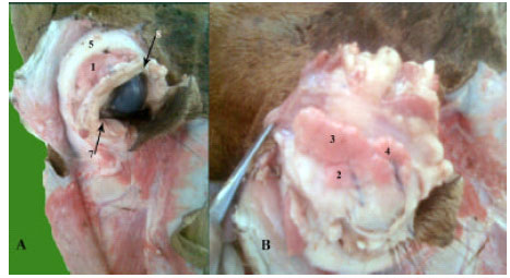 | |
| Fig. 1: | The lacrimal gland of one humped camel. (A) The position of the gland and (B) The shape and lobulation of the gland. 1: The lacrimal gland, 2: The cranial lobe, 3: The caudal lobe, 4: Lateral, 5: The supraorbital process, 6: The medial angle of the eye, 7: The lateral angle of the eye |
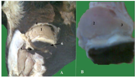 | |
| Fig. 2: | The lacrimal gland of goat. (A) The position of the gland and (B) The shape and lobulation of the gland. 1: The lacrimal gland, 2: The body of the gland, 3: The appendage-like part of the gland, 4: The supraorbital process, 5: The medial angle of the eye, 6: The lateral angle of the eye |
The gland body dimension was 2.5-2.8 cm in length and 2 cm in width, while the appendage was 1cm in length and 0.7 cm in width (Fig. 2A, B).
In donkey, the lacrimal gland was ovoid in shape, light brown in colour. The gland was partially covered with fat. It was situated on the dorsolateral aspect of the eyeball covered by the supraorbital and zygomatic process of frontal bone. The gland dimension was 3.2 cm in length and 2 cm in width (Fig. 3A, B).
The excretory ducts of the lacrimal gland: The gland possessed three excretory ducts in camel, two ducts in goat and donkey emerged from the ventral surface of the gland, ran parallel to each other, penetrated the periorbita and opened at the fornix of the upper eyelid conjunctiva in all studied animals.
The lacrimal apparatus system: The lacrimal apparatus system provided a passage from the eye to the nasal cavity. The system for each eye in all species under study consisted of dorsal and ventral lacrimal opening (puncta), paired canaliculi, lacrimal sac and the nasolacrimal duct.
The dorsal and ventral lacrimal puncta were absent and the lacrimal duct started blindly at the medial part of the upper and lower eye lids in camel, while in goat and donkey, the dorsal and ventral lacrimal puncta appeared slit like openings 0.05 cm in diameter and 0.5 cm away from the medial angle of the eye.
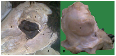 | |
| Fig. 3: | The lacrimal gland of donkey. (A) The position of the gland and (B) The shape of the gland. 1: The lacrimal gland, 2: The supraorbital process, 3: The medial angle of the eye, 4: The lateral angle of the eye |
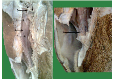 | |
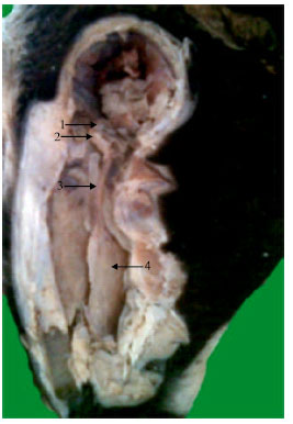
| Fig. 4: | The lacrimal apparatus system of one humped camel. 1: The lacrimal canaliculi, 2: The lacrimal sac, 3: The nasolacrimal duct, 4: Arrow refers to the opening of nasolacrimal duct, 5: Ventral nasal concha |
The dorsal and ventral canaliculi converged to lacrimal sac.
The lacrimal sac: The lacrimal sac was the enlarged funnel shaped beginning of the nasolacrimal duct; it was situated in the lacrimal fossa of lacrimal bone outside the periorbita in all animals under study (Fig. 4-6).
The nasolacrimal duct: The nasolacrimal duct extended from the lacrimal sac to the nostril in the wall of the nasal cavity, the proximal part of the lacrimal duct ran within the osseus lacrimal canal. In camel (Fig. 4), it was measured 20 and 0.2 cm, in goat (Fig. 5), it was measured 7.5 and 0.2 cm, while in donkey (Fig. 6) and it was measured 16 and 0.4 cm in length and diameter, respectively.
The nasolacrimal duct ran in the osseus lacrimal canal rostrally, with a slight curve at its origin. It passed the lacrimal, zygomatic and maxillary bones. It passed through the maxillary sinus and then traversed the nasal cavity in a curved descending fashion, covered only by the nasal mucosa and a thin connective tissue membrane on the lateral surface of ventral nasal concha. The nasolacrimal duct opened at the medial wall of the nasal vestibule at the junction between the mucous membrane and skin by the nasal opening of the nasolacrimal duct that was very minute opening measured about 0.1 cm in camel (Fig. 4), goat and about 0.3 cm in donkey (Fig. 6). The opening located away from the nostril by about 5 cm in camel, 2 cm in goat and 4 cm in donkey.
 | |
| Fig. 5: | The lacrimal apparatus system of goat. 1: The lacrimal canaliculi, 2: The lacrimal sac, 3: The nasolacrimal duct, 4: Ventral nasal concha |
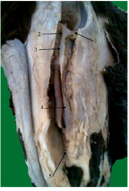 | |
| Fig. 6: | The lacrimal apparatus system of donkey. 1: The lacrimal canaliculi, 2: The lacrimal sac, 3: The nasolacrimal duct, 4: Ventral nasal concha, 5: Arrow refers to the opening of nasolacrimal duct |
DISCUSSION
The lacrimal gland was varied in shape between species. Thus in the camel the gland was irregular flattened and elongated lobular in shape, pink in colour with characteristic indented borders, it was composed by three lobes the main caudal, coma shape lateral and cranial lobes attached together with distinct connective tissue, these findings agree with those of Ibrahim and Abdalla (2007) and Mohammadpour (2008) in camel. While in other studied animals, in goat the lacrimal gland was flattened and oval in shape, light brown in color. The gland possessed two distinct features, a body and appendage-like part, these results is in a line with that denoted by Aslan et al. (2005) in cattle; Mohammadpour (2008) in goat. In donkey it was ovoid in shape, triangular in pig and bipartite in ox (Getty, 1975). In man the lacrimal gland is bipartite, the two parts being connected with each other by the aponeurosis of the levator palpebrae superioris (Romanes, 1986).
It is interesting to note that for so big an animal the gland is so small in size. This has already been commented on by Ibrahim and Abdalla (2007) whose state that the lacrimal gland of the camel is less well-developed than that of either the ox or horse. The lacrimal gland of the camel measured about 3 cm in length and 1.7-1.9 cm in width of the caudal lobe, the lateral lobe was 1.7 cm in length and 1.7 cm width and the cranial lobe was 1.5 cm in length and 1 cm width, these findings were similar to that stated by Ibrahim and Abdalla (2007) whose reported that the dimensions of gland in camel is 40 mm for length and 20 mm for width. Moreover, Awkati and Bagdadi (1971) stated that the lacrimal gland of the camel is 45 mm in length and 24 mm in width.
In the current study, the position of the lacrimal gland of the camel is similar to that reported in the same species by Awkati and Bagdadi (1971), Ibrahim et al. (2006) and Ibrahim and Abdalla (2007).
The position of the lacrimal gland is similar in camel, goat and donkey, the lacrimal gland is situated on the dorsolateral aspect of the eyeball, covered by the zygomatic process of the frontal bone. In both goat and the donkey the gland is partially covered with fat similar results Ibrahim et al. (2006) and Mohammadpour (2008) in camel; Aslan et al. (2005) and Ibrahim et al. (2006) in goat; Getty (1975) in donkey.
The present study reveals that the gland possessed 3 excretory ducts in camel, 2 in goat and donkey emerged from the ventral surface of the gland and the number of excretory ducts of the lacrimal gland is 2-4 denoted by Ibrahim et al. (2006). However, Awkati and Bagdadi (1971), Zaid et al. (1991) claimed that the number of excretory ducts of the lacrimal gland of the camel is two.
In other studied animals there were 2 ducts in goat and donkey as that recorded by Getty (1975), Aslan et al. (2005) and Ibrahim et al. (2006).
In goat and donkey, the two lacrimal ducts starts by a small one upper and one lower opening, the punctum lacrimale, situated close to the medial angle of the respective eyelid. While the puncta of the camel are absent and that the lacrimal ducts start blindly, The dorsal and ventral canaliculi converged to lacrimal sac, the lacrimal sac was the enlarged funnel shaped beginning of the nasolacrimal duct; it was situated in the lacrimal fossa of lacrimal bone outside the periorbita in all animals under study, similar to that recorded by Getty (1975), Aslan et al. (2005) , Ibrahim et al. (2006), Ibrahim and Abdalla (2007) and Mohammadpour (2008).
The nasolacrimal duct extended from the lacrimal sac to the nostril in the wall of the nasal cavity, the proximal part of the lacrimal duct ran within the osseous lacrimal canal. The ducts open into the medial wall of the nasal vestibule at the junction of the mucous membrane and the cutaneous epithelium. The opening is difficult to detect in camel and goat while in donkey it was observed clearly. The nasolacrimal duct of the camel is not functional and, therefore, no lacrimal fluid is carried to the nasal cavity, this in agreement with that stated by Ibrahim and Abdalla (2007) and Mohammadpour (2008).
CONCLUSIONS
In the current study the lacrimal gland shape varied from elongated lobular in camel, oval in goat and ovoid in donkey.
The lacrimal gland size is larger in goat and donkey than in camel in relation to body weight.
The position of lacrimal gland is constant in all studied animals.
The excretory ducts are three in camel, two in goat and donkey.
The dorsal and ventral lacrimal canaliculi openings in the medial angle of the eye clearly distinguished in donkey than goat while in camel they are blind.
The nasolacrimal gland opening at the mucous membrane cutaneous epithelium junction can be clearly observed and catheterized easily in donkey but it is difficult to detect in camel and goat.
REFERENCES
- Marcos, A.H.J. and J.M. Affanni, 2005. Anatomy, histology and fine structure of the harderian gland in the South American armadillo chaetophractus villosus (Xenarthra, mammalia). Anat. Embryol., 209: 409-424.
CrossRefDirect Link - Aslan, K., I. Kurtul, G. Akasy and S. Ozcan, 2005. Gross anatomy of the lacrimal gland (Gl.Lacrimalis) and its arterial vascularization in the fetus of Zavot-Bred cattle. Kafkas Univ. Vet. Fak. Derg., 11: 47-49.
Direct Link - Bigham, A.S. and M. Shadkhast, 2009. Lacrimal apparatus of Iranian river buffalos(bubalis bubalis): Anatomical study. Vet. J., Vol. 4.
Direct Link - Carrington, S.D., P.G.C. Bedford, J.P. Guillon and E.G. Woodward, 1987. Polarised light biomicroscopic observations on the pre-corneal tear film III. The normal tear film of the cat. J. Small Anim. Pract., 28: 821-826.
CrossRefDirect Link - Gilanpour, H., 1979. Anatomic and radiographic studies of the lacrimal drainage system in sheep (Ovis aries). Am. J. Vet. Res., 40: 1177-1179.
PubMed - Hurwitz, J.J. and R.A.N. Welham, 1975. The role of dacryocystorhinography in the management of congenital nasolacrimal duct obstruction. Can. J. Ophthalmol., 10: 346-350.
PubMed - Ibrahim, Z.H.M.A., A.B. Abdalla and D.I. Osman, 2006. A gross anatomical study of the lacrimal apparatus of the camel (Camelus dromedarius). J. Sci. Technol., 9: 1-8.
Direct Link - Latimer, C.A., M. Wyman, C.D. Diesem and J.K. Burt, 1984. Radiographic and gross anatomy of the nasolacrimal duct of the horse. Am. J. Vet. Res., 45: 451-458.
PubMed - Mohammadpour, A.A., 2008. Anatomical characteristic of dorsal lacrimal gland in one humped camel (Camelus dromedaries). J. Biol. Sci., 8: 1104-1106.
Direct Link - Pinard, C.L., M.L. Weiss, A.H. Brightman, B.W. Fenwick and H.J. Davidson, 2003. Normal anatomical and histochemical of the lacrimal glands in the american bison and cattle. Anat. Histol. Embryol., 32: 257-262.
CrossRefDirect Link - Sapienza, J.S., R. Isaza, R.D. Johnson and T.R. Miller, 1992. Anatomic and radiographic study of the lacrimal apparatus of llamas. Am. J. Vet. Res., 53: 1007-1009.
PubMed - Shadkhast, M., A.S. Bigham, N. Vajdi and Z. Shafiei, 2008. Lacrimal apparatus system in goat (Capra aegagrus hircus): Anatomical and radiological study. AJAVA., 3: 457-460.
CrossRefDirect Link - Theoret, C.L., B.H. Grahn and P.B. Fretz, 1997. Incomplete nasomaxillary dysplasia in a foal. Can.Vet. J., 38: 445-447.
Direct Link - Wilkie, D.A. and D.M. Rings, 1990. Repair of anomalous nasolacrimal duct in a bull by use of conjunctivorhinostomy. J. Am. Vet. Med. Assoc., 196: 1647-1650.
PubMed