
N. El-Helaly
66 Mohi El-Deen, Abo El-Ezz Street, Dokki, Guiza, Egypt
A. El-Wan
66 Mohi El-Deen, Abo El-Ezz Street, Dokki, Guiza, Egypt
Y. Kamel
66 Mohi El-Deen, Abo El-Ezz Street, Dokki, Guiza, Egypt
M. Nabih
66 Mohi El-Deen, Abo El-Ezz Street, Dokki, Guiza, Egypt
H. Mahmoud
66 Mohi El-Deen, Abo El-Ezz Street, Dokki, Guiza, Egypt
M. Almasry
66 Mohi El-Deen, Abo El-Ezz Street, Dokki, Guiza, Egypt
H. Hussein
66 Mohi El-Deen, Abo El-Ezz Street, Dokki, Guiza, Egypt
Journal of Biological Sciences
Year: 2009 | Volume: 9 | Issue: 2 | Page No.: 165-169


ABSTRACT
The aim of this research was to assess the clinical utility of Eosinophil Derived Neurotoxin (EDN) and immunoglobulin E (IgE) as biomarkers for bronchial asthma evaluation as regard type (atopic vs non atopic) and severity. The study included 39 atopic asthmatic patients (group 1), 31 non atopic asthmatic patients (group 2) and 20 age and sex matched controls (group 3) with their age ranged from 7-17 years. Eosinophil count, serum level of immunoglobulin E (IgE), EDN and spirometry were done for all cases. A positive correlation between EDN and eosinophil count was found (r = 0.423 and p<0.01) and between serum EDN and IgE (r = 0.401 and p<0.03). Serum EDN and IgE levels showed statistically significant difference between group 1 and 2 (p<0.001 and 0.01, respectively) and between group 1 and 3 (p<0.001 and 0.003, respectively), but no statistically significant difference was found between group 2 and 3 for both parameters. No correlations were found between EDN or IgE and FEV1 (%) predicted. EDN level showed a statistically significant difference between groups when patients classified into 4 groups based on symptoms and drug use in comparison to IgE which showed no statistically significant difference between the same groups. The study suggests that serum EDN may be superior to IgE as a biomarker for evaluation of asthma regarding its type and severity.
PDF Abstract XML References Citation
How to cite this article
N. El-Helaly, A. El-Wan, Y. Kamel, M. Nabih, H. Mahmoud, M. Almasry and H. Hussein, 2009. Eosinophil-Derived Neurotoxin Versus Immunoglobulin E as Biomarkers for Evaluation of Bronchial Asthma. Journal of Biological Sciences, 9: 165-169.
DOI: 10.3923/jbs.2009.165.169
URL: https://scialert.net/abstract/?doi=jbs.2009.165.169
DOI: 10.3923/jbs.2009.165.169
URL: https://scialert.net/abstract/?doi=jbs.2009.165.169
INTRODUCTION
Asthma is a serious public health problem throughout the world, affecting people of all ages. When uncontrolled, asthma can place severe limits on daily life and is sometimes fatal (Guill, 2004). Asthma is chronic inflammatory disorder of the airways in which many cells and cellular elements play a role, in particular mast cells, eosinophils, t-lymphocytes, neutrophils and epithelial cells. In susceptible individuals, this inflammation causes recurrent episodes of wheezing, breathlessness, chest tightness and cough, particularly at night and in the early morning. These episodes are usually associated with widespread but variable airflow obstruction that is often reversible either spontaneously or with treatment. The inflammation also causes an associated increase in the existing bronchial hyperresponsiveness. The characteristic features of airway inflammation in asthma are leukocyte infiltration, epithelial sloughing, basement membrane thickening, edema and hyperplasia of mucous-secreting glands and hypertrophy of bronchial smooth muscle (Amin el al., 2000).
Atopic predisposition is caused by the interaction between genetic and environmental factors as well as by the various clinical manifestations that favor a mainly Thelper type 2 (Th2) response, with interleukins that lead to the formation of immunoglobulin IgE, pro-inflammatory cytokines and bronchial hyperreactivity. T cells derived cytokines, IgE and mast cells initiate an early asthmatic reaction and recruitment and activation of eosinophils. IgE molecules have been found to play a crucial role in allergic respiratory diseases and possibly cause chronic airway inflammation in asthma through activation of effector cells via high-affinity (Fc RI) or low-affinity (Fc RII) IgE receptors (Ozol et al., 2008).
The activated eosinophils release preformed mediators from their granules as Eosinophil Cationic Protein (ECP), Major Basic Protein (MBP) and Eosinophil Derived Neurotoxin (EDN) which are highly toxic to epithelial cells in vitro (Motojima et al., 1989). EDN, also called eosinophil protein X (EPX) is a glycosylated single-chain protein with a molecular weight 18-21 kD and is a member of ribonuclease A super family along with ECP. Although, EDN has high sequence homology to ECP, EDN has a 100 fold greater ribonuclease activity than ECP and neurotoxicity but not cytotoxicity (Silfman et al., 1989). Few studies have focused on the role of EDN in asthma.
The objective of this study is to evaluate the clinical utility of EDN and IgE as biomarkers for evaluation of bronchial asthma regarding type, atopic vs non atopic and severity.
MATERIALS AND METHODS
Subjects: The study was conducted from May till November 2008 and it included 70 children and adolescents (age 7-17 years old) with bronchial asthma and 20 age and sex matched healthy subjects as control group. Cases were recruited from allergy and outpatient clinics, pediatric and internal medicine departments Faculty of Medicine, Cairo University. All cases were subjected to proper history taking and clinical examination with special emphasis on grading of asthma severity and use of medications, total serum IgE and blood EDN measurements and allergy skin prick testing to common allergens. Spirometry was performed for all participants at least six and twelve hours after the use of short-acting or long-acting inhaled bronchodilator, respectively. The patient had to complete at least three forced vital capacity maneuvers and the best one was chosen according to the recommendation of the European Respiratory Society (Miller et al., 1995).
The enrolled asthmatic children and adolescents were defined according to the criteria of the Global Initiative for Asthma (Global Initiative for Asthma, 1995). Asthmatic patients with history of systemic steroids, chest infection in the last month or parasitic infestation and controls with history of wheeze, atopy, recurrent or chronic diseases, infection in the last two weeks, parasitic infestation or high IgE were excluded.
Accordingly, the study subjects were classified into three main groups; group 1 included 39 atopic asthmatic patients, group 2 included 31 non-atopic asthmatic patients and group 3 contained 20 subjects as control group. A positive skin reaction (mean wheal diameter of at least three mm more than with saline control) in response to at least one the tested allergens together with positive family history of other allergic diseases were used to define atopic status. Based on symptoms frequency and the step of the daily medication regimen that the patient is currently on, all the asthmatic patients were classified into four grades of severity according to GINA criteria (Global Initiative for Asthma, 1995).
EDN and total serum IgE measurements: Venous blood samples were obtained from all subjects and Complete Blood Count (CBC) was performed including the absolute eosinophilic count. Assessment of immunoglobulin E (IgE) was done by rate nephlometry, BN ProSpec, DADE BEHRING. The working range for the assay of total IgE determinations was 0 to 2000 IU mL-1. For this analysis, IgE levels up to 120 IU mL-1 were considered within normal range. For measurement of EDN, blood samples were drawn into vacuum tubes and allowed to clot at room temperature for 60 min, followed by centrifugation at 1300 g for 10 min at 4°C and then samples were stored in fresh plastic tubes at 20°C till time of EDN assay. Serum EDN was measured using ELISA kit (7630, MBL, Nagoya, Japan). A 100 μL of each sample were transferred to a microwell coated with anti-human EDN antibody and incubated at room temperature for 1 h. After rinsing with wash solution, 100 μL of conjugated solution were added to the wells and incubation was continued at room temperature for an additional 1 h. After another washing, TMB chromogen solution (100 μL well-1) was added and allowed to incubate for 5 min at room temperature. Enzyme reaction was stopped with 0.2 M sulfuric acid (100 μL well-1) and the absorbance measured at 450 nm with a microplate ELISA reader, STAT Fax 2100, AWARENESS TECHNOLOGY, INC. within 30 min of stopping. All assays were performed in duplicate for each sample, with the mean values reported here.
Statistical analysis: Statistical analysis was performed using software (SPSS 10.0; Chicago, IL). All results are expressed in Mean±SD. The mean values were compared among study groups using student’s t-test or Mann-Whitney test, according to whether the corresponding values followed a normal distribution or not, as tested by Kolmogorov-Smirnov test. Linear regression analysis was used to investigate the potential relationships between variables. p<0.05 was considered statistically significant.
RESULTS AND DISCUSSION
The study included 70 asthmatic children and adolescents, they were divided into two groups according to results of IgE. Group 1 included 39 atopic asthmatic patients (20 males and 19 females) with mean age 12.3±4.5 years. Group 2 included 31 non-atopic asthmatic patients (15 males and 16 females) with mean age 11.5±5.1 years. The control group (group 3) included 30 age and sex matched healthy children and adolescents (16 males and 14 females) with mean age 12.1±4.3 years. The demographic, clinical and laboratory data are shown in Table 1.
A positive correlation was elicited between serum level of both EDN and IgE with eosinophilic count (r = 0.423, p<0.01 and r = 0.431, p<0.01, respectively). Also there was a positive correlation between serum EDN and IgE (r = 0.401, p<0.03). No correlation was found between both IgE or EDN serum levels and FEV1.
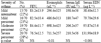
Spirometric and laboratory data of patients categorized according to severity based on symptoms and medication regimen are shown in Table 2.
| Table 1: | The demographic, clinical and laboratory data of study groups |
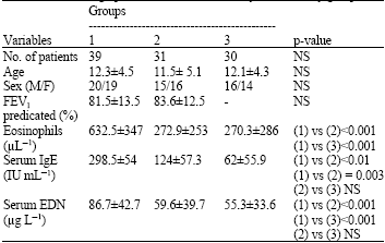 | |
| Table 2: | Spirometric and laboratory data of patients categorized according to severity based on symptoms and medication regimen |
 | |
Bronchial asthma is a common chronic condition that affects up to 10% of adults and 35% of children worldwide and it is an increasing public health problem. Asthma may develop at least in part as a result of genetic or host susceptibility to allergy or atopy and bronchial hyperresponsiveness (Guill, 2004). In present study, we asses both EDN and IgE for their utility as biomarkers for evaluation of asthma as regard type and severity.
The study showed both EDN and IgE were positively correlated with the eosinophilic count. Eosinophils has a crucial role in the pathogenesis and course of asthma, as most allergic and non-allergic asthmatic patients, including those with mild asthma, has a bronchial eosinophilia and there is significant association between eosinophils activation and asthma severity as well as bronchial hyperresponsiveness. Tissue eosinophilia was found to be significantly greater in fatal asthma (Guill, 2004) than in patients with chronic asthma. Eosinophils are recruited and found to be activated during segmental allergen challenge. Soluble vascular cell adhesion molecule-1 (sVCAM-1) levels after segmental antigen challenge correlate with eosinophil influx, IL-4 and IL-5 production and the late-phase response (Zangrilli et al., 1995).
The biological properties of eosinophils include the release of toxic granule proteins, oxygen free radicals, eicosanoids (sulfido-peptide leukotrienes), Th2-like cytokines and growth factors. Once activated, products from eosinophils contract human bronchial smooth muscles, increase vascular permeability and induce airway hyperresponsiveness. Eosinophils are deleterious in asthma by the release of highly toxic products including Major Basic Protein (MBP), Eosinophil Cationic Protein (ECP), Eosinophil-Derived Neurotoxin (EDN) and oxygen free radicals, which induce the shedding of the surface epithelium in keeping with the hypothesis of eosinophil-induced damage of the bronchi (Busse and Sedgwick, 1994).
Eosinophils can be important cells of airways remodeling. Eosinophils can release growth factors, elastase and metalloproteases, involved in the process of tissue remodeling and fibrosis. Eosinophil products stimulate fibroblasts (Pincus et al., 1987). Eosinophils appear to be involved in pulmonary fibrosis or in tropical pulmonary eosinophilia (Schlick, 1993) and have long been associated with endomyocardial fibrosis (Magnaval and Bavel, 2005 ) but the involvement of eosinophils in the fibrotic process is not completely understood. Moreover, in an extensive study, MBP deposition was found to be present in some, but not all, cases of pulmonary fibrosis at the site of the fibrotic lesions (Noguchi et al., 1992).
Regarding evaluation of asthma, first the differentiation of atopic and non atopic type, second, the determination of degree of severity and third the diagnosis of asthma per say in pre-school age.
Concerning the differentiation of atopic and non atopic type, (Amin et al., 2000) reported that in atopic asthmatic subjects, the airway inflammation is characterized by high numbers of eosinophils, mast cells and T lymphocytes, whereas non-atopic asthmatic patients mainly display high numbers of neutrophils and mast cells. There are also distinct structural alterations in the airway mucosa in patients with atopic asthma that are not found in non-atopic asthma. Because there is no clear definition of the asthma phenotype, researchers studying the development of this complex disease turn to characteristics that can be measured objectively. It has been reported that atopic patients with asthma have higher concentrations of exhaled Nitric Oxide (NO) compared with non-atopic patients with asthma who have exhaled NO concentrations similar to normal values (Lúdvíksdottír et al., 1999). Also, atopic patients with asthma are more hyperresponsive to inhaled adenosine-5’-monophosphate (AMP) than non-atopic patients with asthma (Lúdvíksdottir et al., 2000), although skin-prick test and IgE are the widely used tools for this purpose.
The study showed that serum EDN level was significantly higher in atopic asthmatic than non-atopic asthmatic patients and controls. Earlier studies about the relation of EDN to asthma are deficient. Kim et al. (2007) reported that EDN and ECP are significantly higher in atopic asthma than non-atopic asthma and both factors were positively correlated with eosinophilic count. On the other hand, Nuijsink et al. (2007) found weak inverse correlation between urinary EDN and FEV1 and borderline correlation between urinary EDN and PD20 methacholine. No significant correlation was found between urinary EDN and markers of eosinophilic airway inflammation including % eosinophils or ECP levels in induced sputum and so suggested that urinary EDN is not a suitable complementary marker of airway inflammation in asthma.
The importance of differentiating subtypes of asthma specially in pre-school age is critically useful as atopic asthma tends to persist in adult life, furthermore, loss of lung function is more severe and early in atopic asthmatic patients, that is why the atopic patients are more likely to require daily anti-inflammatory medications (Chipps et al., 2007).
Second, assessment of severity of asthma was the scope of many studies. As several eosinophil granular mediators were evaluated for assessment of severity of asthma. Deposition of MBP has been detected around injured epithelium in fatal asthma (Fujisawa et al., 1998). Also, MBP was measured in bronchoalveolar lavage (BAL) and it was found that it alters bronchial hyper-reactivity via activation of C fibers, bradykinins and interaction with muscarinic receptors (Gleich, 2000). Its level was found to be correlated with degree of inflammation, but for ethical purpose, it is unpractical to do BAL on wide base. On the other hand ECP has been widely studied to correlate its serum level to asthma symptoms, pulmonary functions, bronchial hyperresponsiveness and response to therapeutic interventions. Results showed major debate, as some concluded that ECP is a better marker for monitoring and predicting clinical course of asthma than pulmonary function tests (Gleich, 2000; Sorkness et al., 2002), while others concluded that it is a poor indicator of disease activity in chronic asthma and can not differentiate it from nasal inflammation (Ferguson et al., 1995). Regarding respiratory function Bacharier et al. (2004) reported that in children and adolescents, asthma severity classified by symptoms frequency and medications when categorized according to the National Asthma Education and Prevention Program Guidelines does not correlate with forced expiratory volume in 1 sec (FEV1). Another problem in diagnosing and assessing the severity of asthma is that lung function testing is often can not be performed in pre-school children.
In this study, it was found that serum EDN level was significantly correlated with severity of asthma when study groups were categorized based on the frequency of asthma symptoms and medication use. So, ECP might be a supportive biomarker for evaluation of asthma severity. On the other hand, inspite of the wide variations of serum level of IgE in the same groups, they did not mountain to statistically significant differences between them.
Similar results were obtained by Kim et al. (2007), as they found that serum EDN is directly correlated with severity of asthma when study groups are categorized according to both GINA and Joseph-Bowen guidelines and it was more sensitive than eosinophil cationic protein for severity of asthma. Also, Ozol et al. (2008) evaluated the relationships between asthma severity and total IgE in adult female and reported that there is no statistically significant positive relationships between asthma severity and total IgE levels and conclude that atopy does not influence asthma severity in adult female patients.
Lastly, the diagnosis of asthma in children five years and younger presents a particularly difficult problem. This is because episodic wheezing and cough are also common in children who do not have asthma, particularly in those under age of three. Wheezing is usually associated with a viral respiratory illness predominantly respiratory syncytial virus in children younger than two years of age and other viruses in older preschool children. This findings point to the need for a specific biological marker which can diagnose asthma. EDN might be a feasible biomarker for diagnosis of asthma in this age group. A matter which necessitate further studies especially that an interaction between atopy and viral infections appears to be a complex relationship, in which the atopic state can influence the lower airway response to viral infections, viral infections can then influence the development of allergic sensitization and interactions can occur when individuals are exposed simultaneously to both allergens and viruses (Bacharier et al., 2004).
In conclusion, this study suggest that serum EDN may be superior to IgE as a biomarker for evaluation of asthma and its outcome, as it could help to differentiate atopic from non atopic types in addition to its sensitivity to assess the degree of severity of asthma.
REFERENCES
- Amin, K., D. Ludviksdottir, C. Janson, O. Nettelbladt and E. Bjornsson et al., 2000. Inflammation and structural changes in the airways of patients with atopic and non atopic asthma. Am. J. Respir. Crit. Care Med., 162: 2295-2301.
PubMed - Bacharier, L.B., R.C. Strunk, D. Mauger, D. White, R.F. Lemanske, Jr. and C.A. Sorkness, 2004. Classifying asthma in children, mismatch between symptoms, medication use and lung function. Am. J. Respir. Critical Care Med., 170: 426-432.
CrossRefDirect Link - Busse, W.W. and J.B. Sedgwick, 1994. Eosinophil eicosanoid relations in allergic inflammation of the airways. Adv. Prostaglandin Thromboxane Leukotriene Res., 22: 241-249.
PubMed - Chipps, B.E., S.J. Szefler, F.E. Simons, T. Haselkorn, D.R. Mink, Y. Deniz and J.H. Lee, 2007. Demographic and clinical characrteristcs of children and adolescents with severe or difficult to treat asthma. J. Allergy Clin. Immunol., 119: 1156-1163.
PubMed - Ferguson, A.C., R. Vaghan, H. Brown and C. Curtic 1995. Evaluation of serum eosinophilic cationic protein as a marker of disease activity in chronic asthma. J. Allergy Clin. Immunol., 95: 23-28.
PubMed - Fujisawa, T., A. Terada, J. Atsuta, K. Iguchi, H. Kamiya and M. Sakurai, 1998. Clinical utility of serum levels of eosinophil cationic protein (ECP) for monitoring and predicting clinical course in childhood asthma. Clin. Exp. Allergy, 28: 19-25.
Direct Link - Gleich, G.J., 2000. Mechanisms of eosinophil-associated inflammation. J. Allergy Clin. Immunol., 105: 651-663.
PubMedDirect Link - Guill, M.F., 2004. Asthma update: Clinical aspects and management. Pediatr. Rev., 25: 335-344.
PubMedDirect Link - Kim, K.W., K.E. Lee , E.S. Kim, T.W. Song, M.H. Sohn and K.E. Kim, 2007. Serum Eosinophil-Derived Neurotoxin (EDN) in diagnosis and evaluation of severity and bronchial hyperresponsivenees in childhood asthma. Lung, 185: 97-103.
PubMedDirect Link - Lúdvíksdottir, D., H. Hedenström, C. Janson, E. Björnsson and G. Strålenheim et al., 2000. Different airway responsiveness profiles in atopic asthma, nonatopic asthma and Sjögren's syndrome. Allergy, 55: 259-265.
PubMed - Lúdvíksdottír, D., C. Janson M. Högman H. Hedenstöm, E. Björnsson and G. Boman, 1999. Exhaled nitric oxide and its relationship to airway responsiveness and atopy in asthma. Respir Med., 93: 552-556.
PubMed - Magnaval, J.F. and A. Berry, 2005. Tropical eosinophilia. Cli. Infect. Dis., 40: 635-636.
PubMedDirect Link - Motojima, S., E. Frigas, D.A. Loegering and G.J. Gleich, 1989. Toxicicity of eosinophil cationic proteins from guinea pigs tracheal epithelium in vitro. Am. Rev. Resp. Dis., 139: 801-805.
Direct Link - Noguchi, H., G.M. Kephart, T.V. Colby and G.J. Gleich, 1992. Tissue eosinophilia and eosinophil degranulation in syndromes associated with fibrosis. Am. J. Pathol., 140: 521-528.
PubMed - Nuijsink, M., W.C.J. Hop, P.J. Sterk, E.J. Duiverman and P.S. Hiemstra et al., 2007. Urinary eosinophil protein X in children with atopic asthma. Mediators Inflamm., 2007: 49240-49240.
CrossRefDirect Link - Ozol, D., C. Koca, E. Mete and R. Yigitoğlu, 2008. Influence of atopy on asthma severity in adult female patients. J. Investig. Allergol. Clin. Immunol., 18: 36-40.
PubMed - Pincus, S.H., K.S. Ramesh and D.J. Wyler, 1987. Eosinophils stimulate fibroblast DNA synthesis. Blood, 70: 572-574.
Direct Link - Schlick, W., 1993. Current issues in the assessment of interstitial lung disease. Monaldi Arch. Chest Dis., 48: 237-244.
Direct Link - Silfman, N.R., P. Venge, C.G. Peterson and G.J. Gleich, 1989. Human eosinophil protein X and human eosinophil-derived neurotoxin are likely the same protein. J. Immunol., 143: 2317-2322.
Direct Link - Sorkness ,C., K. McGill and W. Busse, 2002. Evaluation of serum cationic protein as a predictive marker for asthma exacerbation in patients with persistent disease. Clin. Exp. Allergy, 32: 1355-1359.
Direct Link - Zangrilli, J.G., J.R. Shaver, R.A. Cirelli, S.K. Cho and C.G. Garlisi et al., 1995. SVCAM-1 levels after segmental antigen challenge correlate with eosinophil influx, IL-4 and IL-5 production and the late-phase response. Am. J. Respir. Crit. Care Med., 151: 1346-1353.
PubMed - Miller, M.R., J. Hankinson, V. Brusasco, F. Burgos and R. Casaburi et al., 2005. Standardisation of spirometry. Eur. Respir. J., 26: 319-338.
CrossRefDirect Link