
S.H. Garba
Department of Veterinary Pharmacology and Physiology,
Faculty of Veterinary Sciences, University ofMaiduguri, Nigeria
J. Prasad
Department of Veterinary Pharmacology and Physiology,
Faculty of Veterinary Sciences, University ofMaiduguri, Nigeria
U. K. Sandabe
Department of Veterinary Pharmacology and Physiology,
Faculty of Veterinary Sciences, University ofMaiduguri, Nigeria
Journal of Biological Sciences
Year: 2007 | Volume: 7 | Issue: 2 | Page No.: 276-281


ABSTRACT
The aqueous root-bark extract of Ficus sycomorus (Linn) was tested for its chemical constituents, acute toxicity and hepatoprotective effect against Carbon tetrachloride (CCl4) induced hepatotoxicity in rats. A total of 78 adult albino rats weighing between 150-320 g were used. The animals were each weighed at the start of the experiment and divided into two segments consisting of 42 rats for the acute toxicity and 36 rats for the hepatoprotective study segments, respectively. In the acute toxicity study the aqueous extract of the root-bark of Ficus sycomorus was administered intraperitoneally (ip) in a dose range of 0.2-12 g kg-1 and the rats were observed for the physical signs of toxicity for 24 h. The hepatoprotective segment involved dosing the negative control rats intraperitonealy with carbon tetrachloride (CCl4) 3 mL kg-1 that was dissolved in corn oil to induce liver damage while the treatments groups were pretreated with 640 mg kg-1 of the extract orally an hour before CCl4 (3 mL kg-1) was administered to observe if the extract has any hepatoprotective effect against CCl4 induced hepatotoxicity At the end of each treatment period, the animals were weighed and blood was obtained from animals administered CCl4 and pre-treated with 640 mg kg-1 of the extract for biochemical analysis with the liver extracted, weighed and processed for histological assessment. Phytochemical analysis of the extract revealed the presence of saponins, flavonoids, alkaloids, tannins and reducing sugar and LD50 was calculated as 3.20�0.6031 g kg-1. Pre-treatment of the rats with the extract was able to reduce though not significantly, changes in the biochemical parameters (decrease in albumin but increase in Aspartate Transaminase (AST), Alanine-Transaminase (ALT), Alkaline Phosphatase (ALP) and bilirubin) and preserved the liver parenchymal architecture against CCl4 induced degenerative changes, fibroplasia and cirrhosis. The results of this study showed that the plant extract had hepatoprotective effect on the parenchymal architecture of the liver against CCL4 induced hepatotoxicity in rats. But further studies to observe its hepatocurative potentials would be useful and is recommended.
PDF Abstract XML References Citation
How to cite this article
S.H. Garba, J. Prasad and U. K. Sandabe, 2007. Hepatoprotective Effect of the Aqueous Root-Bark Extract of Ficus sycomorus (Linn) on Carbon Tetrachloride Induced Hepatotoxicity in Rats. Journal of Biological Sciences, 7: 276-281.
DOI: 10.3923/jbs.2007.276.281
URL: https://scialert.net/abstract/?doi=jbs.2007.276.281
DOI: 10.3923/jbs.2007.276.281
URL: https://scialert.net/abstract/?doi=jbs.2007.276.281
INTRODUCTION
Traditional healers and local people in Africa rely heavily on medicinal plants for curing illnesses and as tonics and Ficus sycomorus (Moracea) is one of such plants and was venerated as a holy tree of outstanding importance in ancient Egypt and is found scattered around Maiduguri, in the Sahel, Sudan and Guinea Savannas and usually grows on sites with high ground water table. Usually it prefers fresh nutrient rich soils, particularly riverbanks and water holes.
Extracts obtained from the fruits, leaves, stem bark and root bark usually administered in the form of infusions, decoctions, tinctures, syrups and lotions have been used in the treatment of a wide range of diseases and disorders in various African countries (NAPRALERT, WHO, 2003). Oral administration of the dried fruit extract has been used in the treatment of tuberculosis. (Arnold and Gulumian, 1984). Abdurrahman (1992) has also reported the use of the plant extract in the treatment of mental illness. Its sedative and anticonvulsive properties have also been documented in rats (Sandabe et al., 2003). Its indigenous names include Tarmu (kanuri), Baure (Hausa) and Kamda (Babur/Bura) In view of its many uses in traditional medicine this study was undertaken to investigate if the plant has any hepatoprotective effect against Carbon tetrachloride (CCl4) induced hepatotoxicity.
MATERIALS AND METHODS
Collection and identification of plant materials: The plant part (root bark) was collected from Maiduguri Metropolis. The plant was identified and authenticated by Dr. S.S. Sanusi (plant taxonomist) of the Department of Biological Sciences, University of Maiduguri, Borno state. A specimen voucher (FS.01) of the plant was prepared and deposited at the herbarium of the Department of Veterinary Physiology and Pharmacology, University of Maiduguri. The root bark was sun-dried for 3 days and pulverized using a pestle and mortar. The pulverized parts were stored in cellophane bags at room temperature.
Preparation of extract: The World Health Organization (WHO, 1992) procedure of extraction was adopted for this study. One hundred grams of the root bark powder of Ficus sycomorus (L) were subjected to exhaustive soxhlet extraction in 500 mL of distilled water for 72 h. The extract obtained was concentrated in a water bath until a constant dark sticky residue was obtained. This was further oven dried and maintained in a desiccator until a constant weight was obtained. This gave a mean extract yield of 8.5 g w/w. The dried root bark extract obtained was stored in a tightly stoperred container in a refrigerator at -4°C until required. Stock solution of the extract was prepared by dissolving 2 g weight of the powdered root bark in 20 mL of normal saline and the concentration used was 0.1 g mL-1.
Phytochemical screening: Phytochemical screening of the extract was performed according to the methods of Clarke (1975), Odebiyi and Sofowora (1978) and Trease and Evans (1989). Tests for carbohydrates, tannins, saponins, flavonoids and alkaloids were carried out.
Experimental animals: This study was carried out in the Departments of Human Anatomy and Human Physiology, University of Maiduguri, Nigeria between March and October, 2004. A total of 78 adult albino rats of the Wistar strain weighing 150 and 320 g and 3-4 months of both sexes were used. They were purchased from the animal house of the Department of pharmacology and pharmaceutical sciences, University of Jos, Plateau State, Nigeria. Following an acclimation period of 2 weeks, the rats were individually identified by colour tattoo and weighed. The rats were kept in plastic cages at room temperature of 32±4°C and <30% relative humidity with a 12 h light/dark cycle. They had access to standard laboratory diet (Sanders Nigeria Limited, Kaduna) and drinking water ad libitum.
Acute toxicity study: For the determination of the median lathal dose (LD50 ), a total of 42 rats were randomly divided into seven groups of 6 rats each. The extract was administered intraperitoneally (ip) in a dose range of 0.2-12 g kg-1 and a maximum of 1.0 mL was administered. All the rats were observed for the physical signs of toxicity for 24 h. The LD50 was calculated by the method of Miller and Tainter (1944).
Hepatoprotective study: A total of 36 rats were used for this study. The rats were divided randomly, into 3 broad groups (A, B and C) of 12 rats each, with each broad group consisting of subgroups I, II and III. Each subgroup was made up of 6 rats. Rats in subgroup I served as control subgroup II were administered intraperitonealy carbon tetrachloride (CCl4) 3 mL kg-1 that was dissolved in corn oil to induce liver damage while rats in subgroup III were administered 640 mg kg-1 of the extract orally an hour before CCl4 (3 mL kg-1) was administered to observe if the extract has any hepatoprotective effect against CCl4 induced hepatotoxicity. At the end of every 2 weeks, rats of one broad group were anaesthetized; blood and liver tissues were collected for biochemical and histological analysis, respectively.
Biochemical analysis: The blood collected from the jugular vein were put into sterile bottles and centrifuged at a rate of 12,000 revolutions per minute (rpm) for 10 min. The clear serum obtained was analyzed for Aspartatetransaminase (AST), Albumin, Alaninetransaminase (ALT), Alkaline phosphatase (ALP) and Bilirubin using Randox Laboratory kits.
Histological analysis: The liver tissues obtained was carefully dissected out, weighed, fixed in Bouins fluid, embedded in paraffin and sectioned at 5 μm. Sections were stained with Haematoxylin and Eosin and mounted in Canada balsam. Light microscopic examination of the sections was then carried out.
Statistical analysis: Numerical data obtained from the study were expressed as the mean value±standard error of mean. Differences among means of control and treated groups were determined using Statistical Package for Social Scientist (SPSS 11.0). A probability level of less than 5% (p<0.05) was considered significant.
RESULTS
Phytochemical screening: The phytochemical screening of the aqueous extract of the root-bark of F. sycomorus (L.) showed that it contained reducing sugars, tannins, alkaloids, saponins and flavonoids.
Acute toxicity study: The physical signs of toxicity observed ranged from decreased motor activity, loss of appetite increased respiratory rate, which was followed by restlessness and gasping for air to death The median lethal dose of the extract was calculated to be 3.20±0.60 g kg-1 body weight.
Hepatoprotective study
Effect of CCl4 and extract on mean body and liver weight: There was a significant (p<0.001) decrease in the body weight of all the rats treated with CCl4 (3 mL kg-1) and those pretreated with 640 mg kg-1 of extract (Table 1). There was also a significant (p<0.001-p<0.05) increase in the liver weight of all the rats that were treated with CCl4. A significant increase (p<0.01) was also observed in the rats that were pretreated with 640 mg kg-1 of extract for 2 weeks (Table 1).
Effect of CCL4 and of extract on biochemical parameters of the liver: There was a significant increase in the levels of AST, ALT, Alkaline phosphatase and bilirubin levels of all the rats that received CCl4 (3 mL kg-1) for 2, 4 and 6 weeks. The levels of Albumin were significantly reduced in rats that were treated with CCl4 (3 mL kg-1). Pretreatment of the rats with 640 mg kg-1 of the extract before CCl4 was induced showed a decrease in the levels of AST, ALT, Alkaline phosphatase and bilirubin levels when compared to that caused by CCl4 (3 mL kg-1). There was also an increase in the albumin level of rats pretreated with the extract as compared to those that received only CCl4 (Table 2).
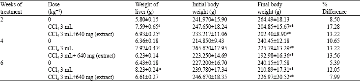
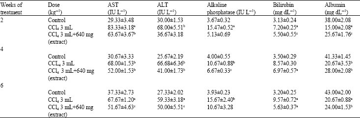
Histological findings: No histological or macroscopic alterations were found in the liver of the control rats (Fig. 1). Rats treated with carbon tetrachloride (CCl4 3 mL kg-1) for 2, 4 and 6 weeks showed few hepatocytes with vacuoles in their cytoplasm (degeneration), fibroplasia of interlobular septae and pseudolobulation (Liver cirrhosis) with macrophages distended with bile pigments (Fig. 2, 3 and 4). The Liver sections of rats pretreated with 640 mg kg-1 of extract before CCl4 (3 mL kg-1) was administered presented a higher level of preservation of the parenchymal architecture. Hepatic cells, sinusoids and nuclei appeared normal and a virtually normal hepatic lobule was also observed (Fig. 5 and 6).
| Table 1: | Effect of carbon tetrachloride (CCl4) and aqueous root bark extract of Ficus sycomorus on the rat mean liver and body weight |
 | |
Significance relative to control a = p<0.001, b = p<0.01, c = p<0.02, d = p<0.05, n = 6, * weight loss. Results are presented as mean±SEM | |
| Table 2: | Effect of carbon tetrachloride (CCl4) and aqueous root bark extract of Ficus sycomorus (L.) on biochemical parameters of the liver |
 | |
Significance relative to control a = p<0.001, b = p<0.01, c = p<0.02, n = 6. Results are presented as Mean±SEM | |
 | |
| Fig. 1: | Light micrograph of a paraffin section of the liver of a control rat showing normal hepatic parenchyma. Central vein (CV), Sinusoid (S) and Hepatocytes (H). H & E stain x 400 |
 | |
| Fig. 2: | Light micrograph of the paraffin section of the liver of a rat treated with CCl4 (3 mL kgG1) for 4 weeks showing few hepatocytes with vacuoles in their cytoplasm (arrow) and marked fibroplasia of interlobular septae (s) extending into the lobules creating pseudolobulation of hepatic lobules (a) (Liver cirrhosis). H & E stain x 200 |
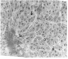 | |
| Fig. 3: | Light micrograph of the paraffin section of the liver of a rat treated with CCl4 (3 mL kgG1) for 6 weeks showing hepatocytes with vacuoles (arrow), marked fibroplasia of interlobular septae (s). H & E stain x 400 |
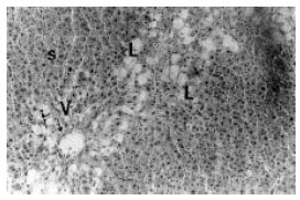 | |
| Fig. 4: | Light micrograph of the paraffin section of the liver of a rat treated with CCl4 (3 mL kgG1) for 6 weeks showing smaller vacuoles (arrow), fibroplasia of interlobular septae (s), large vacuoles (L) and enlarged vesicular nuclei (V). H & E stain x 200 |
 | |
| Fig. 5: | Light micrograph of the paraffin section of the liver of a rat pretreated with 640 mg kgG1 of extract before CCl4 (3 mL kgG1) was administered for 4 weeks showing lesser preservation of parenchymal architecture, Hepatic cell (H), Sinusoids (S) and Nuclei (arrow). H & E stain x 200 |
 | |
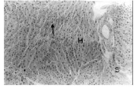
| Fig. 6: | Light micrograph of the paraffin section of the liver of a rat pretreated with 640 mg kgG1 of extract before CCl4 (3 mL kgG1) was administered for 6 weeks showing preservation of parenchymal architecture with the Hepatic cell (H), Sinusoids (S), Central vein (CV), Nuclei (arrow) and hepatic lobulation appearing normal. H & E stain x 200 J. Biol. Sci., 7 (2): 276-281, 2007 279 |
DISCUSSION
The increase in the levels of aminotransferases especially ALT following administration of 3 mL kg-1 of CCl4 is an indication of liver damage. The hepatotoxic effect of CCl4 is due to its conversion by the P-450 mixed function oxidase in the smooth endoplasmic reticulum of the liver and other organs to the highly reactive toxic free radical CCl3. The free radical produced locally, causes auto-oxidation of the polyenic fatty acids present within the membrane phospholipids. There, oxidative decomposition of the lipid is initiated and organic peroxide is formed after reacting with oxygen (lipid peroxidation). This reaction is autocatalytic in that new radicals are formed from the peroxide radicals themselves. Thus, rapid breakdown of the structure and function of the endoplasmic reticulum is due to decomposition of the lipid. There is decline in hepatic protein synthesis and swelling of smooth endoplasmic reticulum and dissociation of ribosomes from the endoplasmic reticulum. Lipid export from the hepatocytes is reduced owing to their inability to synthesize apoprotein to complex with triglycerides and thereby facilitate lipoprotein secretion, mitochondrial injury then occurs and this is followed by progressive swelling of the cells due to increased permeability of the plasma membrane which is followed by a massive influx of calcium and cell death.
It is known that an increase in the enzymatic activity of ALT and AST in the serum directly reflects a major permeability or cell rupture (Wittwer and Bohmwald, 1986; Benjamin, 1978). An increase in AST and ALT, a hepatospecific enzyme that is principally found in the cytoplasm in rats (Benjamin, 1978; Ringler and Dabich, 1979) following administration of CCl4 is attributed to the increased release of the enzymes from damaged liver parenchymal cells (Singh, 1980). The elevated level of alkaline phosphatase an enzyme produced in the liver, bone and placenta indicates liver injury or bile duct obstruction as a result of CCl4 administration. Bilirubin a major breakdown product that results from the destruction of RBC which is removed from the blood by the liver through conjugation and secreted into bile usually becomes elevated as a result of decreased uptake by the liver, decreased conjugation, decreased secretion from the liver or blockage of the bile ducts that is caused in liver damage. The low serum albumin concentration observed in this study following CCl4 administration indicates poor liver function because albumin is synthesized by the liver and secreted into the blood. Serum albumin concentration is usually normal in chronic liver diseases until cirrhosis and significant liver damage is present (Shiff and Schiff, 1982).
Pretreatment of the liver with 640 mg kg-1 of the extract showed that the extract antagonizes the CCl4 induced elevation of AST, ALT, alkaline phosphatase and bilirubin and the decrease in albumin levels. The results appears to confirm the findings of Mandal et al. (2000), which showed that rats pretreated with 400 mg kg-1 of the dried leaf extract of Ficus racemosa showed a decrease in the level of SGOT, alkaline phosphatase and bilirubin values when compared with CCl4 treated rats. This indicates a possible decrease in the CCl4 induced destruction of liver parenchymal cells or it might be possible that the extract may have also modulated the effect of CCl4 on the synthesis and destruction of these biochemical parameters.
Carbon tetrachloride administration to rats causes changes in the lipid profile of both liver and serum. The major lipid components in the plasma, such as cholesterol and triglyceride, do not circulate in the free state but are transported in the form of complexes with lipoprotein. Any disturbance in lipoprotein metabolism is reflected in the lipid profiles of plasma and liver. Since the liver has a major role in the metabolism of lipoprotein, any derangement in its activity leads to alterations in the lipid profile of plasma. Several factors may play a role in the accumulation of lipids in the liver. An increase in the level of free fatty acids in plasma, as a result of mobilization of fat from adipose tissue or from the hydrolysis of lipoprotein, chylomicrons or triglycerides by lipoprotein lipase in extrahepatic tissues (Martin et al., 1983), leads to their increased uptake and esterification in the liver. Since the production of lipoprotein does not keep pace with the increased free fatty acids, triglycerides and cholesterol accumulate in the liver. Fat accumulation in the liver may also occur if there is a derangement in the production of lipoprotein, especially its apoprotein part (Martin et al., 1983).
Pretreatment of the liver with 640 mg kg-1 of the extract was observed to have significantly reduced the destruction of the parenchymal cells and architecture. From the data presented above it is not possible to delineate the exact mechanism of the hepatoprotective effect observed with the extract.
Analysis of the results shows that the extract reversed the majority of the CCl4-induced alterations in liver and serum biochemical parameters. This clearly indicates that it possesses significant hepatoprotective activity against CCl4 induced injury. Histopathological observations further corroborate this inference.
As already mentioned, the extract used in this study contained α-amyrin and a flavonoid fraction. Hepatoprotective activity has been reported for a mixture of α-amyrin and several flavonoid fractions isolated from different plants.
The result of the study suggest that the plant extract produced a significant decrease in body weight, showed hepatoprotective effect on the parenchymal architecture of the liver against CCL4 induced hepatotoxicity in rats. But further studies to observe its hepatocurative potentials would be useful and is recommended.
ACKNOWLEDGMENTS
We wish to acknowledge the technical assistance of Ibrahim Wiam and Justus Jibrin of the Departments of Veterinary Anatomy and Human Pharmacology, University of Maiduguri, Nigeria.
REFERENCES
- Mandal, S.C., B. Saraswathi, C.K.A. Kumar, S.M. Lakshmi and B.C. Maiti, 2000. Protective effect of leaf extract of Ficus hispida Linn. against paracetamol-induced hepatotoxicity in rats. Phytother. Res., 14: 457-459.
CrossRefPubMedDirect Link - Miller, L.C. and M.L. Tainter, 1944. Estimation of the ED50 and its error by means of logarithmic-probit graph paper. Exp. Biol. Med., 57: 261-264.
CrossRefDirect Link - Odebiyi, O.O. and E.A. Sofowora, 1978. Phytochemical screening of Nigerian medicinal plants II. Lloydia, 41: 234-246.
PubMedDirect Link - Sandabe, U.K., A.O. Patrick and A.C. Gregory, 2003. Sedative and anticonvulsant effects of aqueous extract of Ficus syconorus L. stem bark in rats. Vet. Arch., 73: 103-110.
Direct Link - Evans, W.C. and G.E. Trease, 1989. Trease and Evans' Pharmacognosy. 13th Edn., Baillière Tindall, London, ISBN: 9780702013577, Pages: 832.
Direct Link