
M. B. Maina
Department of Human Anatomy, College of Medical Sciences, University of Maiduguri, Borno State, P.M.B, 1069, Nigeria
Y. C. Shapu
Department of Human Anatomy, College of Medical Sciences, University of Maiduguri, Borno State, P.M.B, 1069, Nigeria
S. H. Garba
Department of Human Anatomy, College of Medical Sciences, University of Maiduguri, Borno State, P.M.B, 1069, Nigeria
M. A. Muhammad
Department of Human Anatomy, College of Medical Sciences, University of Maiduguri, Borno State, P.M.B, 1069, Nigeria
A. M. Garba
Department of Human Anatomy, College of Medical Sciences, University of Maiduguri, Borno State, P.M.B, 1069, Nigeria
A. U. Yaro
Department of Human Anatomy, College of Medical Sciences, University of Maiduguri, Borno State, P.M.B, 1069, Nigeria
O. N. Omoniyi
Department of Human Anatomy, College of Medical Sciences, University of Maiduguri, Borno State, P.M.B, 1069, Nigeria
Journal of Applied Sciences
Year: 2011 | Volume: 11 | Issue: 14 | Page No.: 2662-2665


ABSTRACT
Cranial capacity is a measure of the volume of the interior of the cranium which is sometimes used as a rough indicator of the size of the brain and is mostly affected by environmental, geographical, gender, age, nutritional and racial factors. A survey of available literature indicates a lack of study on cranial capacities in adults resident in Maiduguri North Eastern Nigeria, this study was therefore undertaken to determine the Cranial Capacities (CC) of 300 (150 males, 150 females) aged 18-35 years adults resident in Maiduguri Metropolis using a random stratified method. Linear measurements of cranial length, width, height and head circumference were undertaken and their cranial capacities calculated. The Mean (±SD) of cranial capacity was significantly (p<0.0001) higher in males (1424.4±137.9) than in females (1331.3±201.8). Cranial length and height were also found to be significantly higher in males than in females. The results obtained from this study confirms that the cranial volume of males is higher than that of the females and also suggest that the cranial capacity of adult males and females were slightly higher than those of similar studies carried out in some ethnic groups indigenous to the southern part of Nigeria.
PDF Abstract XML References Citation
Received: September 10, 2010;
Accepted: February 18, 2011;
Published: June 07, 2011
How to cite this article
M. B. Maina, Y. C. Shapu, S. H. Garba, M. A. Muhammad, A. M. Garba, A. U. Yaro and O. N. Omoniyi, 2011. Assessments of Cranial Capacities in a North-Eastern Adult Nigerian Population. Journal of Applied Sciences, 11: 2662-2665.
DOI: 10.3923/jas.2011.2662.2665
URL: https://scialert.net/abstract/?doi=jas.2011.2662.2665
DOI: 10.3923/jas.2011.2662.2665
URL: https://scialert.net/abstract/?doi=jas.2011.2662.2665
INTRODUCTION
The influence of genetic, physiological and anatomical differences between males and females had been well documented in various studies (Guyton and Hall, 2006; Moore and Dalley, 1999; Chandrashekar et al., 2005) although, some differences are indistinctive, most are related to body dimensions which are determined by anthropometry (Chamela, 1997; Williams et al., 1995). Craniometric study is an important fraction of anthropometry that can be employed in the determination of the cranial capacity of an individual. Cranial capacity like several bodily dimensions are affected by environmental, ecological, biological, geographical, racial, gender and age factors (Heidari et al., 2006; Golalipour and Kamran, 2004; Golalipour et al., 2003, 2005; Okupe et al., 1984; Tuli et al., 1995; Rajlakshmi et al., 2001). The capacity of the cranium has in many studies been used to indirectly reflect the volume of the brain and to predict mental ability (Gault et al., 1988; Wolf et al., 2003; Mazonakis et al., 2004). Anthropometric studies carried out on the cranium as it relates to gender have shown a lot of variations with males possessing higher values than females in both adults and neonates (Manjunath, 2002b; Acer et al., 2007; Garba et al., 2008; Raji et al., 2010; Odokuma et al., 2010a).
Most studies carried out to determine cranial capacities in a population are done with the aim of detecting the effect of various fundamental parameters such as racial, geographic, ethnic and dietary factors etc. (Manjunath, 2002b). Such studies can also provide useful results in the field of forensic anthropology and paediatrics as an indicator of skull development in both female and male individuals (Pereira et al., 2008). It is also a good determinant of normal or abnormal skull; because, living humans have a cranial capacity ranging from about 950 to 1800 cc, with an average of about 1400 cc (Milner, 1990). Against this background this study was carried out to determine if healthy adults resident in Maiduguri Metropolis of Borno State a very hot and arid region in the North Eastern Region of Nigeria will concur or differ from few studies carried out in other part of Nigeria (Umar et al., 2006; Odokuma et al., 2010b). The objective of the study is to determine the cranial capacities in males and females and to compare our results with similar studies carried out in the southern part of Nigeria.
MATERIALS AND METHODS
Subjects: This study was conducted on 300 (150 males, 150 females) volunteers whose ages ranged between 18 and 33 years. The subjects were randomly selected from persons whose parents and grand parents were of Nigerian origin and showed no obvious physical craniofacial deformity and resident in Maiduguri metropolis, north eastern Nigerian from April to November 2009.
| Table 1: | Comparison of the means of cranial capacity and head measurements between males and females |
| *Significant level p<0.05; **Significant level p<0.001; ***Significant level p<0.0001, n = 150 males, 150 females | |
| Table 2: | Linear Regression between cranial capacity and head measurements |
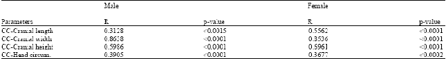 | |
| n = 150 Males, 150 Females | |
| Table 3: | Mean cranial capacity in southern and north eastern nigerian male and female adult population |
| ***Significant level p<0.0001 | |
Prior and informed consent was obtained from the subjects and the study was carried out after dully obtaining clearance from the ethical committee of the College of Medical Sciences, University of Maiduguri, Nigeria.
Craniometric measurements: Standardised measurements of cranial length, cranial width, cranial height and Head circumference were taken with the subject sitting relaxed on a chair, with the head in anatomical position using a spreading calliper and measuring tape.
All measurements were carried out after careful palpation of the head for anatomical landmarks and measurements were taken to the nearest 1mm by a single investigator thrice and the average recorded for computation and subsequent analysis (Umar et al., 2006).
| • | Cranial length: It was taken as the linear length from the glabella to the Inion |
| • | Cranial width: It was taken as the linear distance between the parietal eminences |
| • | Cranial height: It was taken as the linear distance from the root of nose (nasion) to highest point of head (vertex) |
| • | Head circumference: It was measured by passing a measuring tape across a point slightly above the glabella, superciliary arch and superior margin of the external acoustic meatus round via the inion and back to the point above the glabella |
Cranial capacity in males and females were then calculated according to the following formulae (Williams et al., 1995; Manjunath, 2002b).
| • | Males: 0.000337(L-11) (W-11) (H-11)+406.01 |
| • | Females: 0.000400(L-11) (W-11) (H-11)+206.60 |
Statistical analysis: Data obtained from the subjects were recorded in a specially designed form and then transferred into SPSS (2001) 11.0.1 for analysis. The means obtained from this study were subjected to analyses of variance (ANOVA) for comparison within gender and multivariate analyses (MANOVA) was used between this study and similar studies for assessment of statistical significance and linearity with a probability level of less than 0.05 considered significant.
RESULTS AND DISCUSSION
The Mean±SD of cranial volume obtained from this study was 1424.4±137.9 and 1331.3±201.8 cm3 in females (Table 1). Cranial length, width, height and circumference were 191.11±6.4, 135.90±12.9, 145.15±7.5 and 564.51±14.1 cm3, respectively in males while females presented with 183.53±9.9, 135.47±14.9, 141.29±7.6 and 570.03±26.7 cm3, respectively (Table 1). The results indicates that the values in males were significantly (p<0.05-0.0001) higher in males than in females. Analysis for Linear regression between cranial capacity and cranial length, width, height and circumference of males and females revealed a positive correlation in all cases (Table 2).
Comparison between our study and a study carried out by Odokuma et al. (2010a) in three major ethnic groups resident in the southern part of Nigeria is presented in Table 3. Comparison of the male and female population of the three ethnic groups in the study of Odokuma et al. (2010a) with our study showed a significantly (p<0.0001) higher cranial capacity value of 1424.40±137.9 against 1310.65±124.57 and 1208.77±138.65 against 1331.30±201.8 cm3 in males and females, respectively.
Anthropometric studies that have intercultural comparisons as a basis have in many ways given insights into the homogeneity of the human population. The knowledge of the cranium of either a dry skull or of a living being is of significant importance to the study and comparison of populations with various fundamental differences like racial, geographic, ethnic and dietary characteristics. Medically, an analysis of cranial capacity expresses another aspect of growth and development and permits critical evaluation of unusually large, small or misshapen crania (Haack and Meihoff, 1971). Craniometric data is used in mainstream science to compare modern-day animal species and to analyze the evolution of the human species in archeology. Fossil hominids are often found fragmented and are reconstructed upon a paradigm according to the law of correlation (Jerrison, 1979; Odokuma et al., 2010b).
In this study, the Mean (±SD) of cranial capacity were 1424.4±137.9 and 1331.3±201.8 in males and females, respectively with the values significantly (p<0.0001) higher in males than in females. These values were also higher when compared to a study in North Iran by Golalipour et al. (2005), who reported a cranial capacity of 1420.60±85 and 1227.2±120 cc in 17-20 years Turkman males and females and 1369.4±142 and 1215.8±125 cc in 17-20 years Fars males and females, respectively. The findings also revealed that the cranial capacity were higher compared to findings reported by Acer et al. (2007) who revealed a cranial capacity of 1375.67±91.17 and 1237.32±95.12 cm3 in 17-26 years males and females Mugla University students in Turkey. It was also higher than cranial volume of Indian males and females dissecting room cadavers with 1152.813±279.16 and 1117.82±99.09 cc, respectively (Manjunath, 2002a). But a work conducted by Hwang et al. (1995) in Korea reported a cranial volume of 1470±107 in males and 1317±117 cc in females indicating that the male Korean skull had a higher cranial volume than the males of our study but the results obtained from their study on the female skulls were lower than the females in this study.
The observed higher mean cranial capacities of males than that of the females observed in this study tends to agree with similar works conducted on brain weight were the males had significantly higher values than that of the females which could be explained by the generally bigger frame of the average male than the average female. Moreover, it has been shown that gender differences in brain weight could be attributed to activities in which the specific sex excelled (Andy, 1992; Rushton and Osborne, 1995). The higher male values also corresponds with similar findings in North-Iran (Golalipour et al., 2005), in Turkey (Acer et al., 2007), in India (Manjunath, 2002a) and in Korea (Hwang et al., 1995). However, contradictions do exist between the values of the mean cranial capacities in both males and females in this study with other studies by Golalipour et al. (2005), Acer et al. (2007), Manjunath (2002a) and Hwang et al. (1995). This is probably due to ecological, biological, geographical, racial, gender and age factors which have been cited to influence several bodily dimensions (Golalipour et al., 2003; Okupe et al., 1984; Tuli et al., 1995; Rajlakshmi et al., 2001).
CONCLUSION
The values obtained from this study have been able to demonstrate that cranial capacities are higher in males than in females and that the cranial capacity of adult males and females were slightly higher than those of similar studies carried out in some ethnic groups indigenous to the southern part of Nigeria.
ACKNOWLEDGMENT
We thank Yakaka Bukar Maina of Division of General Studies and Mohammad Buba of Department of Human Anatomy for their assistance.
REFERENCES
- Acer, N., M. Usanmaz, U. Tugay and T. Ertekin, 2007. Estimation of cranial capacity in 17-26 years old university students. Int. J. Morphol., 25: 65-70.
Direct Link - Chandrashekar, N., J. Slauterbeck and J. Hashemi, 2005. Sex-based differences in the anthropometric characteristics of the anterior cruciate ligament and its relation to intercondylar notch geometry: A cadaveric study. Am. J. Sports Med., 33: 1492-1498.
PubMedDirect Link - Garba, S.H., A.I. Numan and I.G. Mishara, 2008. Craniofacial classification of normal newborns in Maiduguri metropolis, Nigeria. Int. J. Morphol., 26: 407-410.
Direct Link - Gault, D., F. Brunelle, D. Renier and D. Marchac, 1988. The calculation of intracranial volume using CT scans. Childs Nerv. Syst., 4: 271-273.
Direct Link - Golalipour, M.J. and K. Heydari, 2006. Effect of the ethnic factor on cranial capacity and brain weight of male newborns in northern Iran. Neuroembryol. Aging, 3: 146-148.
CrossRefDirect Link - Golalipour, M.J., M. Jahanshaei and K. Haidari, 2005. Estimation of cranial capacity in 17-20 years old in south east of Caspian Sea Border (North of Iran). Int. J. Morphol., 23: 301-304.
CrossRefDirect Link - Golalipour, M.J., K. Haidari, M. Jahanshahi and RM. Farahani, 2003. The shapes of head and face in normal male newborns in south east of Caspian sea (Iran-Gorgan). J. Anat. Soc. India, 52: 28-31.
Direct Link - Guyton, A.C. and J.E. Hall, 2006. Textbook of Medical Physiology. 11th Edn., Elsevier Saunder, Philadelphia, ISBN: 9788481749267, Pages: 1116.
Direct Link - Haack, D.C. and E.C. Meihoff, 1971. A method for estimation of cranial capacity from cephalometric Roentgnograms. Am. J. Physical Anthropol., 34: 447-452.
CrossRef - Heidari, Z., H.R.M. Sagheb and M.H.N. Mugahi, 2006. Morphological evaluation of head and face in 18-25 years old women in Southeast of Iran. J. Med. Sci., 6: 400-404.
CrossRefDirect Link - Hwang, Y., K.H. Lee, B.Y. Choi, K.S. Lee and H.Y. Lee et al., 1995. Study on the Korean adult cranical capacity. J. Korean Sci., 10: 239-242.
Direct Link - Manjunath, K.Y., 2002. Estimation of cranial volume in dissecting room cadavers. J. Anat. Soc. India., 51: 168-172.
Direct Link - Manjunath, K.Y., 2002. Estimation of cranial volume-an overview of methodologies. J. Anat. Soc. India, 51: 85-91.
Direct Link - Mazonakis, M., S. Karampekios, J. Damilakis, A. Voloudaki and N. Gourtsoyiannis, 2004. Stereological estimation of total intracranial volume on CT images. Eur. Radiol., 14: 1285-1290.
CrossRefDirect Link - Odokuma, E.I., P.S. Igbigbi, F.C. Akpuaka and U.B. Esigbenu, 2010. Craniometric patterns of three Nigerian ethnic groups. Int. J. Med. Med. Sci., 2: 34-37.
Direct Link - Odokuma, E.I., F. Gerald and C. Vhriterhire, 2010. Pattern of brain weight in three West African populations. TAF Prev. Med. Bull., 9: 321-324.
Direct Link - Okupe, R.F., O.O. Coker and S.A. Gbajumo, 1984. Assessment of fetal biparietal diameter during normal pregnancy by ultrasound in Nigerian women. Br. J. Obstet. Gynaecol., 99: 629-632.
PubMed - Pereira, I.M.R., A.A.B. Filho, B.R. Alvares, E.T. Palomari and L. Nanni, 2008. Radiological determination of cranial size and index by measurement of skull diameters in a population of children in Brazil. Radiol Bras., 41: 229-234.
CrossRefDirect Link - Rajlakshmi, C., S.M. Shyamo, T.H. Bidhumukhi and S.L. Chandramani, 2001. Cephalic index of foetuses of manipuri population: A baseline study. J. Anat. Soc. India, 50: 8-10.
Direct Link - Raji, J.M., S.H. Garba, A.I. Numan, M.A. Waziri and M.B. Maina, 2010. Morphological evaluation of head and face shapes in a North-Eastern Nigerian population. Aust. J. Basic Applied Sci., 4: 3338-3341.
Direct Link - Rushton, J.P. and R.T. Osborne, 1995. Genetic and environmental contributions to cranial capacity in black and white adolescents. Intell., 20: 1-13.
CrossRef - Tuli, A., R. Choudhry, S. Agarwal, C. Anand and H. Garg, 1995. Correlation between craniofacial dimensions and foetal age. J. Anat. Soc. India, 44: 1-12.
Direct Link - Umar, M.B.T., R. Singh and A.I. Shugaba, 2006. Cephalometric indicies among nigerians. J. Applied Sci., 6: 939-942.
CrossRefDirect Link - Wolf, H., F. Kruggel, A. Hensel, L.O. Wahlund, T. Arendt and H.J. Gertz, 2003. The relationship between head size and intracranial volume in elderly subjects. Brain Res., 973: 74-80.
PubMedDirect Link