
L. Ouattara
Institut des Sciences de la nature et de la vie, Universite Polytechnique de Bobo-Dioulasso 01 BP 1091 Bobo-Dioulasso 01, Burkina-Faso
J. Koudou
Laboratoire de Chimie des Substances Naturelles, Faculte des Sciences BP908, Universite de Bangui, Centrafrique
C. Zongo
CRSBAN, Laboratoire de Biochimie et Pharmacologie, Universite de Ouagadougou, 03 BP 7131 Ouagadougou, Burkina-Faso
N. Barro
CRSBAN, Laboratoire de Biochimie et Pharmacologie, Universite de Ouagadougou, 03 BP 7131 Ouagadougou, Burkina-Faso
A. Savadogo
CRSBAN, Laboratoire de Biochimie et Pharmacologie, Universite de Ouagadougou, 03 BP 7131 Ouagadougou, Burkina-Faso
I. H. N. Bassole
CRSBAN, Laboratoire de Biochimie et Pharmacologie, Universite de Ouagadougou, 03 BP 7131 Ouagadougou, Burkina-Faso
A. S. Ouattara
CRSBAN, Laboratoire de Biochimie et Pharmacologie, Universite de Ouagadougou, 03 BP 7131 Ouagadougou, Burkina-Faso
Alfred S. Traore
CRSBAN, Laboratoire de Biochimie et Pharmacologie, Universite de Ouagadougou, 03 BP 7131 Ouagadougou, Burkina-Faso
Journal of Applied Sciences
Year: 2011 | Volume: 11 | Issue: 1 | Page No.: 157-162


ABSTRACT
The main goal of this study was to determine the phenol content, the antibacterial and the antioxidant activities of the three species of Lannea largely use in traditional medicine in Burkina Faso. The total phenolic and flavonoid contents of hydro alcoholic extract (70%V/V ethanol/distilled water) from the barks of Lannea acida, Lannea microcarpa and Lannea velutina (Anacardiaceae) were determined by the method of Folin Ciocalteu and AlCl3 by spectrophotometry. These extracts were tested for their antioxidant and antibacterial activities. Antioxidant activity was determined by the method of DPPH and compared with quercetin. Antibacterial activity was performed by disk diffusion and broth microdilution essays against nine reference bacterial strains including gram-positive and gram-negative bacteria. L.acida exhibited the highest total phenolic contents (40.55±0.26 g GAE/100 g) which correlated with better antioxidant activity (IC50 = 345.72±7.76 μg mL-1). Furthermore the highest content of total flavonoids (11.02±0.04 g QE/100 g) and the largest anti bacterial spectrum (7.82 μg mL-1≤MIC≤ 62.5 μg mL-1) were recorded with L.velutina. These results show that the barks of L. acida and L.velutina could be used respectively as a potential natural antioxidant and antibacterial agent.
PDF Abstract XML References Citation
Received: May 05, 2010;
Accepted: October 18, 2010;
Published: November 10, 2010
How to cite this article
L. Ouattara, J. Koudou, C. Zongo, N. Barro, A. Savadogo, I. H. N. Bassole, A. S. Ouattara and Alfred S. Traore, 2011. Antioxidant and Antibacterial Activities of Three Species of Lannea from Burkina Faso. Journal of Applied Sciences, 11: 157-162.
DOI: 10.3923/jas.2011.157.162
URL: https://scialert.net/abstract/?doi=jas.2011.157.162
DOI: 10.3923/jas.2011.157.162
URL: https://scialert.net/abstract/?doi=jas.2011.157.162
INTRODUCTION
In recent decades, the use of medicinal plants has a revival of interest. According to WHO estimations, over 80% of the population in Africa use even traditional medicine to meet their healthcare needs. This is related to toxicity of chemicals, high cost of chemical drugs, removal and / or inadequate health facilities especially in rural areas, which limit a suitable care of public health problems. Furthermore, the control of bacterial and fungal becomes complex because of the emergence of resistant bacteria and fungi to many conventional antibiotics. Many cases of multidrug-resistant bacteria are reported for African countries (Belmonte et al., 2010; Simon et al., 2007; Rebaudet et al., 2007).
Yet bacterial infections and candidiasis are counted among the most dangerous and opportunistic diseases to vulnerable people such as children, elderly and immunocompromised individuals. In HIV/AIDS infections, as in many serious diseases, the involvement of free radicals has been demonstrated (Aderogba et al., 2005; Rabaud et al., 1997; Malorni et al., 1998). These free radicals generate oxidative stress and alter the overall condition of the patient by their paralyzing action on the immune system. These diseases are gaining ground in Africa, even among the young population (Willcox et al., 2004; Kalache et al., 2002; Beckman and Ames, 1998).
With these problems of public health, medicinal plants could provide therapeutic response adapted to the financial resources and socio-cultural environment of populations and thus are a promising way for the development of improved traditional medicines. It has been known that plant extracts which contain phenolic and flavonoid compounds have antioxidant and antibacterial effects (Da-Silva et al., 2006; Majhenic et al., 2007; Pereira et al., 2007).
For these reasons, three plants, Lannea acida A.Rich, Lannea microcarpa Engl and Krause and Lannea velutina.A.Rich (Anacardiaceae) have been studied in this work. These species grow in the sudanian and sahelian savannas from Central African Republic to Senegal and Ghana (Arbonnier, 2002) and are largely used in traditional medicine of Burkina Faso against several ailments and various infections (our own investigations). Earlier studies on these three species have reported the few phytochemical investigations (Picerno et al., 2006) and pharmacological properties (Kone et al., 2004; Kamanzi et al., 2004; Maiga et al., 2006). Preclinical data on traditional uses show that L. acida treats diarrhoea, stomach pains rheumatism, gonorrhea (Kone et al., 2004), traditional healers treats diarrhoea, rachitism, chest pain, gastric ulcer, wounds, skin and respiratory tract diseases by roots and barks of L.velutina (Kerharo and Adams, 1974; Maiga et al., 2006). From our literature survey, no study concerning comparative antioxidant and anti bacterial properties and their relationship with total phenolic contents of these three plants has been done before. The present study reports results on in vitro antioxidant and antibacterial activities of hydroethanolic extracts of Lannea barks with the aim to contributing to the search for beneficial uses of these three species.
MATERIALS AND METHODS
Chemicals: All chemicals used were of analytical grade. Ethanol, methanol and DMSO were obtained from (SdS purex Analytical grade). Folin Ciocalteu reagent, gallic acid, quercetin and 2.2-diphenyl-picrylhydrazyl (DPPH) were purchased from Sigma-Aldrich Chemie, (Steinheim, Germany). Na2CO3, AlCl3 and NaCl from Fluka Chemie (Switzerland). Ciprofloxacin, tetracyclin and erythromycin were supplied from BioMerieux® SA. Muller Hinton agar and Muller Hinton nutrient broth were purchased from Liofilchem S.R.L,( Italy).
Plant material: Barks of L.acida, L.microcarpa and L.velutina were collected in the region of Noumoudara, village at 365 km southern from Ouagadougou, Burkina Faso in June 2009. The plants were authenticated by Professor Millogo of Botany Section, University of Ouagadougou and voucher specimens were deposited.
Preparation of extracts: One hundred gram of dried and powdered barks of each plant were macerated with 500 mL of 70% ethanol for 48 h at room temperature. The extract was filtered using Whatman filter paper No. 1 and then concentrated in vacuo at 40°C using a rotary evaporator Büchi R-200 (Switzerland). The lyophilization of aqueous residue (60 g) was performed on a freeze drying system (Lyovac GT2 ® Germany).
Analysis
Amount of total phenolic compounds: Total phenolic compounds were determined with the Folin Ciocalteu reagent according to the method of Singleton et al. (1999). Hydroethanolic extracts were prepared at concentration of 1 mg mL-1 in water and absorbance measured at 760 nm against a methanol blank using spectrophotometer (μquant, BIO-TEK Instrument, Inc.). One milliliter aliquot of the prepared samples were mixed with 1 mL of Folin Ciocalteu reagent (previously diluted with water 1:1 (v/v) and 2 mL of saturated sodium carbonate (Na2CO3) solution and 10 mL of deionized water. The mixtures were vigorously shaken for 2 h at room temperature. The standard calibration curve was plotted using gallic acid (0-200 mg L-1). All tests were performed in triplicate and the concentration of total phenolic compounds was expressed as mg of gallic acid equivalents (GAE)/100 mg of extract.
Amount of total flavonoids: The determination of total flavonoids was conducted with alumina trichloride (AlCl3) according to the method of Dowd adapted by Arvouet-Grand et al. (1994). Quercetin was used as a standard. We used a microdilution method with 96 well-plates. For each extract, 50 μL of methanolic solution (200 μg mL-1) were mixed with 50 μL of AlCl3 2% in methanol. After 10 min of contact the absorbance was read at 415 nm against a blank sample consisting of a 50 μL of plant extract and 50 μL of methanol without AlCl3 using spectrophotometer (μQuant, BIO-TEK Instrument, Inc.). All tests were performed in triplicate and the results expressed as mg of quercetin equivalents (QE)/100 mg of extract.
Evaluation of antioxidant activity: Antioxidant activity of plant extracts was determined according to the method previously reported (Velazquez et al., 2003). Briefly 1.5 mL of solution of DPPH was added to 0.75 mL of various concentrations of each sample solution (ranged from 3.9 to 500 μg mL-1). The solution of DPPH in methanol (20 mg L-1) was prepared daily before UV measurements. The mixtures were kept in the dark for 15 min at room temperature and the decrease in absorbance was measured at 517 nm against a blank consisting of a 1.5 mL of methanol and 0.75 mL of extract solution. Quercetin and gallic acid were used as positive controls. These were converted to percent DPPH radical scavenging which is calculated with the equation (Montalleb et al., 2005):
where, Ablank is the blank absorbance and Asample the sample absorbance (tested extract solution). The IC50 value of each extract was determined graphically and all tests were performed in triplicate. A lower IC50 value indicates stronger antioxidant activity.
Determination of antibacterial activity
Microorganisms: Reference strains used were: Gram-positive bacteria: Enterococcus faecalis CIP 103907, Bacillus subtilus ATCC 21332, staphylococcus aureus ATCC 9144 and Staphylococcus camorum LMG 13567. Gram-negative bacteria: Enterobacter aerogenes CIP 104725, Proteus mirabilis ATCC 35659, Pseudomonas aeruginosa 19249, Salmonella typhimurium ATCC 13311 and Salmonella enterica CIP 105150. They were collected from laboratory of Microbiology, University of Ouagadougou.
Determination of the strains sensitivity: The tests were performed using Mueller Hinton medium for bacteria strains using disk diffusion method following the National Committee for Clinical Laboratory Standards methods (Kiehlbauch et al., 2000). Overnight broth cultures of each strain were prepared in nutrient Broth (Liofilchem SRL, Italy). The final concentration of each inoculum was obtained by diluting each strain in NaCl 0.9% solution. The turbidity of each inoculum was compared with McFarland 0.5 solution. The final concentration of each inoculum (approximately 5.105 cfu mL-1) was confirmed by viable count on plate Count Agar (Merck, Germany). Ten microliter of each plant extract (1 mg mL-1 concentration) was put on every disk (8 mm diameter).
Positive and negative growth controls were performed for every test. The plates were incubated aerobically at 30 or 37°C for 24 h. The bacterial sensitivity to the three plant extracts was assessed by measuring the diameter of inhibition zone. The inhibition zones were compared with that of ciprofloxacin, tetracyclin and erythromycin (BioMerieux® SA).
Antibacterial activity assay: MICs and MBCs were determined using the Mueller Hinton broth microdilution in 96 well-plates according to the National Committee for Clinical Laboratory Standards (Swenson et al., 2004). The broth from plant extract was only supplemented with DMSO at a concentration of 1% in order to enhance solubility (Coulidiati et al., 2009). The bacterial strains grown on nutrient agar at 37°C for 18 to 20 h were suspended in a saline solution (0.90%, w/v) to a turbidity of 0.5 McFarland standards (108 cfu mL-1). The suspensions were diluted with Mueller Hinton broth to inoculate 96 well-plates containing 2-fold serial dilutions of extracts. Drug concentrations ranged from 1 to 60 mg mL-1. The final volume in wells was 160 μL. The final inocula as determined by colony counts for the growth control wells were approximately 105 cfu per well. Plates were incubated at 37°C for 24 h. MIC was recorded as a lowest extract concentration demonstrating no visible growth in the broth. MBC was recorded as a lowest extract concentration killing 99.9% of bacterial inocula (Michel-Briand, 1986). MBC values were determined by removing 100 μL of bacterial suspension from subculture demonstrating no visible growth and inoculating nutrient agar plates. Plates were incubated at 37°C for 48 h. All tests were performed in triplicate.
Statistical analysis: Data were expressed as Mean±SEM. A one way variance was used to analyse data. p<0.01 represented significant difference between means (Duncan’s multiple range test).
RESULTS AND DISCUSSION
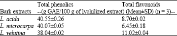
Total phenolic compounds and total flavonoids contents: The results of total phenolic compounds and total flavonoid contents determination by Folin- Ciocalteu method and aluminiun chloride colorimetric essay method are summarized in Table 1. The total phenolic content of hydroethanolic extracts was determined from regression equation of calibration curve (Y = 0.0069X+0.0002, R2 = 0.9977) and expressed in Gallic Acid Equivalents (GAE). The total phenolic compounds amount varied from 38.04 to 40.55.
The total flavonoids content expressed in Quercetin Equivalents (QE) was determined from Y = 0.01X -0.0032, R2 = 09974 and its content varied between 6.45 and 11.02.
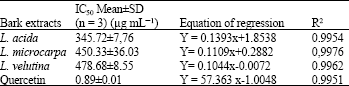
Antioxidant activity: The IC50 values of quercetin and gallic acid were 0.89±0.01 and 0.60±0.01 μg mL-1, respectively. The Lannea acida barks extract exhibited IC50 = 345.72±7.76 μg mL-1 while IC50=478.68± 8.55 was recorded with L. velutina and IC50 = 450.33±36.03 with L.microcarpa. The result of DPPH free radicals scavenging activity is reported in Table 2. The highest amount of phenolic compounds was found in Lannea acida (40.55 ± 0.26 g GAE/100 g).
| Table 1: | Total Phenolic and flavonoid contents of the tree plant extracts |
 | |
| Means of total phenolics and total flavonoids content expressed respectively Gallic Acid Equivalent (GAE) and Quercetin Equivalent (QE)/100 g lyophilized extracts. Each mean value is associated with a standard deviation (SD n = 3) | |
| Table 2: | Antioxidant activities of the tree plant extracts |
 | |
| Each mean value is associated with a Standard Deviation (SD) | |
Lannea velutina barks extract exhibited the highest amount of flavonoids than the other extracts. Lannea acida barks extract demonstrated a highest antioxidant activity among the three plant extracts with a good ability of scavenging DPPH free radicals (Fig. 1).This activity correlated with the high quantity of total phenolic contents (Karou et al., 2005).However all extracts showed lower activity than quercetin and gallic acid used as standard agents.
L. velutina extract exhibited a lowest amount of total phenolic compounds and a larger IC50 value. This result suggests that L. velutina extract possessed a weak DPPH radical scavenging action in comparison with Lannea acida. Concerning the standard deviations, no significant difference was observed between the antioxidant capacity of L. microcarpa and L. velutina barks extracts. However minor compounds as total flavonoids might also exhibit antioxidant activity (Bruneton, 1999; Oliveira et al., 2008). The total flavonoids content level was higher in L. velutina extract but its antioxidant effect is the lowest, this result was not consistent with Oliveira et al. (2008), possible synergetic and antagonist effects of compounds in the three plant extracts should be taken into consideration.
Antibacterial activity: The results in Table 3 recorded by disk diffusion method show that most of microorganisms were sensitive to all plant extracts. MICs and MBCs of the three plant extracts varied from 7.82 to 125 μg mL-1 for all bacterial strains tested (Table 4). MIC and MBC values were different and suggested a selective activity of the three plant extracts. In order to elucidate the antibacterial effect, MBC/MIC ratios were calculated. When the ratio value was lower than 2 the extract exhibited a bactericidal effect (Michel-Briand, 1986).
The antibacterial activity can be considerate when the diameter of inhibition zone observed is 9 mm or more around the paper disk (Kitzberger et al., 2006). The results show that most of germs tested were sensitive to all plant extracts (Table 3). The best sensitivity to the three plant extracts was obtained on Bacillus subtilus ATCC 21332. Enterobacter aerogenes CIP 104725 and Pseudomonas aeruginosa 19249 were resistant strains for L. acida and L. microcarpa extracts respectively. However staphylococcus aureus ATCC 9144 was more sensitive to L. microcarpa while Salmonella typhimurium ATCC 13311 was more sensitive to L. velutina. The most important information was that Bacillus subtilus ATCC 21332 was more sensitive to the three plant extracts (17 mm) than tetracyclin (10 mm), Enterococcus faecalis CIP 103907 was more sensitive to L. microcarpa (14 mm) than Erythromycin (8 mm), Enterobacter aerogenes CIP 104725 was more sensitive to L. velutina (14 mm) than ciprofloxacin and Erythromycin (6 mm; 0 mm).
| Table 3: | Determination of strains sensitivity: diameter of inhibition (mm) |
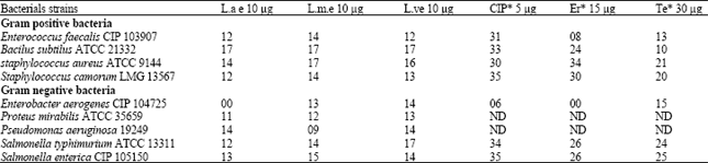 | |
| ND: Not determinated; L.a.e: L. acida; Lm.e: L. microcarpa; ;Lve: L. velutina; CIP: Ciprofloxacin (5 μg); Er: Erythromycin (15 μg); Te: Tetracyclin (30 μg). *Reference disk already producted by Bio Merieux | |
| Table 4: | Antibacterial activity of the tree plant extracts |
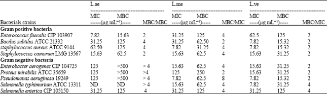 | |
| ND: Not determinate; MIC: Minimum inhibitory concentration, MBC: Minimum bactericidal concentration | |
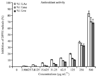 | |
| Fig. 1: | DPPH radicals scavenging activity. %I La e: percentage of free DPPH radicals scavenged by ethanol extract of Lannea acida. %Lme: percentage of free DPPH radicals scavenged by ethanol extract of Lannea microcarpa, %LVe: percentage of free DPPH radicals scavenged by ethanol extract of Lannea velutina |
L. velutina extract was bactericidal for all strains tested: Enterococcus faecalis CIP 103907, Bacillus subtilus ATCC 21332, staphylococcus aureus ATCC 9144, Staphylococcus camorum LMG 13567. Enterobacter aerogenes CIP 104725, Proteus mirabilis ATCC 35659, Pseudomonas aeruginosa 19249 (Table 4). While L. acida and L. microcarpa extracts were respectively bactericidal for Enterococcus faecalis CIP 103907, Staphylococcus camorum LMG 13567 and Bacillus subtilus ATCC 21332, Proteus mirabilis ATCC 35659. This antibacterial activity might be due to the presence of chemical compounds such as tannins, phenolic compounds, polyphenols and flavonoids (Bruneton, 1999; Oliveira et al., 2008).
CONCLUSION
This study shows in vitro high and low antibacterial activities of the three plant extracts. L. velutina is most bactericidal for almost strains tested and demonstrates a large spectrum with the best MICs than L. acida and L. microcarpa. However, L. acida extract possess a high antioxidant activity than others. Because of its higher antioxidant activity, the L. acida barks extract is more useful than the other two plants in medical approach, particularly in case when high activity of preparation is desired during anti-cancer therapy or other degenerative deseases (inflammatory, cardiovascular diseases). Furthermore, the use of L.velutina barks may help to prevent infections such as diarrhoea, dysentery, gastric ulcer, skin deseases or sexual infections while the use L. acida barks may help to prevent oxidative damages such as hypertension, rheumatisms, cancers, prematuring aging and atherosclerosis. These results show that the barks of L. acida and L. velutina could be used respectively as a potential natural antioxidant and antibacterial agent. Further investigations will be performed by i/the isolation and identification of pure compounds in the extracts, ii/testing these compounds against pathogenic bacteria and determining their antioxidant activity, iii/ the comparison of the antibacterial activities of extracts with those of polyphenols of reference.
REFERENCES
- Aderogba, M.A., E.K. Okoh and T.O. Idowu, 2005. Evaluation of antioxidant activity of the secondary metabolites from Poliostigma reticulatum (DC) hochst. J. Biol. Sci., 5: 239-242.
Direct Link - Arvouet-Grand, A., B. Vennat, A. Pourrat and P. Legret, 1994. [Standardization of propolis extract and identification of principal constituents]. J. Pharm. Belg., 49: 462-468, (In French).
PubMedDirect Link - Belmonte, O., D. Drouet, J. Alba, M.P. Morton and B. Kuli et al., 2010. Evolution of enterobacteriaceae resistance to antibiotics in Reunion Island: Emergence of extended spectrum betalactamases. Pathol. Biol., 58: 18-24.
Direct Link - Beckman, K.B. and B.N. Ames, 1998. The free radical theory of aging matures. Physiol. Rev., 78: 547-581.
PubMed - Coulidiati, T.H., H. Millogo-Kone, A. Lamien-Meda, C.E. Lamien and M. Lompo et al., 2009. Antioxidant and antibacterial activities of Combretum nioroense Aubrev. Ex keay (Combretaceae). Pak. J. Biol. Sci., 12: 264-269.
CrossRefPubMedDirect Link - Da-Silva, J.F.M., M.C. De-Souza, S.R. Matta, M.R. De-Andrade and F.V.N. Vidal, 2006. Correlation analysis between phenolic levels of brazilian propolis extracts and their antimicrobial and antioxidant activities. Food Chem., 99: 431-435.
CrossRef - Kalache, A., I. Aboderin and I. Hoskins, 2002. Compression of morbidity and active ageing: Key priorities for public health policy in the 21st century. Bull. World Health Org., 80: 243-244.
Direct Link - Karou, D., H.M. Dicko, J. Simpore and A.S. Traore, 2005. Antioxidant and antibacterial activities of polyphenols from ethnomedicinal plants of Burkina Faso. Afr. J. Biotechnol., 4: 823-828.
Direct Link - Kone, W.M., K.K. Atindehou, C. Terreaux, K. Hostettmann, D. Traore and M. Dosso, 2004. Traditional medicine in North Côte-d’Ivoire: Screening of 50 medicinal plants for antibacterial activity. J. Ethnopharmacol., 93: 43-49.
CrossRefDirect Link - Maiga, A., K.E. Malterud, D. Diallo and B.S. Paulsen, 2006. Antioxidant and 15-lipoxygenase inhibitory activities of the Malian medicinal plants Diospyros abyssinica (Hiern) F. White (Ebenaceae), Lannea velutina A. Rich (Anacardiaceae) and Crossopteryx febrifuga (Afzel) Benth (Rubiaceae). J. Ethnopharmacol., 104: 132-137.
PubMedDirect Link - Majhenic, L., M. Skerget and Z. Knez, 2007. Antioxidant and antimicrobial activity of guarana seed extracts. Food Chem., 104: 1258-1268.
CrossRef - Malorni, W., R. Rivabene, B.M. Lucia, R. Ferrara, A.M. Mazzone, R. Cauda and R. Paganelli, 1998. The role of oxidative imbalance in progression to AIDS: Effect of the thiol supplier N-Acetylcysteine. AIDS Res. Hum. Retroviruses, 14: 1589-1596.
PubMedDirect Link - Montalleb, G., P. Hanachi, S.H. Kua, O. Fauziah and R. Asmah, 2005. Evaluation of phenolic content and total antioxidant activity in Berberis vulgaris fruit extract. J. Biol. Sci., 5: 648-653.
CrossRefDirect Link - Oliveira, I., A. Sousa, I.C.F.R. Ferreira, A. Bento, L. Estevinho and J.A. Pereira, 2008. Total phenols, antioxidant potential and antimicrobial activity of walnut (Juglans regia L.) green husks. Food Chem. Toxicol., 46: 2326-2331.
CrossRefDirect Link - Pereira, J.A., I. Oliveira, A. Sousa, P. Valentao and P.B. Andrade et al., 2007. Walnut (Juglans regia L.) leaves: Phenolic compounds, antibacterial activity and antioxidant potential of different cultivars. Food Chem. Toxicol., 45: 2287-2295.
CrossRefDirect Link - Picerno, P., T. Mencherini, R.D. Loggia, M. Meloni, R. Sanogo and R.P. Aquino, 2006. An extract of Lannea microcarpa: Composition, activity and evaluation of cutaneous irritation in cell cultures and reconstituted human epidermis. J. Pharm. Pharmacol., 58: 981-988.
PubMedDirect Link - Rabaud, C., H. Tronel, S. Fremont, T. May, P. Canton and J.P. Nicolas, 1997. Free radicals during HIV infection. Annal. Biol. Clin., 55: 565-571.
Direct Link - Rebaudet, S., J.J. de Pina, C. Rapp, P. Kreamer, H. Savini, E. Demortiere and F. Simon, 2007. Le risque nosocomial en Afrique intertropicale Partie4: Prevention. Med. Trop., 68: 73-82.
Direct Link - Simon, F., P. Kreamer, J.J. de Pina, E. Demortiere and C. Rapp, 2007. Le risque nosocomial en Afrique intertropicale Partie 2 : Les infections des patients. Med. Trop., 67: 197-203.
Direct Link - Singleton, V.L., R. Orthofer and R.M. Lamuela-Raventos, 1999. Analysis of Total Phenols and Other Oxidation Substrates and Antioxidants by Means of Folin-Ciocalteu Reagent. In: Methods in Enzymology, Burslem, G.L. (Ed.), Academic Press, Cambridge, Massachusetts, ISBN: 9780121822002, pp: 152-178.
CrossRefDirect Link - Swenson, J.M., G.E. Killgore and F.C. Tenover, 2004. Antimicrobial susceptibility testing of Acinetobacter sp. by NCCLS broth microdilution and disk diffusion methods. J. Clin. Microbiol., 42: 5102-5108.
CrossRef - Willcox, J.K., S.L. Ash and G.L. Catignani, 2004. Antioxidants and prevention of chronic disease. Crit. Rev. Food Sci. Nutr., 44: 275-295.
CrossRefPubMedDirect Link - Atindehou, K.K., C. Schmid, R. Brun, M.W. Kone and D. Traore, 2004. Antitrypanosomal and antiplasmodial activity of medicinal plants from Cote d'Ivoire. J. Ethnopharmacol., 90: 221-227.
CrossRefDirect Link - Kiehlbauch, J.A., G.E. Hannett, M. Salfinger, W. Archinal, C. Monserrat and C. Carlyn, 2000. Use of the National Committee for Clinical Laboratory Standards guidelines for disk diffusion susceptibility testing in New York state laboratories. J. Clin. Microbiol., 38: 3341-3348.
CrossRefPubMedDirect Link - Velazquez, E., H.A. Tournier, P.M. de Buschiazzo, G. Saavedra and G.R. Schinella, 2003. Antioxidant activity of Paraguayan plant extracts. Fitoterapia, 74: 91-97.
CrossRefDirect Link - Kitzberger, C.S.G., A. Smania, R.C. Pedrosa and S.R.S. Ferreira, 2006. Antioxidant and antimicrobial activities of shiitake (Lentinula edoles) extracts obtained by organic solvents and supercritical fluids. J. Food Eng., 80: 631-638.
CrossRef