
Yodthong Baimark
Center of Excellence for Innovation in Chemistry, Department of Chemistry, Faculty of Science, Mahasarakham University, Mahasarakham 44150, Thailand
Journal of Applied Sciences
Year: 2009 | Volume: 9 | Issue: 12 | Page No.: 2287-2293


ABSTRACT
Aims of this study were to synthesize methoxy poly (ethylene glycol)-b-poly (ε-caprolactone) diblock copolymers (MPEG-b-PCL) and to prepare their surfactant-free nanospheres by modified-spontaneous emulsification solvent diffusion method for controlled release delivery of hydrophobic drug. Ibuprofen was used as a poorly-water soluble model drug. Influences of PCL block length and drug loading content on nanosphere characteristics and drug release behaviors were evaluated. The prepared nanospheres with and without drug loading were found of spherical shape and smooth surface with size less than 150 nm. FTIR spectroscopy and differential scanning calorimetry studies showed the interactions between ibuprofen and MPEG-b-PCL had occurred, suggesting the well distribution of ibuprofen into nanosphere matrices. An increase in the PCL block length led to a decrease in drug encapsulation efficiency. The drug release profiles were biphasic with a fast burst release followed by a slow one. The drug release rates from the MPEG-b-PCL nanospheres strongly depended on the PCL block length and the drug loading content.
PDF Abstract XML References Citation
How to cite this article
Yodthong Baimark, 2009. Surfactant-Free Nanospheres of Methoxy Poly (Ethylene Glycol)-b-Poly
(ε-Caprolactone) for Controlled Release of Ibuprofen. Journal of Applied Sciences, 9: 2287-2293.
DOI: 10.3923/jas.2009.2287.2293
URL: https://scialert.net/abstract/?doi=jas.2009.2287.2293
DOI: 10.3923/jas.2009.2287.2293
URL: https://scialert.net/abstract/?doi=jas.2009.2287.2293
INTRODUCTION
Poly (∈-caprolactone) (PCL) is a biodegradable and biocompatible polymer that is widely used in medicine and pharmaceutical applications. Diblock copolymers composed of methoxy poly (ethylene glycol) (MPEG) and PCL have been synthesized to attain versatile biodegradable polymers having more water-absorbing capacity because of the inclusion of hydrophilic MPEG segments within the relative hydrophobic PCL segments (He et al., 2004; Shuai et al., 2004; Aliabadi et al., 2005). These diblock copolymers have been used for the preparation of drug-loaded nanoparticles (Kim and Lee, 2001; Shuai et al., 2004; Aliabadi et al., 2005; Zhang and Zhuo, 2005). The nanoparticles have shown potential as drug delivery systems because of their small sizes, improving circulation times in the body and creates more available routes of administration than do microparticles, which are rapidly cleared by the reticulo-endothelial tissues (Kumar, 2000).
The modified-spontaneous emulsification-solvent diffusion method (modified-SESD method) for the preparing surfactant-free nanoparticles of hydrophilic-hydrophobic diblock copolymer was first proposed as previously described by Baimark et al. (2007). MPEG-b-poly (D, L-lactide) was dissolved in volatile water-miscible organic solvents with lower toxicity, acetone and ethanol. Higher energy apparatus, such as a homogenizer or a sonicator (usually applied in larger scale preparation of polymer nanoparticles), was not used for this technique. However, preparation of drug-loaded nanoparticles of MPEG-b-PCL by the modified-SESD method has not been reported.
The aims of present study were to prepare surfactant-free drug-loaded MPEG-b-PCL nanospheres by the modified-SESD method and to investigate the influences of PCL block lengths and drug loading content on the nanosphere characteristics and drug release behaviours. Ibuprofen was used as a poorly-water soluble model drug. Interactions between ibuprofen and nanosphere matrices were also determined.
MATERIALS AND METHODS
Materials: Methoxy poly (ethylene glycol) (MPEG) with a molecular weight of 5,000 g mol-1 (Fluka, Germany) was dried at 120°C under vacuum for 4 h before use. The ∈-caprolactone (CL) monomer (99%, Acro, USA) was purified by drying with CaH2 followed by distillation under reduced pressure before storage over molecular sieves in a refrigerator. Stannous octoate (Sn(Oct)2, 95%, Sigma, USA) was used as received. Acetone (Merck, Germany) and ethanol (Merck, Germany) in analytical grade were used.
Methods
Synthesis of MPEG-b-PCL: MPEG-b-PCL diblock copolymers with different molecular weights of PCL block were synthesized by ring-openning polymerization of CL monomer in bulk at 130°C for 48 h under nitrogen atmosphere. MPEG:CL feed mole ratios of 1:350 and 1:700 were used. Sn(Oct)2 and MPEG were used as the initiating system. Sn(Oct)2 concentration was kept constant at 0.04 mol%. The MPEG-b-PCL products were purified by dissolving them in chloroform before precipitation in cool n-hexane and then drying to a constant weight under reduced pressure at room temperature. The both purified MPEG-b-PCLs were obtained with approximately 95% yields.
Characterization of MPEG-b-PCL: Chemical compositions of the MPEG-b-PCL were determined by 1H-nuclear magnetic resonance (NMR) spectrometry using a Bruker Advanced DPX 300 1H-NMR spectrometer. CDCl3 was used as a solvent at room temperature and tetramethysilane was used as the internal standard. The number-average molecular weight (Mn) and Molecular Weight Distribution (MWD) were determined by Gel Permeation Chromatography (GPC) using a Waters 717 plus Autosampler GPC equipped with a Ultrastyragel® column operating at 30°C. A refractive index detector was employed. Tetrahydrofuran was used as the solvent at a flow rate of 1 mL min-1. The thermal properties of the MPEG-b-PCL were characterized by non-isothermal Differential Scanning Calorimetry (DSC) using a Perkin-Elmer Pyris Diamond DSC. For DSC analysis, approximately 10 mg of the sample was placed in a seal aluminium pan and heated at the rate of 10°C min-1 under helium flow to measure the melting temperature (Tm) and the heat of melting (ΔHm).
Preparation of ibuprofen-loaded nanospheres: Ibuprofen-loaded nanospheres of the MPEG-b-PCL were prepared according to the modified-SESD method without surfactant used (Baimark et al., 2008). Briefly, 0.2 g of ibuprofen/MPEG-b-PCL mixtures with different ratios were dissolved in 20 mL of the 3/3 (v/v) acetone/ethanol mixture solvent. The ibuprofen with 1.25, 2.5 and 5.0% (w/w) were used. These solutions were added drop-wise into 160 mL distilled water in a 250 mL beaker with stirring at 600 rpm. After evaporation of organic solvents at room temperature for 6 h in a fume hood, the nanosphere colloid was centrifuged at 12,000 rpm for 1 h at 4°C before freeze-drying for 48 h. Then dried nanospheres were obtained. The ibuprofen-free nanospheres of both MPEG-b-PCL were also prepared by the same method as controls.
Characterization of ibuprofen-loaded nanospheres: Functional groups of ibuprofen and MPEG-b-PCL of the nanospheres and interactions between them were studied by FTIR spectroscopy using a Perkin-Elmer Spectrum GX FTIR spectrophotometer with air as the reference. The resolution of 4 cm-1 and 32 scans were used. FTIR spectra were obtained from KBr disk of dried nanospheres. Particle sizes and size distributions of the nanospheres with and without ibuprofen loading were directly determined from their nanosphere colloids by light-scattering analysis using a Coulter LS230 light scattering particle size analyzer at 25°C. Morphology of the nanospheres was investigated by Scanning Electron Microscopy (SEM) using a JEOL JSM-6460LV SEM. Before SEM measurement, the dried nanospheres were sputter coated with gold for enhancing the surface conductivity. Thermal properties of the dried nanospheres were measured by DSC as described earlier.
Ibuprofen loading content and encapsulation efficiency: Ibuprofen loaded in nanospheres was determined by UV-Vis spectroscopy using a Perkin-Elmer Lambda 25 UV-Vis spectrophotometer at 264 nm (Borovac et al., 2006). The ibuprofen-loaded nanospheres were dissolved in dichloromethane for this purpose. Ibuprofen loading content and encapsulation efficiency were calculated from Eq. 1 and 2, respectively. The measurement was performed in triplicate.
 | (1) |
(2) |
In vitro ibuprofen release: An exact amount (about 10 mg) of ibuprofen-loaded nanospheres was disclosed into a dialysis bag (molecular weight cut off was 8,000 to 12,000) and immersed into a flask containing 150 mL of Phosphate Buffer Solution (PBS) at pH 7.4. The sample flasks were incubated at 37°C under shaking at the rate of 150 rpm. At predetermined time intervals, 10 mL samples were withdrawn and 10 mL of fresh PBS was added into the flask for continuing the release test. The released ibuprofen was measured by UV-Vis spectrophotometer at 220 nm (Borovac et al., 2006). According to a predetermined ibuprofen concentration-UV absorbance standard curve, ibuprofen concentration of the release medium was obtained and ibuprofen (%) released was calculated. The average value was calculated from the three measurements.
RESULTS
Characterization of MPEG-b-PCL: The chemical compositions of MPEG-b-PCLs were determined from the 1H-NMR spectra by calculating the ratio of the integral peak areas corresponding to the Ethylene Oxide (EO, repeating units of MPEG) methylene protons at δ = 3.6- 3.7 ppm and the CL ∈-methylene protons at δ = 4.0-4.2 ppm. From the peak area integrations of the peaks a and b in Fig. 1a and b, the copolymer compositions can be determined as EO:CL = 24:76 and 14:86 (mol%) corresponding to the MPEG:CL mole ratios of 1:361 and 1:701 for the MPEG-b-PCL40000 and the MPEG-b-PCL80000, respectively.
The Mns of MPEG-b-PCLs obtained from GPC curves were 44,000 and 82,500 g mol-1 for the MPEG-b-PCL40000 and the MPEG-b-PCL80000, respectively. The DSC curve of MPEG in Fig. 2a showed the melting temperature (Tm) and the heat of melting (ΔHm) and they were found to be 61°C and 177.4 J g-1, respectively. The DSC curves of MPEG-b-PCL revealed a semi-crystalline morphology with a single-Tm as shown in Fig. 2b, c. The Tms of MPEG-b-PCL were 58 and 60°C, whereas the ΔHms were 92.8 and 101.2 J g-1 for the MPEG-b-PCL40000 and the MPEG-b-PCL80000, respectively.
 | |
| Fig. 1: | 1H-NMR spectra of (a) MPEG-b-PCL40000 and (b) MPEG-b-PCL80000 |
Characterization of nanospheres: In this study, the drug-loaded nanospheres of MPEG-b-PCL were firstly prepared by the modified-SESD method without any surfactants for used as controlled release drug delivery systems.
Functional groups and interactions of the MPEG-b-PCL nanosphere matrices and the loaded ibuprofen were determined from FTIR spectra, as example of which is shown in Fig. 3a-e for the MPEG-b-PCL80000 nanosphere (No. 5-8) and the ibuprofen. The FTIR spectra of the pure MPEG-b-PCL nanospheres (Fig. 3a) and the ibuprofen (Fig. 3e) showed carbonyl absorption bands at 1761 and 1721 cm-1, respectively. The FTIR spectra of ibuprofen-loaded nanospheres of MPEG-b-PCL40000 with different drug loading contents showed similar evidence (Fig. 3a-e).
The surfactant-free nanosphere colloids with and without ibuprofen loading were clear aqueous suspensions. The sizes of colloidal nanospheres were investigated by the light-scattering analysis.
 | |
| Fig. 2: | DSC thermograms of (a) MPEG, (b) MPEG-b-PCL40000 and 8 MPEG-b-PCL80000 |
 | |
| Fig. 3: | FTIR spectra of nanosphere No. (a) 5, (b) 6, 8 7 and (d) 8 and (e) ibuprofen |
 | |
| Fig. 4: | Particle size graphs of nanosphere No. (a) 5, (b) 6, 8 7 and (d) 8 |
| Table 1: | Particle sizes and thermal properties of ibuprofen-loaded MPEG-b-PCL nanospheres |
 | |
| aNanospheres of MPEG-b-PCL40000, bNanospheres of MPEG-b-PCL80000, cObtained from light-scattering analysis, dObtained from DSC thermograms | |
Their sizes were found in the range of 82 to 97 nm. These data are shown in Fig. 4a-d and summarized in Table 1. The average particle size of surfactant-free nanosphere colloids of the MPEG-b-PCL40000 was slightly larger than the MPEG-b-PCL80000. However, the different drug loading contents did not significantly effect to their average particle sizes.
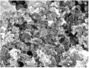
The morphologies of nanospheres were determined from SEM micrographs as examples are shown in Fig. 5a and b for the nanosphere No. 4 and 8. It was found that the colloidal nanospheres have a spherical shape in the nanometer size range and smooth surfaces. The particle sizes significantly decreased as the PCL block length increased according to the light-scattering analysis. However, the nanospheres observed from SEM micrographs were similar in size.
The Tm and the ΔHm of ibuprofen measured from DSC curve were 74°C and 116.4 J g-1, respectively, as shown in Fig. 6.
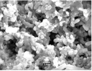 | |
| Fig. 5: | SEM micrographs of nanosphere No. (a) 4 and (b) 8 (bar = 1 μm) |
 | |
| Fig. 6: | DSC thermogram of ibuprofen |
The ibuprofen crystallites entrapped in the nanospheres were not detected as example of which is shown in Fig. 7a-d for the MPEG-b-PCL80000 nanospheres. The thermal properties of nanospheres are also shown in Table 1. In addition, the both Tm and ΔHm of MPEG-b-PCL nanosphere matrices significantly decreased as the drug loading content increased.
 | |
| Fig. 7: | DSC thermograms of nanosphere No. (a) 5, (b) 6, 8 7 and (d) 8 |
 | |
| Fig. 8: | (a, b) Ibuprofen release profiles of nanosphere No. |
Ibuprofen loading content and encapsulation efficiency: The calculated results of ibuprofen loading content and encapsulation efficiency are also shown in Table 1. It was found that the ibuprofen-loaded nanospheres with different ibuprofen loading contents can be prepared by using different ibuprofen feed ratios. The ibuprofen encapsulation efficiencies were in the range of 48-64%. These values of the MPEG-b-PCL80000 were slightly lower than the MPEG-b-PCL40000.
In vitro release of ibuprofen: The ibuprofen release profiles from the nanospheres of MPEG-b-PCL40000 and MPEG-b-PCL80000 investigated in PBS pH 7.4 at 37°C are shown in Fig. 8a and b. The release profiles of ibuprofen-loaded nanospheres were biphasic containing rapid initial burst release and sustaining release. The initial burst releases from the MPEG-b-PCL80000 nanospheres were approximately 60% at the first 3 h of release time (Fig. 8a, b). Then, release profiles were followed by a constant slow release until to 98, 95 and 85% ibuprofen released within 312 h with ibuprofen loading contents of 0.8, 1.6 and 3.0%, respectively for the MPEG-b-PCL40000 nanospheres.
The drug release behaviors from the MPEG-b-PCL80000 nanospheres also showed similar phenomenon. Moreover, Fig. 8a and b showed that the drug loading content was an important factor of the drug release rate. The nanospheres with lower drug loading content showed the higher drug release rate. Finally, the drug release from the nanospheres of MPEG-b-PCL40000 was faster than that of MPEG-b-PCL80000.
DISCUSSION
Characterization of MPEG-b-PCL: As could be expected, the copolymer compositions obtained from the 1H-NMR were very similar to the MPEG:CL feed mole ratios (1:350 and 1:700 for MPEG-b-PCL40000 and MPEG-b-PCL80000, respectively). Therefore, the synthesized reaction was taken to near-quantitative conversion. The Mns obtained from GPC (44000 and 82500 g mol-1 for MPEG-b-PCL40000 and MPEG-b-PCL80000, respectively) were closely similar to that obtained from the feed ratios (45000 and 85000 g mol-1 for MPEG-b-PCL40000 and MPEG-b-PCL80000, respectively). The lower molecular weights obtained from GPC curves may be due to the degradation-side reactions such as hydrolysis and thermal degradation reactions. From DSC results suggested that the both MPEG and MPEG-b-PCL contained semi-crystalline morphology. The MPEG crystallinity was disappeared when the MPEG block was connected with the poly (D, L-lactide) (PDLL) block as previously described by Baimark et al. (2007). Then the obtained MPEG-b-PDLL showed completely amorphous structure. Thus, the crystallinity of MPEG was also suppressed when connected to the PCL block. It can be concluded that the crystallinity of MPEG-b-PCL synthesized in this study can be attributed to PCL crystallites.
Characterization of nanospheres: The carbonyl absorption band at 1721 cm-1 attributed to the crystalline form of ibuprofen (Kazarian and Martirosyan, 2002) which shifting to higher wave number for the ibuprofen loaded in nanospheres as shown in Fig. 3b-d suggested that the ibuprofen-ibuprofen interactions in its crystalline fraction were destroyed when the ibuprofen was entrapped in the MPEG-b-PCL nanosphere matrices. This may be indicated that the ibuprofen molecules were well distributed throughout the nanosphere matrices.
The broader carbonyl absorption bands in the region of 1785-1735 cm-1 were assigned to shift of the carbonyl bands of the both MPEG-b-PCL and ibuprofen indicated the existence of interactions between the MPEG-b-PCL nanosphere matrices and the loaded ibuprofen. These intermolecular interactions were expected as hydrogen bonding between carbonyl groups of MPEG-b-PCL and hydroxyl groups of ibuprofen corresponding to the hydrogen bonding between carbonyl groups of poly (vinylpyrrolidone) matrices and loaded ibuprofen as previously reported by Kazarian and Martirosyan (2002). The intermolecular bonding between MPEG-b-PCL40000 nanosphere matrices and loaded ibuprofen can be also detected from their FTIR spectra. In addition, intensities of the absorption band at 1560 cm-1 assigned to ibuprofen characteristics increased as the increasing ibuprofen content for the all ibuprofen-loaded nanospheres supported that the ibuprofen contents were strongly depended upon the ibuprofen feed ratio.
The sizes of nanospheres with and without ibuprofen loading measured from the both light-scattering and SEM analyses were in the nanometer size ranges. The nanosphere sizes obtained from the SEM looked larger than those obtained from the light-scattering analysis. It was due to the colloidal nanospheres, which were slightly flatten during the centrifugation and drying processes before SEM measurement. The nanospheres of MPEG-b-PCL80000 were slightly smaller than the MPEG-b-PCL40000. The MPEG-b-PCL with higher ΔHm usually leads to higher crystallinity that was larger self-condensed than the lower one during nanosphere solidification. The MPEG-b-PCL80000 contained higher crystallinity than the MPEG-b-PCL40000. Therefore, the nanosphere sizes decreased when the molecular weight of PCL block was increased.
The DSC results indicated that the MPEG-b-PCL nanospheres and the ibuprofen contained crystalline structures. It is significant to note that the ibuprofen crystallizability in the all of drug-loaded nanospheres was suppressed after loaded into the MPEG-b-PCL nanospheres supported that the ibuprofen loaded in the nanospheres had a completely amorphous state corresponding to the FTIR results as described earlier. Meanwhile, the increasing drug content can also inhibited the PCL crystallization due to the ibuprofen interpenetrated between the MPEG-b-PCL chains. It concluded that the ibuprofen was well distributed into the MPEG-b-PCL nanosphere matrices.
Ibuprofen loading content and encapsulation efficiency: The drug loading content of the ibuprofen-loaded MPEG-b-PCL nanospheres was strongly depended upon the drug feed ratio. The obtained ibuprofen loading contents as shown in Table 1 were lower than the ibuprofen feed ratios of 1.25, 2.5 and 5.0% (w/w) for the nanosphere No. 2, 6, 3, 7 and 4, 8, respectively. This can be indicated that the some drug has released out during the nanosphere formation. The changes of ibuprofen encapsulation efficiency directly related to the ibuprofen loading content (Eq. 1, 2). The both ibuprofen loading content and encapsulation efficiency of the drug-loaded nanospheres decreased when the PCL block length increased for the same ibuprofen feed ratios. This could be due to higher PCL crystallinity which could decrease the drug loading content, the drug molecules were squeezed out because crystallization of PCL blocks, as only the amorphous PCL phase is likely to accommodate drug molecules (Shuai et al., 2004).
In vitro release of ibuprofen: The rapid initial burst releases of drug from the nanospheres were probably due to the releasing of drug that was entrapped or adsorbed near to the nanosphere surfaces. After that the slow release may be due to diffusion and matrix erosion mechanisms. The drug release rates increased when the drug loading content increased. This may be due to higher ibuprofen loading content and encapsulation efficiency. Finally, a faster rate of ibuprofen releasing was obtained when a less hydrophobic matrix (MPEG-b-PCL40000) due to the shorter PCL block length was used.
CONCLUSION
The MPEG-b-PCL diblock copolymers with different PCL block lengths were successfully synthesized by ring-opening polymerization of the CL monomer using Sn(Oct)2 and MPEG as the initiating system. They were semi-crystalline diblock copolymers. The surfactant-free nanospheres of the MPEG-b-PCL with and without ibuprofen, a hydrophobic model drug, loading were successfully prepared by the modified-SESD method using an acetone/ethanol mixture as the organic solvent. The average sizes of nanospheres were less than 100 nm. The prepared nanospheres looked spherical, had a smooth surface and a narrow size distribution under SEM. The nanosphere size and drug encapsulation efficiency were found mainly affected by the drug loading and the PCL block length. The drug release rate can be optimally controlled by adjusting these parameters.
ACKNOWLEDGMENTS
The author would like to acknowledge the Research Development and Support Unit, Mahasarakham University and the Center of Excellence for Innovation in Chemistry (PERCH-CIC), Commission on Higher Education, Ministry of Education, Thailand for financial supports.
REFERENCES
- Aliabadi, H.M., A. Mahmud, A.D. Sharifabadi and A. Lavasanifar, 2005. Micelles of methoxy poly(ethylene oxide)-b-poly(ε-caprolactone) as vehicles for the solubilization and controlled delivery of cyclosporine A. J. Control. Release, 104: 301-311.
CrossRefDirect Link - Baimark, Y., M. Srisa-Ard, J. Threeprom and N.A. Narkkong, 2007. Preparation of nanoparticle colloids of methoxy poly(ethylene glycol)-b-poly(D,L-lactide): Effects of surfactant and organic solvent. Colloid Polymer Sci., 285: 1521-1525.
CrossRefDirect Link - Baimark, Y., Y. Srisuwan, N. Kotsaeng and P. Threeprom, 2008. Preparation of surfactant-free and core-shell type nanoparticles of methoxy poly(ethylene glycol)-b-poly ε-caprolactone-co-D, L-lactide diblock copolymers. Asian J. Applied Sci., 1: 237-245.
CrossRefDirect Link - Borovac, T., J.P. Pelage, A. Kasselouri, P. Prognon, G. Guiffant and A. Laurent, 2006. Release of ibuprofen from beads for embolization: In vitro and in vivo studies. J. Controlled Release, 115: 266-274.
CrossRefDirect Link - He, C., J. Sun, C. Deng, T. Zhao, M. Deng, X. Chen and X. Jing, 2004. Study of the synthesis, crystallization and morphology of poly(ethylene glycol)-poly(ε-caprolactone) diblock copolymer. Biomacromolecules, 5: 2042-2047.
CrossRefDirect Link - Kazarian, S.G. and G.G. Martirosyan, 2002. Spectroscopy of polymer/drug formulations processed with supercritical fluids: In situ ATR-IR and raman study of impregnation of ibuprofen into PVP. Int. J. Pharm., 232: 81-90.
CrossRef - Kim, S.Y. and Y.M. Lee, 2001. Taxol-loaded block copolymer nanospheres composed of methoxy poly(ethylene glycol) and poly(ε-caprolactone) as novel anticancer drug carriers. Biomaterials, 22: 1697-1704.
CrossRefDirect Link - Kumar, M.N.V.R., 2000. Nano and microparticles as controlled drug delivery devices. J. Pharm. Pharmaceut. Sci., 3: 234-258.
PubMedDirect Link - Shuai, X., H. Ai, N. Nasongkla, S. Kim and J. Gao, 2004. Micellar carriers based on block copolymers of poly(ε-caprolactone) and poly(ethylene glycol) for doxorubicin delivery. J. Control Release, 98: 415-426.
CrossRefPubMedDirect Link - Zhang, Y. and R.X. Zhuo, 2005. Synthesis and in vitro drug release behavior of amphiphilic triblock copolymer nanoparticles based on poly(ethylene glycol) and polycaprolactone. Biomaterials, 26: 6736-6742.
CrossRefPubMedDirect Link