
Wei Wei-Yi
College of Electrical Engineering and Information Engineering,Lanzhou University of Technology, Lanzhou 730050, China
Li Zhan-Ming
College of Electrical Engineering and Information Engineering,Lanzhou University of Technology, Lanzhou 730050, China
Zhang Gui-Cang
College of Mathematics and Information Science,Northwest Normal University, Lanzhou 730070, China
Zhang Guo-Quan
College of Electrical Engineering and Information Engineering,Lanzhou University of Technology, Lanzhou 730050, China
Information Technology Journal
Year: 2010 | Volume: 9 | Issue: 8 | Page No.: 1682-1685


ABSTRACT
Spatial intensity variations caused by illumination changes have been a challenge for microscopic image segmentation. This study presents a new scheme for color microscopic image segmentation free from the influence of uneven illumination. First extract the most important component in RGB space by the Principal Component Analysis (PCA) which can transform the high dimension to lower dimension effectively, meanwhile, build a new estimating uneven illumination model from the data required from real scene, last find out the model’s parameters in principal component by means of iteration method. Experiment results show that the proposed method can get better segmentation results in microscopic image under the circumstance of uneven illumination.
PDF Abstract XML References Citation
Received: May 11, 2010;
Accepted: July 19, 2010;
Published: August 21, 2010
How to cite this article
Wei Wei-Yi, Li Zhan-Ming, Zhang Gui-Cang and Zhang Guo-Quan, 2010. Novel Color Microscopic Image Segmentation with Simultaneous Uneven Illumination Estimation based on PCA. Information Technology Journal, 9: 1682-1685.
DOI: 10.3923/itj.2010.1682.1685
URL: https://scialert.net/abstract/?doi=itj.2010.1682.1685
DOI: 10.3923/itj.2010.1682.1685
URL: https://scialert.net/abstract/?doi=itj.2010.1682.1685
INTRODUCTION
Automatic color microscopic image segmentation is one of the most important segmentation problems because of both the complex nature of the microscopic cell tissues and problems inherent to microscopy image (Mao et al., 2006; Navarro-Jover, et al., 2009). For instance, object multiplicity, clutter, non-random noise and the intensity inhomogeneity caused by uneven illumination are some main difficulties.
Many microscopic image segmentation methods were presented. Image thresholding segmentation is the simplest scheme (Haralick and Shapiro, 1985; Sahoo et al., 1988), but it is only suitable for those image which has obvious two peak in histogram; other methods such as watershed (Bleau and Leon, 2000) and deformable model can obtain better segmentation results but they often deal with gray image, meanwhile, they often conquer for the influence of noises but omit the intensity inhomogeneity caused by uneven illumination. However, for the color microscopic image, it is important step to choose the feasible color space and better segmentation method. In general, the color image was divided three color components and segmentation it respectively such as RGB space (Cheng et al., 2001), However, the color components have high interdependency in RGB space models and their data size is very large, therefore, it is not suitable for segmentation in RGB space. To reduce data volumes, some methods have presented. In Yuan-Yuan and Xian-Gang (2009), first cluster the data in RGB space then segment it by threshold method, but the efficiency rely on the performance of clustering badly. In Zhi-Qiang et al. (2009), segment every component in HSV space and choose the best result which has maximum entropy, but it is inefficiencies when their entropies are equal in three components. At the same time, they do not take into account the influence of intensity inhomogeneity caused by uneven illumination.
In this study, we propose a novel color microscopic image segmentation approach with simultaneous uneven illumination estimation. First transform RGB space into 3components by PCA (principal component analysis) and choose the first component, then model the intensity inhomogeneity field as a linear combination of smooth basis functions (Powell, 1981) and build an energy function, finally, obtain the segmentation results by means of minimum the energy function (Li et al., 2009). A salient advantage of this method is that it retains the most information in single component and eliminates the influence of intensity inhomogeneity field.
SEGMENTATION METHOD WITH SIMULTANEOUS UNEVEN ILLUMINATION ESTIMATION BASED ON PCA
Principal component analysis: PCA is one of the most common approaches to reduce the dimensionality of data set while retaining those characteristics of the dataset (Yeung and Ruzzo, 2001). It is a linear transformation that maps the data to a new coordinate system so that the greatest variance across the dataset comes to lie on the first coordinate or principal component, the second greatest variance on the second coordinate and so on.
Assume Mmxn is m collection of N-dimension data samples, denotes by X = (x1, x2, ..., xm)T, its covariance is C = cov(X). To calculate its principal components, find a orthogonal matrix E = (E1, E2, L, Ep)T, its column vector Ei = (I = 1, 2, ..., p) is the unit eigenvector corresponding to the eigenvalue λi of C and satisfied λ1≥λ≥...λp, then Z = EX = (z1, z2,...,zp) are new components of data set X that their importance is descend. For the matrix E is orthogonal, the correlation of X was eliminated. So, the main component zi corresponding to the maximum eigenvalue and includes more information. The results of 3 components by PCA with cell image are shown in Fig. 1. It can be found that the first component include the most image information.
The model of uneven illumination field: Spatial intensity variations caused by uneven illumination have been a challenge for image segmentation and many other computer vision tasks. Suppose the real image is Ir, acquired image data from Ir is I, the Spatial intensity variations field is B, noise is N, then the I was denoted by:
| (1) |
Image is the projection of 3-dimensional objects in 2-dimension space. The image data acquired was quantized by 2 value corresponding to objects and background in image segmentation. For this reason the real image is 2 value scenes only include objects and background, the image data acquired is gray or color because of uneven illumination field.
So, the image is denotes by I(x) = B(x) . Ir(x) without noise. Meanwhile, Ir is real image just include objects and background, which can be modeled as piecewise approximately constant, therefore, the real image is divided as:
denote objects and background respectively. So, the real image Ir is denoted as:
 | |
| Fig. 1: | Original image and PCA components |
| (2) |
ui(x) is the member function of Ωi, it satisfied as:
| (3) |
Suppose the influence of uneven illumination field is B, it can estimated by a linear combination of a set of basis function because the field effect is smooth. Let, g1, g2, ... , gM be a set of basis functions defined on Ω. We estimate the field effect by a linear combination of the basis functions:
| (4) |
where, wk ε R, k = 1,..., M, are the combination coefficients, gk(x) is orthogonal polynomial.
So, the image data acquired is model as:
| (5) |
Energy minimize method: It is difficult to estimate the wk, ci, ui in the condition that only the image data is known. So we formulate the problem of segmentation and intensity inhomogeneity field estimation as a task of seeking the better coefficient of wk, ci, ui. The fitting error is defined as follows:
| (6) |
The formula is convex in each of its variables; therefore it can be minimized by an iterative process of interleaved minimization with respect to each variable. Solve the partial derivative of F and let them equal to 0, so these coefficient are solved respect as follows Eq. 10:
| (7) |
 | (8) |
 | (9) |
Let ∂F/∂w = 0, solve the w is:
 | (10) |
G(x) = (g1(x), ..., gM(x))T, Ir(x) has the form of Eq. 2.
Microscopic image segmentation scheme:
| • | Transform the image data by PCA method, choose the first component |
| • | Iterate solve Eq. 7-10 in main component data, obtain the c1u1(x), c2u2(x) which corresponding to objects and background. The number of iteration is decided by the threshold of fitting error |
| • | Utilize the result c1u1(x) and c2u2(x), segment the color microscopic image |
EXPERIMENTAL RESULTS
In the experiments, we select some images with the intensity inhomogeneity field such as T shape image I1 (size is 640, 480), the cell image of rat’s urinary tissues I2 (size is 500, 400) and blood cell image.
To verify the validity and efficiency of this segmentation method, we compare it with traditional Otsu (1979) segmentation method and PCNN (pulse coupled neural networks) (Bi and Qiu, 2005) method for image I1, the experimental results as show in Fig. 2a-d. It is obvious that proposed method has better result with the influence of intensity inhomogeneity field effect.
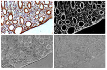
Meanwhile, we compare the run time of PCNN method and proposed method for color microscopic image. In PCNN method, segments every component in RGB with PCNN then combine 3 segmentation results, as show in left column in Fig. 3a-d. The proposed method segments the main component in RGB space directly and then achieves the color segmentation result, which is showed in right column in Fig. 3.
From the experiments results, it’s obvious that the proposed method is better than classical Ostu and PCNN image segmentation method for those color microscopic image with the influence of uneven illumination field, meanwhile, the efficiency is developed largely, Table 1 shows the efficiency between this method and color image segmentation based on PCNN.
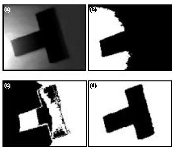 | |
| Fig. 2: | Comparisons of segmentation results with different method (a) Original image; (b) OSTU method; (c) PCNN method and (d) Proposed method |
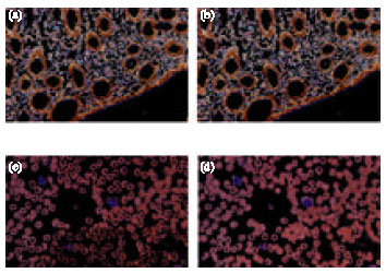 | |
| Fig. 3: | Comparisons of segmentation results in different space (a) PCNN segmentation result in RGB for I2; (b) Segmentation result with proposed method for I2 main component; (c) PCNN segmentation result in RGB for I3 and (d) segmentation result with proposed method for I3 main component |
| Table 1: | The comparison of running time with different methods |
CONCLUSION
We have presented a new scheme for microscopic image segmentation under the circumstances of uneven illumination. The proposed scheme is based on PCA, moreover, it can estimate the uneven illumination field simultaneously. Our method is able to obtain more details in segmentation results and free from the influence of uneven illumination. Comparisons with two well-know color image segmentation methods demonstrate the advantages of the proposed method.
ACKNOWLEDGMENT
The authors would like to thank the reviewers for their valuable comments and suggestions.
REFERENCES
- Mao, K.Z., P. Zhao and P.H. Tan, 2006. Supervised learning-based cell image segmentation for p53 immunohistochemistry. IEEE Trans. Biomed. Eng., 53: 1153-1163.
CrossRef - Navarro-Jover, J.M., M. Alcaniz-Raya, V. Gomez, S. Balasch, J. R. Moreno, V. Grau-Colomer and A. Torres, 2009. An automatic colour-based computer vision algorithm for tracking the position of piglets. Spanish J. Agric. Res., 7: 535-549.
Direct Link - Haralick, R.M and L.G. Shapiro, 1985. Image segmentation techniques. Comput. Vision Graphics Image Process, 29: 100-132.
CrossRef - Bleau, A. and L.J. Leon, 2000. Watershed-based segmentation and region merging. Comput. Vision Image Understanding, 77: 317-370.
CrossRef - Cheng, H.D., X.H. Jiang, Y. Sun and J. Wang, 2001. Color image segmentation: Advances and prospects. Pattern Recognition, 34: 2259-2281.
CrossRef - Yuan-Yuan, C. and J. Xian-Gang, 2009. Research on the adhering segmentation based on the local distribution characteristics of cell image. J. East China JiaoTong Univ., 26: 52-57.
Direct Link - Yeung, K.Y. and W.L. Ruzzo, 2001. Principal component analysis for clustering gene expression data. Bioinformatics, 17: 763-774.
Direct Link - Otsu, N., 1979. A threshold selection method from gray-level histogram. IEEE Trans. Syst. Man Cybern., 9: 62-66.
CrossRefDirect Link