
Nader Goodarzi
Department of Basic Sciences, Faculty of Veterinary Medicine, Razi University, Kermanshah, Iran
Toraj Shah Hoseini
Faculty of Veterinary Medicine, Islamic Azad University, Sanandaj Branch, Sanandaj, Iran
International Journal of Zoological Research
Year: 2015 | Volume: 11 | Issue: 4 | Page No.: 160-168


ABSTRACT
The aim of this study was to investigate the morphostructure of lingual papillae in Markhoz goat. Tissue samples were taken from the dorsal surfaces of the apex, body and root of the tongues of five adult goats. After routine procedure of preparation, the samples were observed under the scanning electron microscope. Three mechanical and tow gustatory types of papillae were seen. Filiform papillae were distributed on the dorsal surface of the anterior two-thirds of the tongue. A bulb-like structure formed the basal portion of the papilla. Between six and eight secondary papillae with sharp pointed tips emerged at the base of filiform papillae. The conical papillae were found on the torus linguae and elongated with a round base while their tips were blunt and sharp, differing structurally from the filiform papillae which posses larger size, a tip without projections and lack of secondary papillae. The round-shaped type I lenticular papillae had a flat surface whereas, those of the leaf-shaped type II lenticular papillae were protruded from the lingual surface with a blunt apex. The round to oval shaped vallate papillae on both rims of the torus linguae were encircled by a prominent gustatory furrow and a thick annular pad. The fungiform papillae were scattered among filiform papillae in the anterior two-thirds of the dorsal surface and each papilla was surrounded by a distict papillary groove. Accordingly, despite of some specific morphological features in lingual papillae, the tongue of markhoz goat is well equipped for mechanical functions similar to that of the other ruminants.
PDF Abstract XML References Citation
Received: May 08, 2015;
Accepted: July 27, 2015;
Published: September 01, 2015
How to cite this article
Nader Goodarzi and Toraj Shah Hoseini, 2015. Fine Structure of Lingual Papillae in the Markhoz Goat (Iranian Angora): A Scanning Electron Microscopic Study. International Journal of Zoological Research, 11: 160-168.
DOI: 10.3923/ijzr.2015.160.168
URL: https://scialert.net/abstract/?doi=ijzr.2015.160.168
DOI: 10.3923/ijzr.2015.160.168
URL: https://scialert.net/abstract/?doi=ijzr.2015.160.168
INTRODUCTION
The tongue as a taste organ in the oral cavity with its species-specific lingual papillae on the dorsal surface plays an important role in food intake and digestion in many mammals (Iwasaki, 2000; Pastor et al., 1993). An important aspect of morphological studies of the tongue is the structure of lingual papillae on the dorsal surface of the tongue and their distribution.
The mammalian lingual papillae differs largely in their occurrence, distribution and structure (Liu and Lee, 1982; Witt and Miller Jr., 1992; Emura et al., 2000) due to the animal’s diet, feeding habit and taxonomy. Based on their function the lingual papillae are grouped into mechanical papillae (filiform, conical and lenticular) which are cornified and aid in licking while, protecting the deeper structures from injury and gustatory papillae which have taste bud (Konig and Leibich, 2004).
Markhoz goat, known as Angora goat in other places, is a dwarf breed indigenous mainly in kordish region of Iran such as Kurdistan province and many arid and semi-arid areas (Rashidi, 2000; Zarkawi and Al-Masri, 2002; Zarei et al., 2009). The potential for commercial mohair, meat production and leather industry makes Markhoz goat a popular candidate for breeders (Zarei et al., 2009).
Much work has been published on the three-dimensional structures of the lingual surface in ruminant species including camel (Qayyum et al., 1988; Eerdunchaolu et al., 2001), cattle (Steflik et al., 1983; Scala et al., 1995), goat (Kumar et al., 1998; Kurtul and Atalgin, 2008) and lamb (Tadjalli and Pazhoomand, 2004).
To the authors’ knowledge, there is no previous report on the morphological characteristics of the lingual papillae in Markhoz goat, so the objective of this study was to reveal the morphostructural differences of the lingual papillae in the Markhoz goat (Iranian Angora).
MATERIALS AND METHODS
Tongues were collected from five (2 males and 3 females with mean age of 12 months) mature Markhoz breed goats, (approximately 30 kg in weight and with a known pedigree) immediately after slaughter in a local slaughterhouse. Tissue samples were taken from the dorsal surfaces of the apex, body and root of the tongue and were fixed in 2.5% glutaraldehyde in 0.1 M cacodylate buffer at 4°C for 24 h. After rinsing in cold 0.1 M cacodylate buffer, samples were post-fixed with 1% osmium tetroxide solution at 4°C. Following dehydration in graded ethanol series (50-99.8%) at room temperature, the samples were dried with liquid CO2 with critical point drier and mounted on the aluminium stubs, sputter-coated with gold-palladium. The mounted specimens were observed and photographed at various angles under the scanning electron microscope (S-4160, Hitachi, Japan) at 15 KV.
RESULTS
On the dorsal surface of the lingual mucosa three types of mechanical papillae (filiform, conical and lenticular) and two types of gustatory papillae (circumvallate and fungiform) were seen (Fig. 1-8).
Filiform papillae: The filiform papillae were the most numerous type of lingual papillae densely distributed on the dorsal surface of the anterior two-thirds of the tongue (Fig. 1). Their number decreased caudally and no filiform papillae were seen on the root. The height of these papillae varied between 275 and 368 μm. A bulb-like structure formed the basal portion of the papilla. Between six and eight secondary papillae with sharp pointed tips emerged at the base of the papillae (Fig. 2). The height of some secondary papillae (accessory branches which detach from the basal part of the papillae) was nearly identical to that of the main filiform papillae. Some projections were seen on each tip and appeared to be transformed into hard scales due to keratinization. Several layers of shingle-like flattened cells of a stratified squamous epithelium covered the surface. These cells provided a rough surface, as dead cells from the superficial epithelial layer were constantly shed.
Conical papillae: The conical papillae were found on the torus linguae, were elongated with a round base while their tips were blunt and sharp (Fig. 3). They were 455-704 μm in length and appeared to emerge from the lingual mucosa with a distict groove at their base. These papillae differed from filiform papillae in terms of their larger size, a tip without projections and lack of secondary papillae. The scales were superimposed irregularly on the surface.
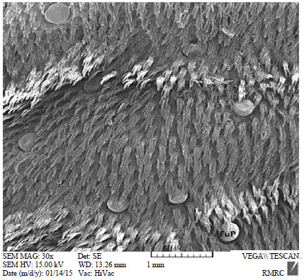 | |
| Fig. 1: | Scanning electron micrograph of the filiform papillae densely distributed on the dorsal lingual mocusa, closely surrounding the fungiform papillae (×30), FUP: Fungiform papillae |
 | |
| Fig. 2: | Scanning electron micrograph of the filiform papillae with projections at the free end and secondary papillae at the base of them at higher magnification (×350), sep: Secondary papillae |
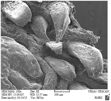 | |
| Fig. 3: | Scanning electron micrograph of the conical papillae with sharp and blunt tips on the rims of the torus linguae (×100), Cp: Conical papillae |
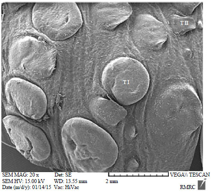 | |
| Fig. 4: | Scanning electron micrograph of the round-shaped type I lenticular papillae (T I) and type II (T II) lenticular papillae with blunt apex (×20) |
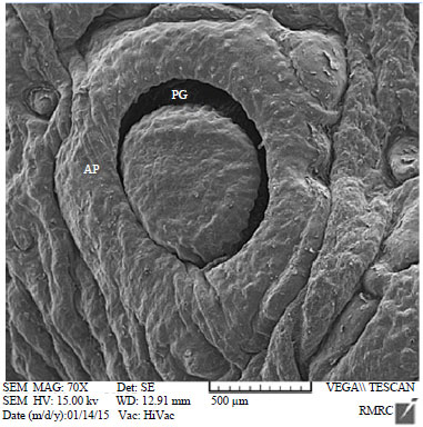 | |
| Fig. 5: | Scanning electron micrograph of the vallate papillae in round to oval shape on both rims of the torus linguae with distict papillary groove and annular pad (×70), AP: Annular pad and PG: Papillary groove |
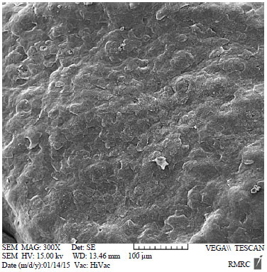 | |
| Fig. 6: | Scanning electron micrograph showing stratified scales on the dorsal surface of the vallate papilla (×300) |
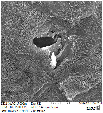 | |
| Fig. 7: | Scanning electron micrograph of the taste pore on the surface of the vallate papilla (×500) |
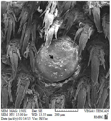 | |
| Fig. 8: | Scanning electron micrograph of the fungiform papilla with a taste pore (arrow) surrounded by a papillary groove (×150) |
Lenticular papillae: The lenticular papillae were limited to the torus linguae and situated in close association with the vallate papillae. These papillae are presented in various diameters (1100-1530 μm) and classified into two types based on their appearance. The predominantly occurring round-shaped type I had a flat surface whereas that of the leaf-shaped type II lenticular papillae were protruded from the lingual surface and possessed a blunt apex (Fig. 4).
Circumvallate papillae: The round to oval shaped vallate papillae were present on both rims of the torus linguae and encircled by a prominent gustatory furrow and a thick annular pad or valium of lingual mucosa (Fig. 5). Occasionally, two adjacent vallate papillae were surrounded by a common valium being separated by a secondary groove. The largest vallate papilla was 1005 μm in diameter while the smallest one was 530 μm. At higher magnification, the papillary surface revealed the presence of stratified scales (Fig. 6) and the opening of the taste pores were seen on the surface of the papillae (Fig. 7).
Fungiform papillae: The fungiform papillae were round and convex in shape, i.e., mushroom-like and were scattered among the filiform papillae in the anterior two-thirds of the dorsal surface and on the both lateral borders of the tongue (Fig. 8). The diameter of these papillae varied between 310 and 426 μm. At higher magnification each fungiform papillae was encircled by a papillary groove and the free surface seemed to be rough due to desquamated epithelial cells. Taste pores were also recognizable.
DISCUSSION
In general, scanning electron microscopy has identified some degree of structural variation in size and shape of lingual papillae on the surface of the tongue (Tadjalli and Pazhoomand, 2004). Lingual papillae are species-specific, differing in shape, size, number, orientation and distribution among mammalian species, so these differences may depend on dissimilarities in diet, feeding habits and handling of food in mouth (Emura et al., 2002). The morphological structure of filiform papillae differs from simple in rodents to compound structure in artiodactyls. The filiform papillae observed in our study in the presence of secondary papillae, caudally direction were similar to those reported in the Jamunapari goat (Kumar et al., 1998), lamb (Tadjalli and Pazhoomand, 2004), buffalo (Scala et al., 1995) and sannen goat (Kurtul and Atalgin, 2008). Likewise, the papillary surface was rough similar to those seen in the Jamunapari goat (Kumar et al., 1998) and Sannen goat (Kurtul and Atalgin, 2008) but it was reported to be smooth in lamb (Tadjalli and Pazhoomand, 2004) and ox (De Paz Cabello et al., 1988).
The height of these papillae measured in this study (275-368 μm) was shorter than those reported in the sannen goat (510 and 620 μm) (Kurtul and Atalgin, 2008) and higher than those measured in the Jamunapari goat (Kumar et al., 1998). These differences could be due to the different genetic factors and feeding habits between various breed. It should be considered that the animals used in our study were a special dwarf breed which were fed like other ruminants.
The lenticular papillae, as it was reported in other ruminants, were located on the surface of the torus linguae that could serve as a complementary protection of the tongue surface (Scala et al., 1995; Kumar et al., 1998; Kurtul and Atalgin, 2008). A papillary groove was present as it was reported in other animals (Scala et al., 1995; Kurtul and Atalgin, 2008; Tadjalli and Pazhoomand, 2004). But, in contrast to Jamunapari goat (Kumar et al., 1998) and sannen goat (Kurtul and Atalgin, 2008) no pyramidal papilla was seen in our study. Kurtul and Atalgin (2008) reported pyramidal type I lenticular papillae with double apices in the sannen goat which were not seen in this study.
The morphology of conical papillae was similar to those reported in Sannen goat (Kurtul and Atalgin, 2008), ox (Chamorro et al., 1986) and Jamunapari goat (Kumar et al., 1998) with except the fact that some of the conical papillae in our study had a sharp tip.
Vallate papillae in Markhoz goat had a papillary groove and a thick annular pad as observed in lamb (Tadjalli and Pazhoomand, 2004), cow (Chamorro et al., 1986; De Paz Cabello et al., 1988), Sannen goat (Kurtul and Atalgin, 2008) and buffalo (Scalaet al., 1995). It was documented that in the Jamunapari goat (Kumar et al., 1998) the valium was divided into 2-3 portions by small furrows that led into the papillary groove of the papilla.
Fine structure of the fungiform papillae observed in this study were similar to those demonstrated in the sannen goat (Kurtul and Atalgin, 2008), ox (Chamorro et al., 1986) and Jamunapari goat (Kumar et al., 1998). They were distributed among the filiform papillae with a distict papillary groove. The stratified scale was seen on the free surface of the fungiform papillae as it was reported in lamb (Tadjalli and Pazhoomand, 2004) and sannen goat (Kurtul and Atalgin, 2008). Furthermore, in contrast to the Jamunapari goat (Kumar et al., 1998) and Sannen goat (Kurtul and Atalgin, 2008), taste pores were seen on the surface of fungiform papillae in our study. In conclusion, although there were some specific morphostructure characteristics in lingual papillae of Markhoz goat similar to other ruminants, the tongue was equipped with more mechanical papillae in respect of better development and variety than gustatory papillae.
REFERENCES
- Chamorro, C.A., P.C. de Paz, J. Sandoval and J.G. Fernandez, 1986. Comparative scanning electron-microscopic study of the lingual papillae in two species of domestic mammals (Equus caballus and Bos taurus). 1. Gustatory Papillae. Acta Anat., 125: 83-87.
CrossRefPubMedDirect Link - De Paz Cabello, P., C.A. Chamorro, J. Sandoval and F. Fernandez, 1988. Comparative scanning electron microscopic study of the lingual papillae in two species of domestic mammals (Equus caballus and Bostaurus). II. Mechanical papillae. Acta Anat., 132: 120-123.
CrossRefDirect Link - Eerdunchaolu, K. Takehana, E. Yamamoto, A. Kobayashi and G. Cao et al., 2001. Characteristics of dorsal lingual papillae of the Bactrian camel (Camelus bactrianus). Anat. Histol. Embryol., 30: 147-151.
CrossRefPubMedDirect Link - Emura, S., A. Tamada, D. Hayakawa, H. Chen and S. Shoumura, 2000. Morphology of the dorsal lingual papillae in the black rhinoceros (diceros bicornis). Anat. Histol. Embryol., 29: 371-374.
CrossRefPubMedDirect Link - Emura, S., D. Hayakawa, H. Chen, S. Shoumura, Y. Atoji and H. Wijayanto, 2002. SEM study on the dorsal lingual surface of the large flying fox, pteropus vampyrus. Okajimas Folia Anat. Japn., 79: 113-119.
CrossRefPubMedDirect Link - Iwasaki, S.I., 2002. Evolution of the structure and function of the vertebrate tongue. J. Anat., 201: 1-13.
CrossRefDirect Link - Kumar, P., S. Kumar and Y. Singh, 1998. Tongue papillae in goat: A scanning electron-microscopic study. Anat. Histol. Embryol., 27: 355-357.
PubMedDirect Link - Kurtul, I. and S.H. Atalgin, 2008. Scanning electron microscopic study on the structure of the lingual papillae of the Saanen goat. Small Rumin. Res., 80: 52-56.
CrossRefDirect Link - Liu, H.C. and J.C. Lee, 1982. Scanning electron microscopic and histochemical studies of foliate papillae in the rabbit, rat and mouse. Acta Anat. (Basel), 112: 310-320.
PubMedDirect Link - Pastor, J.F., J.A. Moro, J.A.G. Verona, A. Gato, J.J. Represa and E. Barbosa, 1993. Morphological study by scanning electron microscopy of the lingual papillae in the common european bat (pipistrellus pipistrellus). Arch. Oral Biol., 38: 597-599.
CrossRefDirect Link - Qayyum, M.A., J.A. Fatani and A.M. Mohajir, 1988. Scanning electron microscopic study of the lingual papillae of the one humped camel, Camelus dromedarius. J. Anat., 160: 21-26.
PubMedDirect Link - Steflik, D.E., B.B. Singh, R.V. McKinney and J.L. Boshell, 1983. Correlated TEM, SEM and histological observations of filiform papillae of the cow tongue. Acta Anat., 117: 21-30.
CrossRefPubMedDirect Link - Tadjalli, M. and R. Pazhoomand, 2004. Tongue papillae in lambs: A scanning electron microscopic study. Small Ruminant Res., 54: 157-164.
CrossRefDirect Link - Witt, M. and I.J. Miller Jr., 1992. Comparative lectin histochemistry on taste buds in foliate, circumvallate and fungiform papillae of the rabbit tongue. Histochemistry., 98: 173-182.
CrossRefDirect Link - Zarei, M.A., A. Farshad and S. Akhondzadeh, 2009. Variations in thyroidal activity during estrous cycle and natural breeding season in markhoz goat breeds. Pak. J. Biol. Sci., 12: 1420-1424.
CrossRefPubMedDirect Link - Zarkawi, M. and M.R. Al-Masri, 2002. Use of Radioimmunoassay to measure progesterone levels during different reproductive stages in female Damascus goats. Trop. Anim. Health Prod., 34: 535-539.
CrossRefDirect Link