
Shaimaa M. Badr-Eldin
Department of Pharmaceutics, Faculty of Pharmacy, King Abdulaziz University, Jeddah 21589, Saudi Arabia
LiveDNA: 20.35365
Hibah Mubarak Aldawsari
Department of Pharmaceutics, Faculty of Pharmacy, King Abdulaziz University, Jeddah 21589, Saudi Arabia
LiveDNA: 966.25108
Sabna Kotta
Department of Pharmaceutics, Faculty of Pharmacy, King Abdulaziz University, Jeddah 21589, Saudi Arabia
LiveDNA: 966.35350
Nabil Abdulhafiz Alhakamy
Department of Pharmaceutics, Faculty of Pharmacy, King Abdulaziz University, Jeddah 21589, Saudi Arabia
International Journal of Pharmacology
Year: 2021 | Volume: 17 | Issue: 1 | Page No.: 15-27


ABSTRACT
Background and Objectives: Ciprofloxacin (CIP) is a fluoroquinolone antibiotic used for the management of eye bacterial infections. To overcome the challenges like poor ocular residence time and poor bioavailability, this study aimed at formulating and optimizing ciprofloxacin Self-assembled supramolecular hydrogel based on α-cyclodextrin/Poloxamer polypseudorotaxanes to enhance the ocular availability of the drug. Materials and Methods: A Box-Behnken experimental design with three formulation factors including the concentration of poloxamer, cyclodextrin and their ratio were studied using Design-Expert software. In vitro permeation, ex vivo permeation, anti-bacterial studies and in vivo of precorneal concentration of drug was determined. Results: Both the viscosity and drug release were found to be dependent on poloxamer and cyclodextrin concentrations, while PLX:CD ratio was found to have a significant effect on the viscosity only. The optimized system with maximized viscosity and drug release exhibited a higher CIP permeation compared to conventional Carbopol® gel. The antibacterial studies with various bacterial strains indicated a significant improvement in the antibacterial activity of CIP when presented in the optimized formulation. Further, a higher amount of CIP was retained in the precorneal area for a prolonged period following the instillation of supramolecular gel as compared to the conventional gel. Conclusion: Overall, the proposed supramolecular gel could be regarded as an effective platform for improving the ocular retention and availability of ciprofloxacin. Further studies and clinical trials would help to explore this formulation for therapeutic use.
PDF Abstract XML References Citation
Copyright: © 2021. This is an open access article distributed under the terms of the creative commons attribution License, which permits unrestricted use, distribution and reproduction in any medium, provided the original author and source are credited.
How to cite this article
Shaimaa M. Badr-Eldin, Hibah Mubarak Aldawsari, Sabna Kotta and Nabil Abdulhafiz Alhakamy, 2021. Self-Assembled Supramolecular Hydrogel Based on α-Cyclodextrin/Poloxamer Polypseudorotaxanes for Ocular Delivery of Ciprofloxacin. International Journal of Pharmacology, 17: 15-27.
DOI: 10.3923/ijp.2021.15.27
URL: https://scialert.net/abstract/?doi=ijp.2021.15.27
DOI: 10.3923/ijp.2021.15.27
URL: https://scialert.net/abstract/?doi=ijp.2021.15.27
INTRODUCTION
The complexity of the eye structure is unparalleled with peculiar and unique anatomy and physiology. The unique anatomy of the human eye renders barriers to the protection of eye tissues against toxic substances. These barriers are either static, e.g. corneal epithelium, or dynamic, e.g., tear dilution1. The uniqueness of this vital organ and the barriers it affords for molecule passage presents great challenges for ocular drug delivery2. Furthermore, blinking of the eyes, wash out by tear fluid and draining of drugs through nasolacrimal duct also represent further challenges for conventional ophthalmic formulations that warrant frequent administration for maintenance of adequate drug concentration to treat acute ocular disorders like bacterial infections3,4. Thus, advancement in the arena of ocular delivery has been targeted towards surpassing the ocular barriers to enhance ocular availability of the drugs, maintaining drug concentration at the target site and reducing instillation frequency for improving patient compliance1. To achieve these targets, several strategies have been explored for enhancing ocular drug availability. These strategies include the utilization of mucoadhesive agents, viscosity enhancers and in situ gels for prolonging eye residence5. Further, the utilization of prodrugs, colloidal systems, permeation enhancers and cyclodextrin complexation was proven to be effective for enhancing drugs' permeation6,7.
Ciprofloxacin (CIP) is a fluoroquinolone antibiotic used for the management of eye bacterial infections such as conjunctivitis, keratitis and endophthalmitis8. It is a broad-spectrum antibiotic with low toxicity, however, most of the available commercial CIP formulations suffer from a poor ocular residence that compels for frequent administration9. Besides, the bioavailability of CIP at the target tissue in the anterior segment of the eye is usually combated by two major factors, namely, the low solubility in the tear fluid pH that could lead to precipitation and the anatomical and physiological ocular barriers10,11. Therefore, the selection of an appropriate ocular delivery system for CIP is crucial for efficient action.
Cyclodextrins (CDs) have been extensively utilized for drug delivery via Host-guest interactions. In ocular delivery, CDs provide the advantages of improving drugs' solubility, enhancing corneal tolerability and permeability and alleviating toxicity12. Poloxamer (Pluronic®) is a thermosensitive triblock copolymer with polyethylene oxide (PEO)-polypropylene oxide (PPO)-polyethylene oxide (PEO) structure and a wide range of molecular weights13. Poloxamer (PLX) has been utilized for the formulation of in situ gels, however, the hydrophilic character of the formed gel usually leads to rapid erosion resulting in limited residence time in the application site. Using high concentrations of poloxamer could be helpful to overcome this problem, however, it could represent a pitfall from both technological and safety point of view14. Other approaches for improving the gelling behaviour of poloxamer include mixing with other polymers for enhancing the rigidity of the formed gel15 or forming supramolecular architectures with cyclodextrin16.
The Self-association of macromolecules into highly organized and specific nanostructured functional architectures through supramolecular interactions has gained marked attention for application in various areas. CDs represent one of the most widely used hosts for macromolecular spontaneous assembly17,18. Recently, CD/ PEO-based poly(pseudo)rotaxanes, composed of ring structure (CD) threaded onto a block polymer chain, have given rise to advancement in the biomaterials area. In polyrotaxanes (PRs), the CDs are anchored to particular sites of the polymer chains owing to the existence of bulky functional groups that limit their movement. On the other hand, in the case of polypseudorotaxanes (PPRs), CDs are freely mobile along or off the polymer backbone due to the absence of stoppers19,20. The reversible reinforced ring/string interaction in PPRs markedly enhances the viscoelastic behaviour of the system21. The triblock copolymer, PLX, has been reported as a promising candidate for interaction with CDs to yield a supramolecular hydrogel that provides improved gelation compared to polymeric hydrogels22-24. The previously described biocompatible CD-based supramolecular systems, especially those based on α-CD, have been deployed successfully for the controlled delivery of a plethora of therapeutic moieties25,26.
Accordingly, Self-assembled supramolecular hydrogels could be a promising approach for CIP ocular delivery because the amphiphilic polymer and the cyclodextrin (CD) may improve the drug solubility while the enhanced gelling property could promote the residence in the eye.
Thus, this study aimed at exploring the potential of α-CD/PLX supramolecular self-assemblies for enhancing the ocular bioavailability of CIP. A Box-Behnken design was applied for optimization of the proposed formulation. The optimized system with maximized viscosity and drug release was then subjected to ex vivo permeation studies, microbiological assessment and in vivo evaluation in rabbits.
MATERIALS AND METHODS
Study area: The study was carried out in Research Laboratory, Department of Pharmaceutics, Faculty of Pharmacy, King Abdulaziz University, Jeddah, Saudi Arabia from May-November, 2020.
Materials: Ciprofloxacin; CIP, was supplied as a gift by the Saudi Arabian Japanese Pharmaceutical Company Limited (SAJA, Jeddah, KSA), poloxamer 407; PLX, (Poly (ethylene glycol)-block-poly (propylene glycol)-block-poly (ethylene glycol)) and α-cyclodextrin; α-CD (MW 972.84) were purchased from Sigma Company (St. Louis, MO, USA). All other reagents were of analytical grade.
Preparation of molecular dispersions: The aqueous dispersion of poloxamer 407 was prepared in different concentrations ranging from 2-5% (w/v) by adding the polymer into cold water under magnetic stirring for 24 hrs until a clear solution was formed. Similarly, α-cyclodextrin solutions were also prepared in concentrations ranging from 8-10% with the help of a magnetic stirrer14. CIP solution (in 0.9% sodium chloride) was added to the poloxamer 407 dispersions27. Definite concentrations of dispersions of poloxamer 407 and α-cyclodextrin were mixed in the volume ratio suggested for PLX: CD by the design to obtain the final supramolecular hydrogel (SMG).
Formulation and optimization of CIP self-assembled supramolecular hydrogels (SMGs)
Experimental design: In this work, a Box-Behnken experimental design composed of 3 factors at 3 levels was implemented for the formulation of CIP Self-assembled supramolecular hydrogels (SMG) for ocular delivery using Design-Expert software (Version 12; Stat-Ease Inc., Minneapolis, MN, USA). Three formulation factors were studied, namely, PLX concentration (X1, %), CD concentration (X2, %) and PLX: CD ratio (X3, v/v). Unlike the reported studies which use final concentrations of PLX and CD in the SMG15,28, in the present study, we used the concentrations of PLX and CD dispersions used to mix to result in the desired PLX: CD ratio (third independent factor) in the final SMG. This modification was aimed to develop a scalable manufacturing process for the SMG by defining the concentrations of components for the SMG preparation process. The standardization of reactant concentrations is far simple and predictive than defining a final concentration in the SMG.
| Table 1: Independent variables and responses used in Box Behnken design for the formulation of CIP self-assembled supramolecular hydrogels | |||||
| Levels | |||||
| Variables | (-1) | (0) | (+1) | ||
| A: PLX (%) | 2.00 | 3.50 | 5.00 | ||
| B: α-CD (%) | 8.00 | 9.00 | 10.00 | ||
| C: PLX:CD | 1.00 | 1.25 | 1.50 | ||
| Responses | Desirability constraints | ||||
| Y1: Viscosity (cP) | Maximize | ||||
| Y2: Drug release after 24 hrs (%) | Maximize | ||||
| CIP: Ciprofloxacin, PLX: Poloxamer, α-CD: Alpha-cyclodextrin | |||||
The design is intended for statistically optimizing all formulation factors and evaluating the main as well as interaction effects and also the quadratic effects of the components used in formulations on the viscosity (Y1, cP) and CIP percentage released after 24 hrs (Y2, %)29. Fit statistics was used to choose the best fitting sequential model for each response. The data of Table 1 compiles the dependent and independent variables with their levels. The design yielded 17 runs including five centre points as presented in Table 2. Analysis of Variance (ANOVA) was then performed to analyze responses data at a 95% level of significance. Three-dimensional surface plots were generated to demonstrate the interaction between the explored variables.
Preparation of self-assembled supramolecular hydrogels based on polypseudorotaxanes: Polypseudorotaxanes were prepared by mixing the drug-loaded dispersions of poloxamer 407 with the α-cyclodextrin solution under constant stirring for 4 hrs. The concentration of dispersions and mixing ratio was as per the Box-Behnken experimental design, Table 221. The formation of polypseudorotaxane was observed visually. Changes in turbidity and phase separation were evaluated by visual inspection21.
Rheological characterization: The viscosity of formulations was determined using Brookfield's viscometer, DV model (Massachusetts, USA). Angular velocity was increased gradually from 10, 20, 50 and 200 rpm and the viscosity of the formulation was measured at a constant temperature. The average of three readings was taken to calculate the viscosity. All measurements were performed in triplicate30.
In vitro release study: A total of 2 mL of each of the formulations was taken in test tubes and Simulated Tear Fluid (STF) was slowly added to the surface of gels. The test tubes were kept for shaking for 24 hrs.
| Table 2: Variables levels and observed responses for CIP self-assembled supramolecular hydrogels formulated according to Box-Behnken design | |||||||
| Independent variables | |||||||
| Experimental run # | PLX (%) | α-CD (%) | PLX:CD | Viscosity (cP)±SD | Percent CIP released after 24 hrs (%)±SD | ||
| F1 | 5.0 | 8.0 | 2.0 | 18.88±0.23 | 44.70±1.95 | ||
| F2 | 2.0 | 10.0 | 2.0 | 36.16±1.78 | 83.60±3.71 | ||
| F3 | 3.5 | 9.0 | 2.0 | 32.96±1.99 | 24.78±1.56 | ||
| F4 | 2.0 | 9.0 | 3.0 | 21.70±1.13 | 56.72±2.31 | ||
| F5 | 2.0 | 9.0 | 1.0 | 22.72±1.89 | 79.65±2.99 | ||
| F6 | 5.0 | 10.0 | 2.0 | 58.24±2.95 | 42.27±3.11 | ||
| F7 | 3.5 | 9.0 | 2.0 | 32.14±1.17 | 25.12±1.78 | ||
| F8 | 3.5 | 8.0 | 1.0 | 16.29±1.11 | 45.93±2.11 | ||
| F9 | 3.5 | 10.0 | 3.0 | 34.01±1.79 | 62.89±2.89 | ||
| F10 | 3.5 | 8.0 | 3.0 | 12.48±0.98 | 62.72±2.11 | ||
| F11 | 3.5 | 9.0 | 2.0 | 32.87±1.92 | 25.77±1.89 | ||
| F12 | 3.5 | 9.0 | 2.0 | 32.06±1.87 | 24.34±2.11 | ||
| F13 | 5.0 | 9.0 | 1.0 | 43.78±2.24 | 50.52±2.99 | ||
| F14 | 2.0 | 8.0 | 2.0 | 15.36±0.99 | 38.39±2.11 | ||
| F15 | 3.5 | 9.0 | 2.0 | 32.88±2.19 | 25.59±1.78 | ||
| F16 | 3.5 | 10.0 | 1.0 | 52.16±2.78 | 93.98±3.56 | ||
| F17 | 5.0 | 9.0 | 3.0 | 26.24±1.54 | 64.35±3.11 | ||
| CIP: Ciprofloxacin, PLX: Poloxamer, α-CD: Alpha-cyclodextrin | |||||||
Samples of the medium were taken and diluted with STF and after filtration concentration of CIP in each tube was determined by UV-VIS spectrophotometer (UV-2600 Shimadzu, Tokyo, Japan) at 275 nm. The experiment was done in triplicate14.
Numerical optimization: Numerical optimization and desirability approach was implemented for predicting the levels of the variables that provide maximized responses according to the aim of the process. The predicted optimized formulation was then prepared for performing further characterization.
Evaluation of optimized CIP self-assembled supramolecular hydrogel
In vitro permeation: CIP diffusion from the optimized CIP SMG was examined using automated vertical Franz diffusion (MicroettePlus; Hanson research, CA, USA) with a volume of 7 mL and area of 1.766 cm2 in Simulated Tear Fluid (STF) fitted with dialysis tubing cellulose membrane with a molecular cut off 12000 Da (Sigma-Aldrich, USA). A total of 100 μL of the formulation was located in the donor compartment. The temperature of the medium was kept at 37±0.5°C and the stirring rate was set to 400 rpm. Aliquots of 1 mL were automatically withdrawn at time intervals of 0.5, 1, 2, 4 and 8 hrs and replaced with STF to keep the volume constant31,32. The samples were analyzed using a previously reported HPLC method33. The experiment was done in triplicate.
Ex vivo permeation: Ex vivo corneal permeation studies were carried out using goat's cornea. The whole eyeballs of a goat were collected from a slaughterhouse. The cornea along with 4-5 mm surrounding sclera was carefully removed. The method of dissection of the cornea was the same as those described previously34,35. The corneas were then washed with cold saline and then transferred to STF in Petri dishes. Then the tissue was mounted into a horizontal corneal diffusion cell. The donor compartment was filled with optimized CIP gel in Simulated Tear Fluid (STF). The receiver compartment was filled with an STF pH of 7.4. The medium in both the chambers was slightly stirred with a magnetic stirrer to avoid any boundary layer effect. The samples were withdrawn at different time intervals and analyzed for drug content using the previously mentioned HPLC method. The receptor phase was replenished with an equal volume of STF at each time interval for a period of 6 hrs. The percentage of drug permeated was plotted against time10. CIP loaded in Carbopol® gel was used as a control. Results are expressed as the percentage of drug permeated at each sampling time.
Antibacterial studies: The antimicrobial efficiency of the optimized formulation was tested on Staphylococcus aureus, Escherichia coli and Pseudomonas aeruginosa. Agar diffusion test was used to study the inhibitory effect of optimized gel using. Wells were made in agar plates after surface inoculation of the test organism. The bacterial count of 105 CFU mL–1 was adjusted by the McFarland standard. Each well was inoculated with 100 μL of either the optimized formulation or CIP loaded in Carbopol® gel and the plates were kept in an incubator for 24 hrs at 37°C. After incubation, the zone of inhibition was measured and compared to previous srudies27.
In vivo determination of precorneal concentrations: Six healthy albino rabbits weighing between 1.5-2 kg were divided into two groups of three rabbits. About 50 μL of optimized CIP SMG was instilled into the lower conjunctival sac of both eyes cautiously to guard against spilling. Similarly, 50 μL of conventional CIP gel was given to the other group of animals. Tear fluid samples (1 μL) were obtained without anaesthesia using microcapillaries at 0, 0.25, 0.5, 1, 2, 3 and 4 hrs. The rabbits were kept in restraining boxes during the experiment where they were able to move their heads and eyes freely. The collected samples were transferred into centrifuge tubes and the microcapillaries were flushed with 10 μL methanol and then the mobile phase was added up to 1 mL. The centrifuged supernatant after filtration was analyzed for CIP content in HPLC30.
The concentrations of CIP in the samples were determined on a UFLC Shimadzu system with an SPD-M20A prominence diode array detector. (Shimadzu, Kyoto, Japan) LC solution was used for the data collection and control of the overall system. A PhenomenexC18 analytical column (250×4.6 mm) (Phenomenex, Torrance, CA) was used for the separation with a mobile phase consisting of acetonitrile-2% acetic acid aqueous solution (16: 84, v/v), at a flow rate of 1 mL min–1. Samples were filtered through a 0.20 μm pore-size nylon membrane filter The injection volume was 10 μL for all samples. The elutes were analyzed at 280 nm33.
Statistical analysis: Results are expressed as Mean±SD. The Student's t-test was used to identify differences that were considered to be statistically significant at p<0.05.
RESULTS AND DISCUSSION
The traditional hit and trial method requires a large number of independent trials which is time-consuming. The design of the experiment saves time by analyzing a large number of factors simultaneously. Here, the optimization procedure was done by selecting objective functions and finding the most relevant factors.
In this study, we used the Box-Behnken design selected because it needs less number of runs when compared to a central composite design for 3 or 4 variables. The experimental design contains several points located in the middle of every edge and also the simulated centre point of the multidimensional cube which defines the region of interest. Response surface modelling is meant to build a regression model by approximation which is closest to the original regression model. Since the experiment investigates the effects of different blends of more than one factor and its levels on different response variables, a multiple regression analysis is required.
Sequential model selection and diagnostic analysis: Both viscosity and percentage drug released after 24 hrs data were fitted to the quadratic model, Table 3. As per the fit statistics of the quadratic model the R2 value was found to be 0.9987 and 0.9953 with an adequate precision value of 87.7 and 40.94 for viscosity and drug release (%), respectively.
The model was selected based on the greatest multiple correlation coefficient (R2) and the smallest Predicted Residual Error Sum of Squares (PRESS). The predicted and adjusted R2 for each response coincide reasonably with each other ensuring the model validity. Besides, adequate precision of greater than 4 indicates an appropriate signal to noise ratio that consequently implies that the chosen model is appropriate for exploring the experimental design space36. Diagnostic plots for both responses were developed to assess the goodness of fit of the model and ascertain its reliability. The result of Fig. 1a shows the externally studentized residuals vs. run number plots for viscosity and Fig. 1b shows predicted vs. actual values plots for viscosity. Similarly, Fig. 1c shows the externally studentized residuals vs. run number plots for % drug release after 24 hrs and Fig. 1d shows predicted vs. actual values plots for % drug release after 24 hrs. The randomly scattered point presented in the externally studentized residuals versus run plots showed is an indication that both responses were not affected by any lurking variable. Besides, the good correlation between the observed responses, supported by the good linearity of the predicted versus actual values plots of both responses, confirmed the predictability of the selected model37.
Influence of variables on viscosity (Y1): The viscosity of ocular preparations is a crucial characteristic related to ocular retention time and consequent clinical benefit. The prepared SMGs exhibited average viscosity ranging from 12.48±0.98-58.24±2.95 cP, Table 2.
| Table 3: Fit statistics of CIP self-assembled supramolecular hydrogels according to the quadratic model | |||||||
| Responses | Sequential p-value | R2 | Adjusted R2 | Predicted R2 | Adequate precision | PRESS | Significant terms |
| Y1: Viscosity (cP) | <0.0001 | 0.9987 | 0.9971 | 0.9843 | 87.70 | 39.58 | X1, X2, X3, X1X2, X1X3, X2X3, X32 |
| Y2: Percent CIP released after 24 hrs (%) | <0.0001 | 0.9953 | 0.9906 | 0.9387 | 40.94 | 483.21 | X1, X2, X3, X1X2, X1X3, X2X3, X12, X22, X32 |
| CIP: Ciprofloxacin, R2: Coefficient of determination | |||||||
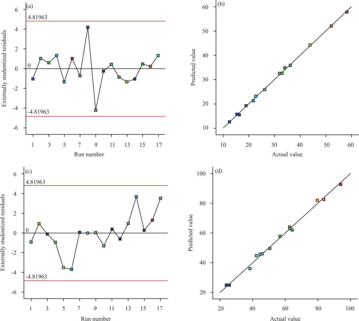 |
| Fig. 1: | Diagnostic plots for the quadratic model for viscosity and percentage CIP released after 24 hrs from CIP self-assembled supramolecular hydrogel (a) Externally studentized residuals vs. run number for viscosity, (b) Predicted vs. actual values plots for viscosity, (c) Externally studentized residuals vs. run number for percentage CIP released after 24 hrs and (d) Predicted vs. actual values plot for percentage CIP released after 24 hrs |
The f-value of 617.71 computed in the ANOVA analysis for the viscosity ensures that the selected quadratic model is significant (p<0.0001). There is a little chance of 0.01% that an f-value this large could be ascribed to noise. The sequential quadratic model equation presenting the effect of variables influencing the viscosity was suggested by the experimental design in terms of coded factor as follows38,39:
Y1 = 32.74+6.16X1+13.88X2-5.64X3+4.64X1X2-4.00X1X3-3.45X2X3-0.49X12-0.09X22-3.78X32
ANOVA results revealed that all the linear terms have a significant influence on the viscosity (p<0.0001). Also, all the interaction terms, in addition to the quadratic term X32 were significant at the same level. From the equation, it could be inferred that the α-CD percentage (X2) has the most significant effect on the viscosity of the SMG as evidenced by its highest coefficient. Also, the positive sign for the coefficient indicates a positive effect, i.e., using a higher concentration of α-CD leads to an increase in the viscosity. PLX percentage (X2) also exhibited a significant positive effect on viscosity, but to a lesser extent. On the other hand, the PLX:CD ratio (X3) was the least significant variable as depicted by its lowest coefficient and it exhibited a negative effect on viscosity. It is reported that higher α-CD concentration results in a thicker gel phase when added to a PLX-containing system14,40. In the present study also, we have seen that a higher concentration of α-CD leads to an increase in the viscosity. Conversely, a lower α-CD concentration could lead to a thinner gel system with low viscosity. A higher value of PLX:CD implies a low α-CD content compared to PLX. Thus, the observation of lower viscosity of supramolecular gels at high values of PLX:CD ratio is reasonable.
The result of Fig. 2 illustrates the Two-dimensional contour and Three-dimensional response surface plots for the effect of the studied variables on the viscosity of the SMGs. The influence could be evaluated by the Iso-value curves in the contour plot or height/elevation of the response surface. It can be seen that in all the plots the Iso-value curves are near to a perpendicular position to the axis of α-CD%. Similarly, the elevation of the response surface increases as the concentration of α-CD% is increased. This observation implies that the X1 has the most influence on the supramolecular gel as previously mentioned. In Fig. 2a-b, it is observed that the effect of PLX% was greater than that of PLX:CD ratio. The elevation of the response surface plot shows an increase for PL. At the same time the PLX:CD ratio was shown to have less influence. In Fig. 2c-d, pronounced effects for both PLX and α-CD were noted. The Iso-value curves show higher values at higher values for both α-CD% and PLX%. The elevation of the response surface was more at the corner point representing the highest values for both CD and PLX and vice versa. Thus, both the CD and PLX% were influential in increasing the value of the viscosity of the gel. From the elevation of the other two corner points, it could be inferred that α-CD% has more influence on the viscosity of the SMG than the PLX (%). In Fig. 2e-f, the effect of α-CD% and PLX:CD ratio could be observed. The Iso-value curves are almost perpendicular to the α-CD (%) axis and therefore parallel to the PLX:CD axis. The elevation of the response surface was greater at a higher concentration of α-CD% while not much influenced by the PLX:CD ratio. This could confirm the more pronounced effect of α-CD% when compared to PLX:CD ratio on the viscosity of the SMG.
Influence of variables on drug release (Y2): The prepared SMGs showed average drug released ranging from 24.34±2.11-93.98±3.56% after 24 hrs, Table 2. The f-value of 188.41 computed in the ANOVA analysis affirms the significance of the quadratic model (p<0.0001). There is a little chance of 0.01% that an f-value this large could be due to noise. The sequential quadratic model equation presenting the effect of variables influencing the percentage released was generated in terms of coded factor as follows38,39:
Y1 = 1.18+0.63X1+0.10X2-0.01X3+0.08X1X2-0.09X1X3-0.05X2X3-0.21X12+0.07X22-0.09X32
ANOVA results revealed that all the linear terms have a significant influence on drug release (p<0.0001 for X1 and X2, p = 0.0064 for X3). Also, all the interaction and quadratic terms were significant at the same level. In contrast to viscosity, PLX percentage (X1) has the most significant effect on the drug release rather than α-CD percentage followed by the α-CD percentage, while the PLX:CD ratio exhibited the least effect as evidenced by their terms' coefficients.
The contour and response surface plots for drug release from the SMGs are presented in Fig. 3. The result of Fig. 3a-b show the pronounced effect of PLX% compared to α-CD% on drug release. In this case, too, Iso-value curves were almost perpendicular to the axis of PLX% indicating its higher influence. At higher values of PLX, the influence was more pronounced. In the case of α-CD%, the influence was much less compared to that of PLX%. In the present study, the concentration range selected for PLX was larger than that selected for α-CD. The PLX:CD ratio of values higher than 1 might also have exaggerated this effect. The elevation of the response surface remained almost the same throughout at various percentages of PLX. The effects of PLX:CD ratio and PLX% in combinations are shown in Fig. 3c-d. In this case, the Iso-value curves were almost perpendicular to the axis of PL and therefore parallel to the axis of PLX:CD. Also, the elevation of the response surface at the corners representing higher values for PL was more. This implied a marked effect of PLX% on drug release compared to that of PLX:CD. Comparing the influence of α-CD% and PLX:CD as presented in Fig. 3e-f, it was observed that, in contrast to viscosity, both α-CD% and PLX:CD showed a similar magnitude of effects. This was confirmed from the Iso-value curves and the elevation of the response surface. The elevation was almost the same throughout the design space. Based on the previous findings, it could be concluded that the influence of PLX % was more pronounced than the other two factors.
Optimization: The viscosity and drug release after 24 hrs constraints were applied to anticipate the optimal levels of the investigated factors. The desirability function was computed as 0.922. The optimized formulation was prepared and subjected to characterization. The variables levels predicted and observed responses for the optimized formulation are presented in Table 4. The residual error presented by the difference between the predicted and observed data was considerably small indicating that the numerical optimization was valid for this study.
| Table 4: Variables levels and the predicted and observed responses of the optimized CIP self-assembled supramolecular hydrogel | |||||
| Variables | X1:PLX (%) | X2:α-CD (%) | X3:PLX:CD | ||
| Optimum levels | 3.43 | 10.00 | 1:1.1 | ||
Predicted value | Observed value | Error (%) | |||
| Viscosity (cP) | 51.38 | 53.19 | 3.40 | ||
| Percent CIP released at 24 hrs (%) | 93.97 | 92.17 | 1.95 | ||
| CIP: Ciprofloxacin, PLX: Poloxamer, α-CD: Alpha-cyclodextrin | |||||
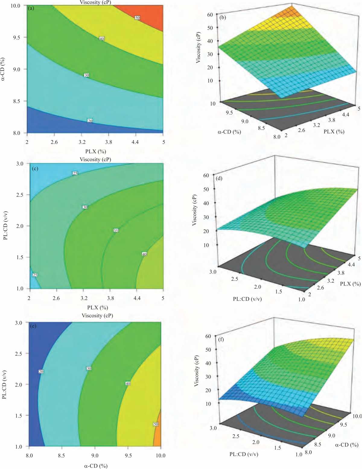 |
| Fig. 2(a-f): | Contour D-plots and response surface 3D-plots for the effect of PLX% (X1), α-CD% (X2) and PLX:CD ratio (X3) on the viscosity of self-assembled supramolecular hydrogels (a) Contour plot for the effect of PLX and α-CD%, (b) Response surface for the effect of PLX and α-CD%, (c) Contour plot for the effect of PLX (%) and PLX:CD, (d) Response surface plot for the effect of PLX (%) and PLX:CD, (e) Contour plot for the effect of α-CD% and PLX:CD and (f) Response surface plot for the effect of α-CD% and PLX:CD. CIP: Ciprofloxacin, PLX: Poloxamer, α-CD: Alpha-cyclodextrin |
 |
| Fig. 3(a-f): | Contour D-plots and response surface 3D-plots for the effect of PLX% (X1), α-CD% (X2) and PLX:CD ratio (X3) on % CIP released at 24 hrs from self-assembled supramolecular hydrogels (a) Contour plot for the effect of PLX and α-CD%, (b) Response surface for the effect of PLX and α-CD%, (c) Contour plot for the effect of PLX (%) and PLX:CD, (d) Response surface plot for the effect of PLX (%) and PLX:CD, (e) Contour plot for the effect of α-CD% and PLX:CD and (f) Response surface plot for the effect of α-CD % and PLX:CD. CIP: Ciprofloxacin, PLX: Poloxamer, α-CD: Alpha-cyclodextrin |
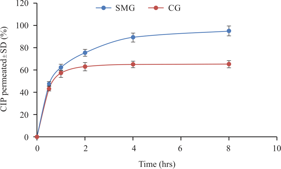 |
| Fig. 4: | In vitro CIP permeation profile from optimized self-assembled supramolecular hydrogel compared to control gel in Simulated Tear Fluid at 37±0.5°C SMG: Supramolecular hydrogel, CG: Control gel |
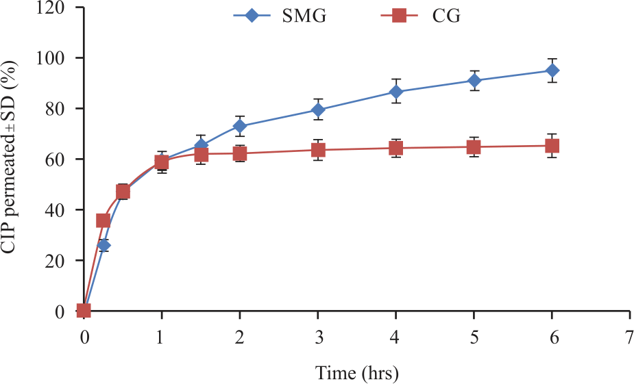 |
| Fig. 5: | Ex vivo CIP permeation profile from optimized self-assembled α-CD/PLX-based supramolecular hydrogel compared to control gel in simulated tear fluid at 37±0.5°C SMG: Supramolecular hydrogel, CG: Control gel |
The optimum values prescribed by the model was 3.43% of poloxamer solution 10% α cyclodextrin solution in a ratio of 1:1.1. The prepared optimized SMG formulation was subjected to further evaluation.
Evaluation of optimized CIP self-assembled supramolecular hydrogel
In vitro permeation study: The in vitro CIP diffusion from the optimized CIP SMG was studied in comparison to the diffusion from Carbopol®-based (1%) gel as a control, Fig. 4. The dialysis tube membrane was used as the permeation barrier and STF as the permeation medium for this purpose and the results showed better in vitro permeation from the optimized SMG than the Carbopol®-based gel. The CIP permeation was comparable for both formulations till 1 hr. At later time intervals, the permeation profile of CIP shows a considerable difference with a higher percentage CIP release from the optimized SMG. After 8 hrs, the percentage of CIP permeation was 94.9±4.4% from the SMG while the Carbopol® gel showed 65.02±3.2%. Thus, the optimized SMG demonstrated a better CIP permeation profile.
Nevertheless, both the systems showed a sustained in vitro CIP permeation. This type of slow and concentration-dependent drug permeation is reported for Carbopol®-based systems41. Thus, the results observed in the present study were comparable to reported data.
Ex vivo permeation study: The results of ex vivo corneal permeation studies using goat's cornea are shown in Fig. 5. The permeation of the optimized SMG was compared with Carbopol®-based (1%) gel. From the results, it could be seen that the SMG enhances the CIP permeation and more percentage drug permeation was observed after 6 hrs. The corneal CIP permeation reached up to 94.46±4.6% from the supramolecular gel while the Carbopol® gel could reach a maximum of only 64.8±4.8% after 6 hrs.
Interestingly, the CIP permeation was similar until 1 hr from both the gels. Later the permeation from the supramolecular gel continued. The extensive increase in permeation of the CIP could also be a result of the ability of CD to enhance ocular drug permeation42. Now, considering the combined effect of PLX and α-CD as an SMG, the suitability of such a system in maximizing and controlling drug permeation is demonstrated24,26. In the case of SMG formation with α-CD and PLX, the hydrophobic interactions outside PPO blocks could be responsible for such an effect26.
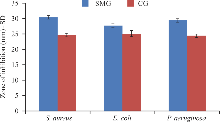
Antibacterial studies: The application of SMGs for antimicrobial therapy is established43. The antibacterial activity of optimized CIP SMG was compared with a conventional CIP Carbopol based gel as a control. A cylinder-plate method was carried out for the evaluation of the antibacterial activity. The results obtained are shown in Fig. 6. It was observed that the zone of inhibition was more for the SMG gel when compared to the conventional gel of CIP. The enhancement of antibacterial activity by SMG was significant (p<0.05) in all the selected bacterial strains. The zone of inhibition for S. aureus reached 30.33±0.58 mm for the SMG. At the same time, it was only 24.67±0.58 mm for the conventional gel. The activity was similar in P. aeruginosa too. In the case of E. coli, the difference in the zone of inhibition was low (2.6 mm), however, the enhancement was still significant. The enhanced zone of inhibition of SMG could be ascribed due to the enhanced diffusion of CIP from SMG. This result was in agreement with the ex vivo corneal permeation study.
 |
| Fig. 6: | Antibacterial activity of optimized CIP self-assembled α-CD/PLX-based supramolecular hydrogel compared to control gel against different bacterial strains SMG: Supramolecular hydrogel, CG: Control gel |
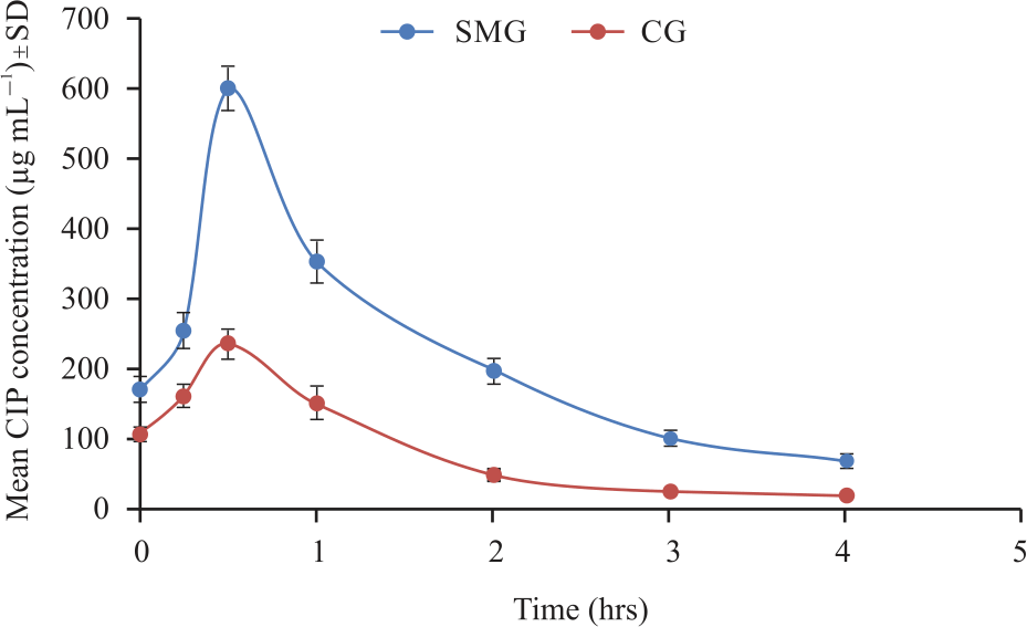 |
| Fig. 7: | In vivo precorneal concentrations of CIP from optimized self-assembled α-CD/PLX-based supramolecular hydrogel compared to control gel SMG: Supramolecular hydrogel, CG: Control gel |
In vivo precorneal concentrations: The data of Fig. 7 illustrates the CIP concentrations in the tear fluid to time. Even though the general trend of all these CIP concentrations versus time curves was similar in both SMG and Carbopol® gel, the CIP concentration from the SMG was significantly higher than that from Carbopol® gel. It was found that the maximum concentration of CIP was observed after 30 min of instillation for both formulations. From the results, it could be seen that the amount of CIP was almost similar after 3 hrs of instillation. This observation might be due to the exhaustion of the releasable drug from the Carbopol® gel. This similar pattern of drug concentration was reported by Liu et al.30 in there in situ gelling gelrite/alginate formulations.
The Area Under the Curve (AUC) was calculated from the Concentration-time curve and it was found that the difference between the AUC following administration of the SMG and Carbopol® gel was statistically significant (p<0.05). The AUC resulted from the supramolecular gel was 2.57 times higher than that from Carbopol® gel. These results indicated that a greater amount of CIP was retained in the precorneal area for a prolonged period following the instillation of supramolecular gel as compared to Carbopol® gel. The area under the curve value works as an indicator of the precorneal exposure to the drug and thus for the therapeutic efficacy of the formulation.
CONCLUSION
A Three-factor Three-level Box-Behnken study model was used successfully to optimize an SMG for the maximized and controlled ocular delivery of CIP. PLX, α-CD% and PLX:CD ratio were chosen as the independent factors while viscosity and CIP released after 24 hrs were taken as the responses in the design. Both responses were found to be dependent on PLX and CD. The PLX:CD ratio was found to have a significant effect on viscosity but not on CIP release. Both the in vitro and the ex vivo permeation studies showed an enhanced and higher CIP release from the supramolecular gel compared to that from the Carbopol® gel. The ex vivo corneal CIP permeation reached up to 94.46±4.6% from the supramolecular gel and was much higher than the permeation from Carbopol® gel. The antibacterial studies S. aureus, E. coli and P. aeruginosa indicated a significant improvement in antibacterial activity of CIP when presented in an SMG. SMG gel was better when compared to Carbopol® gel in providing prolonged retention of CIP in the precorneal area.
SIGNIFICANCE STATEMENT
Overall, the research proved that optimized SMG performed satisfactorily in the ocular delivery of CIP. The formulation could be able to prove the superiority over previous researches on ciprofloxacin formulations both in terms of drug release and antibacterial efficacy. The performance of the SMG for ocular drug delivery could be further exploited to tailor them for a clinically usable system.
ACKNOWLEDGMENT
This project was funded by the Deanship of Scientific Research (DSR), King Abdulaziz University, Jeddah, under grant No. (G: 657-249-1441). The authors, therefore, gratefully acknowledge DSR technical and financial support.
REFERENCES
- Gote, V., S. Sikder, J. Sicotte and D. Pal, 2019. Ocular drug delivery: Present innovations and future challenges. J. Pharmacol. Exp. Ther., 370: 602-624.
CrossRefDirect Link - Chen, H., Y. Jin, L. Sun, X. Li and K. Nan et al., 2018. Recent developments in ophthalmic drug delivery systems for therapy of both anterior and posterior segment diseases. Colloid Interface Sci. Commun., 24: 54-61.
CrossRefDirect Link - Garhwal, R., S.F. Shady, E.J. Ellis, J.Y. Ellis and C.D. Leahy et al., 2012. Sustained ocular delivery of ciprofloxacin using nanospheres and conventional contact lens materials. Invest. Ophthalmol. Vis. Sci., 53: 1341-1352.
CrossRefDirect Link - Morrison, P.W.J. and V.V. Khutoryanskiy, 2014. Advances in ophthalmic drug delivery. Ther. Delivery, 5: 1297-1315.
CrossRefDirect Link - Wu, Y., Y. Liu, X. Li, D. Kebebe and B. Zhang et al., 2019. Research progress of in situ gelling ophthalmic drug delivery system. Asian J. Pharmaceut. Sci., 14: 1-15.
CrossRefDirect Link - Kompella, U.B., R.S. Kadam and V.H.L. Lee, 2010. Recent advances in ophthalmic drug delivery. Ther. Delivery, 1: 435-456.
CrossRefDirect Link - Irimia, T., M.V. Ghica, L. Popa, V. Anuţa, A.L. Arsene and C.E. Dinu-Pîrvu, 2018. Strategies for improving ocular drug bioavailability and corneal wound healing with chitosan-based delivery systems. Polymers, Vol. 10.
CrossRefDirect Link - Balguri, S.P., G.R. Adelli, K.Y. Janga, P. Bhagav and S. Majumdar, 2017. Ocular disposition of ciprofloxacin from topical, PEGylated nanostructured lipid carriers: Effect of molecular weight and density of poly (ethylene) glycol. Int. J. Pharm., 529: 32-43.
CrossRefDirect Link - Alharbi, W.S. and K.M. Hosny, 2020. Development and optimization of ocular in situ gels loaded with ciprofloxacin cubic liquid crystalline nanoparticles. J. Drug Delivery Sci. Technol., Vol. 57.
CrossRefDirect Link - Hosny, K.M., 2010. Ciprofloxacin as ocular liposomal hydrogel. AAPS PharmSciTech, 11: 241-246.
CrossRefDirect Link - Youssef, A., N. Dudhipala and S. Majumdar, 2020. Ciprofloxacin loaded nanostructured lipid carriers incorporated into in situ gels to improve management of bacterial endophthalmitis. Pharmaceutics, Vol. 12.
CrossRefDirect Link - Abdelkader, H., Z. Fathalla, H. Moharram, T.F.S. Ali and B. Pierscionek, 2018. Cyclodextrin enhances corneal tolerability and reduces ocular toxicity caused by diclofenac. Oxid. Med. Cell. Longevity, 2018: 1-13.
CrossRefDirect Link - Batrakova, E.V. and A.V. Kabanov, 2008. Pluronic block copolymers: Evolution of drug delivery concept from inert nanocarriers to biological response modifiers. J. Controlled Release, 130: 98-106.
CrossRefDirect Link - Marcos, X., S. Pérez-Casas, J. Llovo, A. Concheiro and C. Alvarez-Lorenzo, 2016. Poloxamer-hydroxyethyl cellulose-α-cyclodextrin supramolecular gels for sustained release of griseofulvin. Int. J. Pharm., 500: 11-19.
CrossRefDirect Link - Simões, S.M.N., F. Veiga, J.J. Torres-Labandeira, A.C.F. Ribeiro, M.I. Sandez-Macho, A. Concheiro and C. Alvarez-Lorenzo, 2012. Syringeable pluronic–α-cyclodextrin supramolecular gels for sustained delivery of vancomycin. Eur. J. Pharm. Biopharm., 80: 103-112.
CrossRefDirect Link - Li, J. and X.J. Loh, 2008. Cyclodextrin-based supramolecular architectures: Syntheses, structures, and applications for drug and gene delivery. Adv. Drug Delivery Rev., 60: 1000-1017.
CrossRefDirect Link - Gonzalez-Gaitano, G., J.R. Isasi, I. Velaz and A. Zornoza, 2017. Drug carrier systems based on cyclodextrin supramolecular assemblies and polymers: Present and perspectives. Curr. Pharm. Des., 23: 411-432.
CrossRefDirect Link - Tan, S., K. Ladewig, Q. Fu, A. Blencowe and G.G. Qiao, 2014. Cyclodextrin-based supramolecular assemblies and hydrogels: Recent advances and future perspectives. Macromol. Rapid Commun., 35: 1166-1184.
CrossRefDirect Link - Zheng, Y. and I.W. Wyman, 2016. Supramolecular nanostructures based on cyclodextrin and poly(ethylene oxide): Syntheses, structural characterizations and applications for drug delivery. Polymers, Vol. 8.
CrossRefDirect Link - Huang, F. and H.W. Gibson, 2005. Polypseudorotaxanes and polyrotaxanes. Prog. Polym. Sci., 30: 982-1018.
CrossRefDirect Link - Lorenzo-Veiga, B., H.H. Sigurdsson, T. Loftsson and C. Alvarez-Lorenzo, 2019. Cyclodextrin–amphiphilic copolymer supramolecular assemblies for the ocular delivery of natamycin. Nanomaterials, Vol. 9.
CrossRefDirect Link - Collins, C.J., L.A. McCauliff, S.H. Hyun, Z. Zhang and L.N. Paul et al., 2013. Synthesis, characterization and evaluation of pluronic-based β-cyclodextrin polyrotaxanes for mobilization of accumulated cholesterol from Niemann-Pick type C fibroblasts. Biochemistry, 52: 3242-3253.
CrossRefDirect Link - Larrañeta, E. and J.R. Isasi, 2012. Self-assembled supramolecular gels of reverse poloxamers and cyclodextrins. Langmuir, 28: 12457-12462.
CrossRefDirect Link - Ni, X., A. Cheng and J. Li, 2009. Supramolecular hydrogels based on self-assembly between PEO-PPO-PEO triblock copolymers and α-cyclodextrin. J. Biomed. Mater. Res., 88A: 1031-1036.
CrossRefDirect Link - Higashi, T., K. Motoyama and H. Arima, 2013. Cyclodextrin-based polyrotaxanes and polypseudorotaxanes as drug delivery carriers. J. Drug Delivery Sci. Technol., 23: 523-529.
CrossRefDirect Link - Domiński, A., T. Konieczny and P. Kurcok, 2020. α-cyclodextrin-based polypseudorotaxane hydrogels. Materials, Vol. 13.
CrossRefDirect Link - Al-Kassas, R.S. and M.M. El-Khatib, 2009. Ophthalmic controlled release in situ gelling systems for ciprofloxacin based on polymeric carriers. Drug Delivery, 16: 145-152.
CrossRefDirect Link - Simões, S.M.N., F. Veiga, A.C.F. Ribeiro, A.R. Figueiras, P. Taboada, A. Concheiro and C. Alvarez-Lorenzo, 2014. Supramolecular gels of poly-α-cyclodextrin and PEO-based copolymers for controlled drug release. Eur. J. Pharm. Biopharm., 87: 579-588.
CrossRefDirect Link - Kotta, S., A.W. Khan, S.H. Ansari, R.K. Sharma and J. Ali, 2015. Formulation of nanoemulsion: A comparison between phase inversion composition method and high-pressure homogenization method. Drug Delivery, 22: 455-466.
CrossRefDirect Link - Liu, Y., J. Liu, X. Zhang, R. Zhang, Y. Huang and C. Wu, 2010. In situ gelling gelrite/alginate formulations as vehicles for ophthalmic drug delivery. AAPS PharmSciTech., 11: 610-620.
CrossRefDirect Link - Kanoujia, J., P.S. Kushwaha and S.A. Saraf, 2014. Evaluation of gatifloxacin pluronic micelles and development of its formulation for ocular delivery. Drug Deliv. Transl. Res., 4: 334-343.
CrossRefDirect Link - Badr-Eldin, S.M. and O.A.A. Ahmed, 2016. Optimized nano-transfersomal films for enhanced sildenafil citrate transdermal delivery: Ex vivo and in vivo evaluation. Drug Des. Devel. Ther., 10: 1323-1333.
CrossRefDirect Link - Wu, S.S., C.Y. Chein and Y.H. Wen, 2008. Analysis of ciprofloxacin by a simple high-performance liquid chromatography method. J. Chromatogr. Sci., 46: 490-495.
CrossRefDirect Link - Malhotra, M. and D.K. Majumdar, 1997. In vitro transcorneal permeation of ketorolac from oil based ocular drops and ophthalmic ointment. Indian J. Exp. Biol., 35: 1324-1330.
Direct Link - Vodithala, S., S. Khatry, N. Shastri and M. Sadanandam, 2010. Formulation and evaluation of ion activated ocular gels of ketorolac tromethamine. Int. J. Curr. Pharm. Res., 2: 33-38.
Direct Link - Ahmed, O.A.A. and S.M. Badr-Eldin, 2019. Development of an optimized avanafil-loaded invasomal transdermal film: Ex vivo skin permeation and in vivo evaluation. Int. J. Pharmaceutics, Vol. 570.
CrossRefDirect Link - Fahmy, U.A., S.M. Badr-Eldin, O.A.A. Ahmed, H.M. Aldawsari and S. Tima et al., 2020. Intranasal niosomal in situ gel as a promising approach for enhancing flibanserin bioavailability and brain delivery: In vitro optimization and ex vivo/in vivo evaluation. Pharm., Vol. 12.
CrossRefDirect Link - Tayeb, A.M., M.A. Tony and S.A. Mansour, 2018. Application of Box–Behnken factorial design for parameters optimization of basic dye removal using nano-hematite photo-Fenton tool. Appl. Water Sci., Vol. 8.
CrossRefDirect Link - Montgomery, D.C., 1991. Design and Analysis of Experiments. 3rd Edn., John Wiley & Sons Inc., New York, Pages: 500.
Direct Link - Erdoğar, N., G. Esendağlı, T.T. Nielsen, M. Şen, L. Öner and E. Bilensoy, 2016. Design and optimization of novel paclitaxel-loaded folate-conjugated amphiphilic cyclodextrin nanoparticles. Int. J. Pharm., 509: 375-390.
CrossRefDirect Link - Abou-el-Ela, A.E.S.F. and M.M. El-Khatib, 2014. Formulation and evaluation of new long acting metoprolol tartrate ophthalmic gels. Saudi Pharm. J., 22: 555-563.
CrossRefDirect Link - Loftsson, T. and E. Stefánsson, 2002. Cyclodextrins in eye drop formulations: Enhanced topical delivery of corticosteroids to the eye. Acta Ophthalmol. Scand., 80: 144-150.
CrossRefDirect Link - Hu, B., C. Owh, P.L. Chee, W.R. Leow and X. Liu et al., 2018. Supramolecular hydrogels for antimicrobial therapy. Chem. Soc. Rev., 47: 6917-6929.
CrossRefDirect Link