
Sarah I. Othman
Department of Biology, College of Science, Princess Nourah Bint Abdulrahman University, Riyadh, Saudi Arabia
May Bin- Jumah
Department of Biology, College of Science, Princess Nourah Bint Abdulrahman University, Riyadh, Saudi Arabia
LiveDNA: 966.25342
International Journal of Pharmacology
Year: 2019 | Volume: 15 | Issue: 4 | Page No.: 449-456


ABSTRACT
Background and Objective: Although mono-sodium glutamate (MSG) commonly used as food additive, application of higher doses or prolonged uses significantly leads to over accumulations in living cells and finally produces cellular toxicity. The aim of this study was to examine changes in the liver and kidneys of mice due to exposure to mono-sodium glutamate. Methodology: Cytotoxicity of aspartame was investigated histologically by using hematoxylin and eosin (H and E) stains. The animals received a mono-sodium glutamate with a dose of 360 mg kg–1 b.wt., in drinking water for one month. Both liver and kidney tissues were subjected for histological analysis. Results: In mono-sodium Glutamate (MSG), treated mice at doses of 360 mg kg–1/day for one month, significantly increase in weight and fatty tissue around the bowels of mice was reported compared to normal controls (28.5 g vs. 19.7 g). Cellular toxicity of MSG showed significant changes in both liver and kidney tissues. Mild disturbance of liver architecture, small necrotic areas with mild vacuolation, enlarged and congested central vein with disturbed endothelial lining which and more lymphocytic infiltration and few inflammatory cells and accumulation of fat droplets were significantly reported in liver tissues treated with MSG. Also, hemorrhage, areas of necrosis and increased vacuolation with most atrophied cellular nuclei. In kidney tissues, glomeruli appeared with narrowing of the cavity, congestion, shrinkage, hemorrhage, decomposition, disappearance and swelling in most of tubules. In addition, large areas of destruction with visible bleeding were estimated in most areas of renal tissue. Conclusion: This study concluded that mono-sodium glutamate produces more toxic effects on liver and kidney and considered as a major caustic effective agent against human health.
PDF Abstract XML References Citation
Received: November 12, 2018;
Accepted: December 15, 2018;
Published: April 26, 2019
Copyright: © 2019. This is an open access article distributed under the terms of the creative commons attribution License, which permits unrestricted use, distribution and reproduction in any medium, provided the original author and source are credited.
How to cite this article
Sarah I. Othman and May Bin- Jumah, 2019. Histomorphological Changes in Mono-sodium Glutamate Induced Hepato-renal Toxicity in Mice. International Journal of Pharmacology, 15: 449-456.
DOI: 10.3923/ijp.2019.449.456
URL: https://scialert.net/abstract/?doi=ijp.2019.449.456
DOI: 10.3923/ijp.2019.449.456
URL: https://scialert.net/abstract/?doi=ijp.2019.449.456
INTRODUCTION
Mono-sodium Glutamate (MSG) or Chinese salt, a chemical added to many foods such as processed meat, canned food, chips, luncheon, crispy chicken, chicken noodles, spices and fast food in major restaurants as taste and flavor enhancers. Several studies showed that MSG has many serious risks on human health1. Previously; it was shown that MSG has a devastating effect on brain cells2, on the optic nerve which causes headaches3 and increase heartbeat and numbness in the limbs and cause Alzheimer's4.
This salt was received to children via their foods and was reported that it causing more risks, such as decrease in the mental abilities of the child, IQ decreases and affects the kidneys and liver. In addition, it causes allergies in children and obesity in both children and adults5. Although, this MSG salt was used efficiently in various industrial food manufacturing6-14, it was reported that this chemical compound causes severe human health problems especially addiction in human targets, due to its ability to act on brain chemistry5,15,16.
Extensive studies were conducted to clarify the role, benefits and safety of MSG14,17,18. Thus, international and national bodies responsible for the safety of food additives, considered MSG to be safe for human consumption as a fragrance enhancer in recommended safety doses of LD50 for salt (3 g kg–1)18-20. In normal circumstances, human body cells have the ability to metabolize glutamate, which contains a very low percentage of acute toxicity21. For in mice and rodents, the LD50 dose ranges from 15-18 g kg–1 b.wt., in mice and rodents, respectively, which is 5 times higher than LD50 for salt (3 g kg–1 in mice) were shown to be toxically less effective21. Thus, ingestion of MSG as an additive to food and eating the normal level of it in foods does not represent a risk of poisoning in humans20,21.
Cellular toxicity of MSG appears during application of higher doses or prolonged uses of MSG; this leads to over accumulations in cells and finally initiates toxicological effects on living cells. Normally, it was reported that glutamate is essential for better neurotransmission in the human brain, which is an essential element of learning and memory, there is a continuous study by neuroscientists about the possible side effects of MSG in food as its increase leads to over stimulation and apoptosis. Studies have shown that ingestion of mono-sodium glutamate MSG for 10 days reduced the number of neurons in the cochlea, affecting neonatal hearing function, lowering antioxidant capacity and increase of oxidative stress free radicals and nitric oxide2,3,22-25. Also, it was reported that MSG as food additive significantly responsible for the incidence of obesity and type 2 diabetes5. Especially, when it was ingested during pregnancy. It was shown to produce a sharp increase in fat and cholesterol levels for pregnant mothers and maternal obesity during pregnancy3,5,26.
In addition, more studies showed that MSG affects severely on liver and kidney cells. Animal studies indicate that treatment (MSG) was induced by renal failure by its effect on the kidney oxidation system27. The intake of MSG orally at a dose of 4 g kg–1 for 180 days changed the renal oxidation system in addition to tissue changes. Whereas, significant changes in glomerular glands and a swelling occurred in renal tubules were appeared28-32.
Also, administration of MSG greatly effects on the liver function of pregnant females as well as new progeny after birth, whereas MSG at a dose of 4 g kg–1 when injected under the skin, causes a defect in the liver function and reduction in the activity of the system of oxidation due to a defect in the mitochondria caused by MSG33 and prolonged exposure to MSG for 29-32 weeks, leads to obesity, the emergence of type 2 diabetes, liver hepato-toxicity, hepatic fibrosis, degeneration and cellular hypertrophy, as fibrosis was observed in the liver and the onset of cellular patterns mimicking liver cancer and the development of Nash disease34,35. In addition, the exposure to six concentrations of MSG (250, 500, 2000, 4000 and 8000 μg mL–1) resulted in DNA damage in the isolated human lymphocytes as previously reported36.
From the previously mentioned studies relating to cellular toxicity of MSG21-36 and due to the spread of MSG uses around the world as flavors in all types of foods2-22.
The aim through this research was to confirm previous mono-sodium glutamate cellular toxicity in mice at new given dose of 360 mg kg–1 b.wt. Therefore, the changes in kidney and liver tissues in mice treated with mono-sodium glutamate were investigated by histological and morphological analysis.
MATERIALS AND METHODS
Materials: In this experiment, female adult Albino mice were used. It was obtained from the Animal House of the Faculty of Pharmacy, King Saud University, Riyadh. The animals were distributed in special cages equipped with drinking water in their ventilated rooms subjected to the appropriate natural factors of moisture, light and temperature between 25 and 35°C.
Feeding: The animals were provided with the appropriate food, the feed of animals (No. 648) was obtained from the General Establishment of Grain Silos and Flour Mills in Riyadh. The daily diets contain; raw protein 20% phosphorus 6.0%; raw fat 5.3%, vitamin A 20% International unit/g; Ash 6%, vitamin D 2.2% International unit/g; Calcium 1%, vitamin E 70% International unit/g. In addition to the rare mineral elements: Cobalt, copper, iodine, iron, manganese and zinc, water was left as needed. The experimental animals were selected with an average age of 12-15 weeks and an average body weight of 50-60 g.
Sodium glutamate uses: A single sodium glutamate was used and obtained from a commercial center for the sale of foodstuffs. It contained white powder packed in bags and was dissolved in drinking water. The water was taken daily for 1 month for female rats at a dose29 of 360 mg kg–1 b.wt.
Experimental design: Animals were divided into two groups:
| • | Group 1 (Control group; n = 5): Female rats received natural water |
| • | Group 2 (Treatment group, n = 15): Female rats received mono-sodium glutamate with a dose of 360 mg kg–1 b.wt., in drinking water for one month. At the end of treatment periods, all female’s rats in the studied groups were anesthetized, dissected and kidney and liver samples were taken from the rats preserved in a stabilization buffer solutions until reused for analysis |
Assessment of histological analysis: All samples of the liver and kidney were taken from pregnant mice and kept in a 10% neutral formalin solution for the preparation of histological sections using Hematoxylin and eosin (H and E) stains.
RESULTS
Morphological studies
Animal weight and relative changes in weights of organs (liver and spleen): The data showed significant increase (p<0.001) in the weight of mice treated with MSG compared to control group (19.7 vs. 28.5 g) (Table 1). In addition, significant decrease in the ratio of liver index (2.65 vs. 1.65) and increase in the ratio of spleen index (0.75 vs. 1.28) were reported in MSG treated rats (p = 0.001) compared to those of control rats as shown in Table 1.
Internal shape of the viscera: In the mono-sodium glutamate group, the amount of fatty tissue was very clearly observed around the viscera of the mice compared to control mice as shown in Fig. 1a-f.
Histological analysis
Histological changes in liver structure: Sections of mice livers were analyzed histologically by using Hematoxylin and eosin (H and E) stains. Normal liver sections showed normal liver architecture. The liver of mice consisted of several liver lobules confined by the connective tissue and the bovine region.
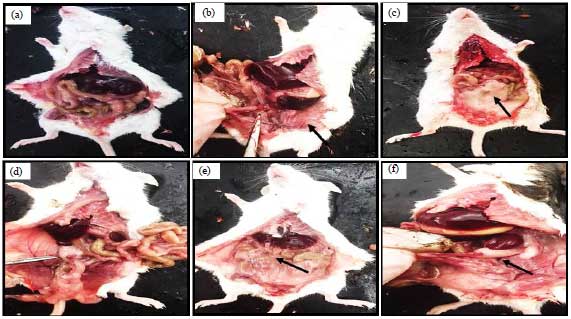 | |
| Fig. 1(a-f): | Shape of the internal organs of mice, (a) Mice of control group are free of any increase in fatty tissue around the bowel, (b) Ovary, (c-d) MSG treated mice showed an increase in the amount of fatty tissues around both the bowels (arrows) and (e-f) Ovary |
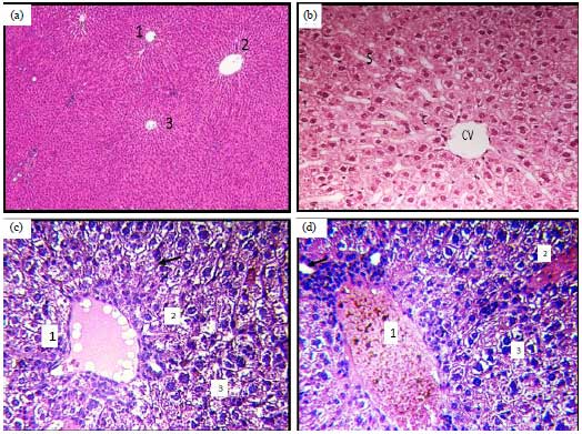 | |
| Fig. 2(a-d): | (a-b) Normal and (c-d) Glutamate treated liver tissues |
1: Normal hepatic tissues showed normal hepatic lobes, 2: Each hepatic segment of a central vein, surrounded by ribbons of interstitial and branching hepatic cells. Each hepatic placenta consists of a central vein (CV) surrounded by ribbons of interstitial and covalent cells (c). In glutamate treated, central venous infiltration (1) narrowing of the blood pockets (arrow), the death of most hepatic cells and 3: Appearance of many fatty cavities within cells were significantly shown after treatment with glutamate | |
| Table 1: | Relative changes in organ weights (liver and spleen) in rats with hepatic fibrosis induced by MSG-toxicity |
 | |
Liver index: Liver weight/body weight×100, Spleen index: Spleen weight/body weight×100, *Student’s t-test, Control vs. MSG treated group (350 mg g–1 b.wt.) | |
Each hepatic placenta consists of a central vein surrounded by ribbons of interstitial and interconnected hepatic cells forming a complex network that encloses the hepatic sinusoids. Multi- polygon hepatic cells appeared with a granular cytoplasm containing a spherical nucleus which appeared with one or more nuclei as shown in Fig. 2a and b.
The effect of mono-sodium glutamate was evident on the hepatic tissue, where tissue degradation and change in the structure of liver cells showed signs of cellular death (necrosis). The concentration of nuclear chromatin and nucleic degradation was observed in most liver cells, endothelial lining rupture with congestion in the central vein, narrowing of most of the hepatic pockets, presence of hemolysis and the emergence of multiple cavities within the cells. This indicated the accumulation of lipid droplets, which indicated the onset of liver lipid, invasion of inflammatory cells in some areas around the central vein (Fig. 2c, d, Fig. 3a, b).
Histological changes in kidney structure: In normal group, histological sections showed that the kidney is made up of two cortex cortices and appears granular because it contains renal pellets, renal tubules, medulla, the universal tubules and henal hoops. The kidney is made up of the Bowman's capsule, which consists of an outer mantle layer and an inner mantle that surrounds a mass of blood capillaries known as glomerulus. The renal tubules are characterized by proximal close-edged tubules with a narrow cavity lined with low epithelial cells based on a basement membrane. The brush edge, a spherical core nucleus and an acid cytoplasm and distant spores have a large cavity and are characterized by cubic epithelial cells based on a basal membrane with acidic cytoplasm and a large spherical nucleus (Fig. 3c, d).
In glutamate treated group, the effect of glutamate mono-sodium was evident on the kidney tissue where a change was observed in the histological structure where the glomeruli had a narrowing of the cavity with some of them, swelling in the tubules, narrowness of the cavity and congestion in the capillaries, as well as the appearance of cell death and necrosis of most of them.
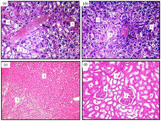 | |
| Fig. 3(a-d): | Liver and kidney tissues treated with glutamate, (a-b) Tissues of liver showed severe congestion and bleeding in the central (1), decomposition and death of most hepatic cells (2) and the emergence of many intracellular fatty cavities (3) attribution of inflammatory cells around the central vein (Arrow) and (c-d) In kidney tissues, showed normal kidney structures, which consists of cortex (1) and marrow (2) and convoluted tubules and the far-flung tubes (3) |
Cells, in addition to the concentration of nuclear chromatin in others and shrinkage and disappearance in the renal pellets. Shrinkage of renal glomeruli and the emergence of bleeding and also observed erosion in the walls of renal tubules and blood congestion, resulting in the emergence of large areas of destruction and bleeding in some areas of renal tissue alive, only some debris cells remain (Fig. 4a-d).
DISCUSSION
The effect of mono-sodium glutamate on health and structure of body organs, especially liver and kidney was investigated in this study. There was a significant increase in body weight (28.5 g vs. 19.7 g for control) and accumulations of fatty tissues around the viscera were observed in rats treated with mono-sodium glutamate compared to normal control group. Histological examinations showed significant alteration in the normal structure of both liver and kidney in rats treated with mono-sodium glutamate compared with those of control rats. It was observed that the toxic effects of mono-sodium glutamate occurs via inflammatory and apoptotic inducing pathways. Cellular cell death was observed in all sections of both kidney and liver such as the concentration of nuclear chromatin, DNA fragmentation and finally shrinkage and nucleic lysis was observed in most liver and kidney cells which confirmed necrotic cell death. The whole cellular architectures of both liver and kidney tissue were significantly changed up on treatment with mono-sodium glutamate. Our results were in accordance with previously reported studies which showed that an increase in the levels of glutamate leads to over stimulation and cellular apoptosis22-24.
Because glutamate is an important neurotransmitter in the human brain, which is a key element in learning and memory, there is a continuous study by neuroscientists about the possible side effects of MSG in food as its increase leads to over stimulation and apoptosis. Studies have shown that mono-sodium glutamate MSG for 10 days reduced the number of neurons in the cochlea, affecting neonatal hearing function23. The intake of MSG with a dose of 10, 20, 30, 40 and 80 mg kg–1 b.wt., for oral male rats for 28 days led to increase brain weight with damage to brain neurons decreased in level of Catalase with an increase in nitric oxide level22.
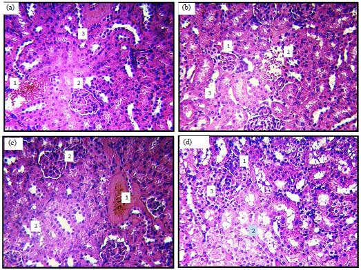 | |
| Fig. 4(a-d): | Structures kidney tissues treated with glutamate, (a-b) Kidney tissues showed, blood haemorrhage, dissolution and disappearance of some renal pellets in the tissues (1), death, narrowness in narrow glomerular cavity (1, 2) and (c-d) Decomposition of most of the nephropathic cells (2) |
The MSG also weakens brain function and causes oxidative damage. This is what scientists proved when treating experimental animals at a dose of 2 g kg–1 b.wt., for 7 days24. It also affected the kinetic coordination and a number of neurons in the cerebellum if a dose of 2 g g–1 b.wt., was given for 10 days orally2. It was found that the administration of MSG for Wister mice at a dose of 10 mg kg–1 b.wt., for 30 days showed an imbalance in the antioxidant balance and an increase in the level of choline in the brain25. The administration of male white glutamate (MSG) with a dose of 8 mg kg–1 b.wt., in drinking water for one month resulted in a defect in the level of neurotransmitters and oxidative stress indicators in the brain tissue16.
The MSG was classified as a cause of headache in the International Classification of Headaches and Disorders, where 45 patients with migraines were followed and analyzes were conducted during the occurrence of migraine headaches. Glutamate levels were significantly increased compared to control group3. The MSG was the study of obesity and type 2 diabetes. A study found that ingestion of MSG for pregnant mice at a dose of 60 mg/day from the 5th day of pregnancy to the last day before birth showed a sharp increase in fat and cholesterol levels for mothers. For pregnant women and maternal obesity during pregnancy5. The administration of pregnant women with MSG at a dose of 4 mg g–1 b.wt., on 2, 4, 6, 8 and 10 days of pregnancy and the killing of animals was observed that the mice were obesed with high levels of triglycerides and total cholesterol with increased proportion of deformed embryos with significant obesity of the survived offsprings26.
Animal studies indicated that chronic treatment with MSG leads to renal failure by its effect on the renal oxidation system27 and ingestion of mono-sodium glutamate MSG at a dose of 4 g kg–1 for 180 days to change the system of oxidation in the kidney as well as changes in the tissue in the kidney where the glomeruli crowded and the occurrence of swelling in the tubules and congestion in the capillaries as well as the emergence of death of cells and necrosis for most of them28. The exposure to the substance of mono-sodium glutamate MSG In a dose of 3 and 6 g kg–1 oral for one month to the occurrence of a defect in the thyroid gland where there was an increase in the layer of epithelial cells lining the glands with a decrease in the diameter of the vesicles and irregularity of the form with the enlargement of some of the vesicles and the density of the nuclei and the emergence of cells multi-nuclei29. Testicular rats was affected by exposure to the substance of mono-sodium glutamate MSG, which led to the inhibition of testosterone as the weight and size of testes and diameter of sperm and sperm cells, as a result of obesity caused by the use of mono-sodium glutamate MSG, which led to the rise of leptin hormone30.
In contrast, MSG was found to affect the ovary, where female newborns were given a dose of 2 mg g–1 b.wt., for 75 days in drinking water. Histological sections of the ovary showed an increase in the number of cells primary ovaries without an increase in the number of follicular vesicles, with a multitude of pathological changes in the cells such as the nucleus with the appearance of some fibrous tissue in the ovarian stomas30-32. This was due to the effect of MSG on the work of the pituitary and adrenal gland. In a study, 10 female Sprague dawley were given a dose of 0.08 mg kg–1 b.wt., of MSG orally for 14 days and mice were dissected and the fallopian tubes were extracted and investigated with microscopy. The data showed a rise in the epithelial layer of the fallopian tube and atrophy and necrosis in the cells, due to the increase in the amount of nutrients consumed by rats and increase the level of estrogen in the blood, which may lead to infertility in women32.
In a study, aimed at evaluating liver function in the model of obesity caused by the use of MSG during the first 10 days after birth at a dose of 4 g kg–1 by subcutaneous injection, the results showed a defect in the liver was evident in the incidence of ADP accompanied by a defect in the oxidation system due to malfunction in the mitochondria which was (MSG). The treatment with MSG after birth and for 29-32 weeks led to an increase in body mass index and blood sugar and the detection of oral glucose tolerance test of type 2 diabetes mellitus, (Cirrhosis of the liver and inflammation of the liver and inflammation of the liver with changes). Satisfactorily in hepatic tissue if given with a dose (4 g kg–1) under the skin, where liver lipid and inflammation of the liver cells and inflammation of fatty cells and fatty lipids were significantly observed in the liver tissue, confirming non-alcoholic fatty liver disease (NASH)35.
Previous research study, showed that exposure to 6 concentrations of mono-sodium glutamate (250, 500, 2000, 4000 and 8000 μg mL–1) resulted in DNA damage in isolated human lymphocytes at all concentrations36.
CONCLUSION
The present study concluded that mono-sodium glutamate produces more toxic effects on liver and kidney and considered as a major caustic effective agent against human health. It was recommended that the addition of monosodium glutamate to children's food should be avoided. Also, children should be avoided from having fast foods containing the mono-sodium glutamate, which has been shown to affect children directly.
ACKNOWLEDGMENT
The authors thank the Princess Nourah Bint Abdulrahman University (Riyadh, Saudi Arabia) for supporting this study.
SIGNIFICANCE STATEMENT
This study confirmed that exposure to mono-sodium glutamate at doses of 360 mg kg–1 b.wt., produces more toxic effects on liver and kidney and considered as a major caustic effective agent against both animal and human health when it received in drinking water for prolonged periods. The study contributes to the effective monitoring of studies mono-sodium glutamate consumption and the cellular risk effects that may lead to cancer or severe complications.
REFERENCES
- Miranda, R.A., C.C. da Silva Franco, J.C. de Oliveira, L.F. Barella and L.P. Tofolo et al., 2017. Cross-fostering reduces obesity induced by early exposure to monosodium glutamate in male rats. Endocrine, 55: 101-112.
CrossRefDirect Link - Prastiwi, D., A. Djunaidi and G. Partadiredja, 2015. High dosage of monosodium glutamate causes deficits of the motor coordination and the number of cerebellar Purkinje cells of rats. Hum. Exp. Toxicol., 34: 1171-1179.
CrossRefDirect Link - Obayashi, Y. and Y. Nagamura, 2016. Does monosodium glutamate really cause headache?: A systematic review of human studies. J. Headache Pain, Vol. 17.
CrossRef - Lopez-Miranda, V., M.L. Soto‐Montenegro, J.A. Uranga‐Ocio, G. Vera and E. Herradon et al., 2015. Effects of chronic dietary exposure to monosodium glutamate on feeding behavior, adiposity, gastrointestinal motility and cardiovascular function in healthy adult rats. Neurogastroenterol. Motil., 27: 1559-1570.
CrossRefDirect Link - Afifi, M.M. and A.M. Abbas, 2011. Monosodium glutamate versus diet induced obesity in pregnant rats and their offspring. Acta Physiol. Hung., 98: 177-188.
CrossRefDirect Link - Walker, R. and J.R. Lupien, 2000. The safety evaluation of monosodium glutamate. J. Nutr., 130: 1049S-1052S.
CrossRefDirect Link - Mahieu, S., M. Klug, N. Millen, A. Fabro, A. Benmelej and M. del Carmen Contini, 2016. Monosodium glutamate intake affect the function of the kidney through NMDA receptor. Life Sci., 149: 114-119.
CrossRefDirect Link - Abu-Taweel, G.M., M.A. Zyadah, J.S. Ajarem and M. Ahmad, 2014. Cognitive and biochemical effects of monosodium glutamate and aspartame, administered individually and in combination in male albino mice. Neurotoxicol. Teratol. J., 42: 60-67.
CrossRefDirect Link - Onaolapo, O.J., A.Y. Onaolapo, M.A. Akanmu and O. Gbola, 2016. Evidence of alterations in brain structure and antioxidant status following ‘low-dose’ monosodium glutamate ingestion. Pathophysiology, 23: 147-156.
CrossRefDirect Link - Foran, L., K. Blackburn and R.J. Kulesza, 2017. Auditory hindbrain atrophy and anomalous calcium binding protein expression after neonatal exposure to monosodium glutamate. Neuroscience, 344: 406-417.
CrossRefDirect Link - Calis, I.U., D.T. Cosan, F. Saydam, U.K. Kolac and A. Soyocak et al., 2016. The effects of monosodium glutamate and tannic acid on adult rats. Iran. Red Crescent Med. J., Vol. 18.
CrossRef - Sadek, K., T. Abouzed and S. Nasr, 2015. Lycopene modulates cholinergic dysfunction, Bcl-2/Bax balance and antioxidant enzymes gene transcripts in monosodium glutamate (E621) induced neurotoxicity in a rat model. Can. J. Physiol. Pharmacol., 94: 394-401.
CrossRefPubMedDirect Link - Suarez-Roman, G., T. Fernandez-Romero, A.J. Perera-Calderin, V.M. Rodriguez-Sosa, C. Arranz and S.C. Hernandez, 2016. Pregestational obesity-induced embryopathy. Reprod. Sci., 23: 1250-1257.
CrossRefDirect Link - Sharma, A., 2015. Monosodium glutamate-induced oxidative kidney damage and possible mechanisms: A mini-review. J. Biomed. Sci., Vol. 22.
CrossRef - Paul, M.S., M. Abhilash, M.V. Varghese, M. Alex and R.H. Nair, 2012. Protective effects of α-tocopherol against oxidative stress related to nephrotoxicity by monosodium glutamate in rats. Toxicol. Mech. Methods, 22: 625-630.
CrossRefDirect Link - Khalaf, H.A. and E.A. Arafat, 2015. Effect of different doses of monosodium glutamate on the thyroid follicular cells of adult male albino rats: A histological study. Int. J. Clin. Exp. Pathol., 8: 15498-15510.
PubMedDirect Link - Yuan, M., G. Huang, J. Li, J. Zhang and F. Li et al., 2014. Hyperleptinemia directly affects testicular maturation at different sexual stages in mice and suppressor of cytokine signaling 3 is involved in this process. Reprod. Biol. Endocrinol., Vol. 12.
CrossRef - Das, R.S. and S.K. Ghosh, 2011. Long-term effects in ovaries of the adult mice following exposure to monosodium glutamate during neonatal life-a histological study. Nepal. Med. Coll J., 13: 77-83.
PubMedDirect Link - Wahdan, R.A. and Z.M. Alazouny, 2015. Effect of monosodium glutamate on the fallopian tubes of adult albino rats and the possible protective role of vitamin C: A histological and immunohistochemical study. Egypt. J. Histol., 38: 68-76.
CrossRefDirect Link - Quines, C.B., P.M. Chagas, D. Hartmann, N.R. Carvalho, F.A. Soares and C.W. Nogueira, 2017. (p-ClPhSe)2 reduces hepatotoxicity induced by monosodium glutamate by improving mitochondrial function in rats. J. Cell. Biochem., 118: 2877-2886.
CrossRefDirect Link - Tsuneyama, K., T. Nishida, H. Baba, S. Taira and M. Fujimoto et al., 2014. Neonatal monosodium glutamate treatment causes obesity, diabetes and macrovesicular steatohepatitis with liver nodules in DIAR mice. J. Gastroenterol. Hepatol., 29: 1736-1743.
CrossRefDirect Link - Takai, A., K. Kikuchi, Y. Kajiyama, A. Sugiura and M. Negishi et al., 2014. Serological and histological examination of a nonalcoholic steatohepatitis mouse model created via the administration of monosodium glutamate. Int. Schol. Res. Not., Vol. 2014.
CrossRef - Ataseven, N., D. Yuzbasioglu, A.C. Keskin and F. Unal, 2016. Genotoxicity of monosodium glutamate. Food ChemToxicol., 91: 8-18.
CrossRefPubMedDirect Link