
Nevin Turan
Department of Chemistry, Faculty of Arts and Sciences, Mus Alparslan University, Mus 49250, Turkey
Ragip Adiguzel
Department of Chemical Engineering, Faculty of Engineering, Tunceli University, Tunceli 62000, Turkey
LiveDNA: 90.1425
Kenan Buldurun
Department of Chemistry, Faculty of Arts and Sciences, Mus Alparslan University, Mus 49250, Turkey
Ercan Bursal
Department of Nursing, School of Health, Mus Alparslan University, Mus 49250, Turkey
International Journal of Pharmacology
Year: 2016 | Volume: 12 | Issue: 2 | Page No.: 92-100


ABSTRACT
The novel mixed ligand-metal complexes with a general formula [M(Sac)2(H2O)2L2].nH2O] [M = Mn(II), Co(II), Ni(II), Cu(II) and Cd(II), L = 5-Methly-4-(4-nitrophenylazo)-2H-pyrazole-3-ylamine (mnppa)] have been synthesized and characterized by elemental analyses, IR, UV-VIS, magnetic susceptibility mass spectra and thermogravimetry-differential thermal analysis (TGA-DTA). Each metal (II) ion is octahedral coordinated by two sac, two mnppa and two aqua ligands. The antioxidant properties of mixed ligand-metal complexes were determined by different in vitro methods such as ABTS, DPPH, FRAP and CUPRAC assays. The results of metal complexes were compared with standard antioxidant compounds.
PDF Abstract XML References Citation
How to cite this article
Nevin Turan, Ragip Adiguzel, Kenan Buldurun and Ercan Bursal, 2016. Spectroscopic, Thermal and Antioxidant Properties of Novel Mixed Ligand-Metal Complexes Obtained from Saccharinate Complexes and Azo Dye Ligand (mnppa). International Journal of Pharmacology, 12: 92-100.
DOI: 10.3923/ijp.2016.92.100
URL: https://scialert.net/abstract/?doi=ijp.2016.92.100
DOI: 10.3923/ijp.2016.92.100
URL: https://scialert.net/abstract/?doi=ijp.2016.92.100
INTRODUCTION
The coordination chemistry of saccharin (sacH, also named 1,1-dioxo-1,2-benzothiazol-3-one or o-benzosulfimide C7H5NO3S) is interesting. The SacH is a weak acid with a pKa value of 2 and readily be deprotonated by water and forms the corresponding monoanion, saccharinate (sac). Recently, the design and synthesis of mixed-ligand metal (II) complexes of sac have attracted great attention because of their interesting structural and solid-state properties (Hamamci et al., 2008; Yilmaz et al., 2008, 2006).
Due to its possible carcinogenesis and DNA-alerting ability (Nabors, 2001; Allen et al., 1957; Munro et al., 1975; Cohen-Addad et al., 1986; West et al., 1986; Suzuki and Suzuki, 1995), saccharin has been the subject of extensive research in the last three decades. On the other hand, these studies simultaneously indicate that the monoanion of sacH, namely saccharinate (sac), readily interacts with the biologically relevant cations. Owing to the presence of the imino, carbonyl and sulfonyl donor sites, sac acts as a mono-, bi- or tridentates polyfunctional ligand and coordinates many transition metal ions and forms complexes from mononuclear species to coordination polymers (Baran, 2005; Baran and Yilmaz, 2006).
Studying the coordination nature of saccharin and determining the binding site (s) to metal ions is perhaps a key to understand the bioinorganic chemistry of saccharin. The azo dyes are used widely in different areas with high dyeing strength and good fastness properties and they have the antimicrobial activity and absorption characteristics (Karci et al., 2008) but any study related to the mixed ligand complexes of azo dyes and saccharin was not found in the literature.
Antioxidant molecules can scavenge radicals and reactive oxygen species, so they prevent cell damage and other metabolic disorders. The scavenging mechanism was explained with transferring electrons through the donation of hydrogen (Isik et al., 2015). The objective of this study is to determine the antioxidant activities of the mixed-ligand metal complexes of saccharin. For this aim, four in vitro spectrophotometric method i.e., DPPH (1,1-diphenyl 2-picrylhydrazyl) free radical scavenging, ABTS (2,2'-azino-bis 3-ethylbenzthiazoline-6-sulfonic acid) cation radical scavenging, FRAP (Ferric ion reducing power) and CUPRAC (Cupric ion reducing power) assays, were applied to determine the antioxidant activity of the complexes.
The preparation and characterization of the mixed ligand–metal saccharin complexes were studied. Saccharin, itself, does not coordinate metal ions but its deprotonated form (saccharinate) readily forms complexes with a large number of metal ions (Jovanovski, 2000).
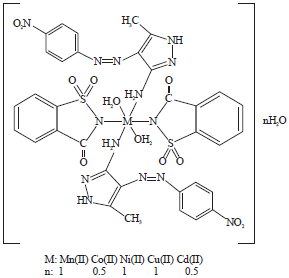
In the course of the synthesis and structural characterization of the mixed ligand-metal complexes of saccharin-aqua complex, recently Mn(II), Co(II), Ni(II), Cu(II) and Cd(II) complexes of saccharin-aqua complex [M(sac)2(H2O)4]·2H2O with azo dye ligand (5-methly-4-(4-nitrophenylazo)-2H-pyrazole-3-ylamine (mnppa)) were reported. In these complexes, both the sac and ligands (mnppa) coordinate the metal ions. The structure of the ligand and saccharin-aqua complex is shown in Fig. 1. These novel complexes may have a very promising future in biomedical and pharmaceutical application such as antimicrobial, antipyretic and analgesic etc. Because of all these reasons, it is aimed to prepare these novel mixed-ligand saccharinate complexes and to characterize those thoroughly using different spectrophotometric techniques as well as studying their biological activities.
MATERIALS AND METHODS
Materials and measurements: All reagents were commercially obtained and used without any further purification. Used solvents were of analytical grade and no further purifications were performed.
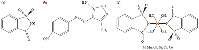 | |
| Fig. 1(a-c): | Structure of saccharin, azo dye (mnppa) and saccharin-aqua-complex (a) Saccharin, (b) Azo dye (Neutral ligand) and (c) Saccharin-aqua-complex |
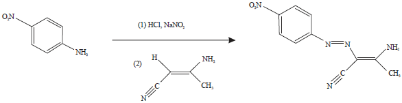 | |
| Fig. 2: | Coupling reaction in the synthesis of the neutral ligand (mnppa) |
 | |
| Fig. 3: | Ring closure reaction in the synthesis of the neutral ligand (mnppa) |
The metal salts MnSO4·H2O, NiSO4·6H2O, CdSO4·6H2O, CuSO4·5H2O, CoSO4·6H2O and starting materials for the ligand were Merck (Darmstadt, Germany), Sigma-Aldrich (GmbH, Sternheim, Germany) and Alfa Aesar (Karlsruh, Germany) products. The starting complex [M(sac)2(H2O)4]·2H2O (M: Mn(II), Co(II), Ni(II), Cu(II) and Cd(II)) was prepared by the method described in the literature (Baran and Yilmaz, 2006). In the same manner the secondary neutral ligand was obtained according to the literature methods (Elnagdi et al., 1976; Tsai and Wang, 2007).
The materials used for antioxidant activity such as ABTS, ascorbic acid (99%), butylated hydroxyanisole (BHA), butylated hydroxytoluene (BHT), DPPH, neocuproine (2,9-dimethyl-1,10-phenanthroline) and trichloroacetic acid (TCA) were obtained from Sigma-Aldrich (GmbH, Sternheim, Germany).
Elemental analyses were carried out with a Leco CHNS-O model 932 elemental analyzer. The IR spectra were recorded with a Perkin Elmer Precisely Spectrum One spectrometer using KBr discs in the wave number range of 4000-400 cm–1. Electronic spectral studies were conducted with a Shimadzu model UV-1800 spectrophotometer in the wavelength 1100-200 nm. Magnetic susceptibility measurements were performed using the standard Gouy tube technique using Hg[Co(SCN)4] as a calibrate. LC/MS-API-ES mass spectra were recorded using an AGILENT model 1100 MSD mass spectrophotometer. Thermogravimetric analysis (TGA) and differential thermal analysis (DTA) were carried out in nitrogen atmosphere with a heating rate of 10°C min–1 using Shimadzu DTG-60 AH (Shimadzu DSC 60 A) thermal analyzers. A sample size of 5-10 mg was used and sintered α-alumina was used as the reference material.
Synthesis of neutral ligand (mnppa): The secondary ligand (mnppa) was synthesized according to the procedure given at Fig. 2 and 3 and (Tsai and Wang, 2007; Koksal et al., 2011). This synthesis consists of two steps. In other words in the synthesis of the azo dye, a neutral ligand has been formed by azo coupling and ring closure reactions.
Synthesis of the mixed ligand Mn(II), Co(II), Ni(II), Cu(II) and Cd(II) complexes: The neutral ligand mnppa 0.30 g (1.25 mmol), was dissolved in 20 mL hot absolute EtOH in a 100 mL round-bottom flask. A solution of [Mn(sac)2(H2O)4]·2H2O 0.33 g (0.625 mmol) in 5 mL EtOH and 0.5 mL DMF was added drop wise in 5 min. periods with continuous stirring about at 50-55°C. In the meantime, tile red solution of the ligand was turned into light cherry mixture. After the addition of 5 drops of glacial acetic acid, it was left under reflux for 8 h and the brownish yellow mixture was left at room temperature. The solvent was removed completely from the mixture. The resulting precipitate in hexane and chloroform mixture was filtered and then washed with hot EtOH and H2O mixture. The product was dried at room temperature. Similar synthesis was performed for the mixed ligand complexes of mnppa using Co(sac)2(H2O)4]·2H2O (0.33 g, 0.625 mmol), [Ni(sac)2(H2O)4]·2H2O (0.33 g, 0.625 mmol), Cu(sac)2(H2O)4]·2H2O (0.34 g, 0.625 mmol), Cd(sac)2(H2O)4]·2H2O (0.36 g, 0.625 mmol), L:mnppa (0.30 g, 1.25 mmol). Analysis results of the neutral ligand and all complexes are as follows:
| • | 5-Methly-4-(4-nitrophenylazo)-2H-pyrazole-3-ylamine (mnppa); m.p.: 201-203°C, Characteristic IR bands (KBr, cm–1): ν(NH): 3445, 3366; ν(NH2): 3342, 3256; ν(Ar CH): 3172, 3134; ν(C=C): 1566; ν(N=N): 1616; ν(C=N): 1506; ν(Ar-NO2): 1578; δ(NH2): 1598, 1585; ν(C-N): 1325, 1268; Characteristic 1H-NMR peaks (DMSO-d6, δ ppm): 6.6 (-NH2, 4H, s), 7.3-8.0 (Ar-CH, m), 10.9 (-NH, s) |
| • | [Mn(sac)2(mnppa)2(H2O)2]·H2O, Yield: (76.0-76%). FW: 964.97 g mol–1. μeff (B.M.): 5.20 Anal Calcd for C34H34N14O12S2Mn: C, 42.28, H, 3.52, N, 20.31, S, 6.63. Found: C, 41.92, H, 3.45, N, 20.75, S, 6.41. Selected IR data (KBr, cm–1): ν(H2O)broad: 3428; ν(NH), ν(NH2)broad: 3316, 3220; ν(Ar C-H): 3116; ν(N=N): 1607; ν(C=O) 1669; ν(C=C), ν(C=N) broad: 1518; ν(C-N): 1378, 1205; ν(Ar-NO2): 1332; ν(SO2): 1155, 1103; ν(M-N): 565, 510; ν(M-O): 470. The UV-VIS (in ethanole): λ max (ε, L mol–1 cm–1) 275 (130), 434 (937), 735 (2) nm; MS [ES]: m/z 965.97 (calc), 965.90 (found) [M+H]+. Color: Brick red |
| • | [Co(sac)2(mnppa)2(H2O)2]·0.5H2O, Yield: (66%). FW: 959.96 g mol–1. μeff (B.M.): 4.25 Anal Calcd for C34H32N14O12S2Co.0.5H2O: C, 42.50, H, 3.43, N, 20.41, S, 6.67. Found: C, 42.47, H, 3.38, N, 20.38, S, 6.70. Selected IR data (KBr, cm–1): ν(H2O): 3450; ν(NH): 3366; ν(NH2): 3307; ν(Ar C-H): 3104; ν(N=N): 1613; ν(C=O) 1610; ν(C=C): 1574; ν(C=N): 1510; δ(NH2): 1574; ν(C-N): 1382, 1284; ν(Ar-NO2): 1325; ν(SO2): 1150, 1105; ν(M-N): 569, 531; ν(M-O): 456. UV-VIS (in ethanole): λ max (ε, L mol–1 cm–1) 279 (410), 438 (235), 782 (5) nm. MS [ES]: m/z 732.96 (calc), 732.88 (found) [M-2.5H2O-Saccharinate]+. Color: Ecru |
| • | [Ni(sac)2(mnppa)2(H2O)2]·H2O, Yield: (80%), FW: 968.74 g mol–1. μeff (B.M.): 2.99 Anal Calcd for C34H34N14O12S2Ni: C, 42.11, H, 3.51, N, 20.23, S, 6.60. Found: C, 42.24, H, 3.74, N, 20.20, S, 6.75. Selected IR data (KBr, cm–1), ν(H2O)broad: 3457; ν(NH), ν(NH2)broad: 3324; ν(Ar C-H): 3167; ν(N=N): 1611; ain ν(C=O) 1633; ν(C=C): 1575; ν(C=N): 1514; ν(C-N): 1382, 1284; ν(Ar-NO2): 1336; ν(SO2): 1152, 1105; ν(M-N): 570-510; ν(M-O): 484-472. UV-VIS (in ethanole): λ max (ε, L mol–1 cm–1) 277 (16), 432 (96), 776 (60) nm. MS [ES]: m/z m/z 969.74. (calc), 970.69 (found) [M+H]+. Color: Brick red |
| • | [Cu(sac)2(mnppa)2(H2O)2]·H2O, Yield: (68%), FW: 973.57 g mol–1. μeff (B.M.): 1.93 Anal Calcd for C34H34N14O13S2Cu: C, 41.91, H, 3.49, N, 20.13, S, 6.57. Found: C, 42.00, H, 3.63, N, 20.22, S, 6.58. Selected IR data (KBr, cm–1), ν(H2O): 3564, 3502; ν(NH): 3412; ν(NH2)broad: 3325; ν(Ar C-H): 3097; ν(N=N): 1610; ν(C=O) 1618; ν(C=C): 580; ν(C=N): 1470; ν(C-N): 1353, 1284; ν(Ar-NO2): 1330; ν(SO2):1156, 1126; ν(M-N): 550, 524; ν(M-O): 472. UV-VIS (in DMF): λ max (ε, L mol–1 cm–1) 274 (100), 336 (294), 438 (337) nm, MS [ES]: m/z 729.57 (calc), 729.80 (found) [M-mnppa+2H]2+. Color: Brown |
| • | [Cd(sac)2(mnppa)2(H2O)2].0.5H2O, Yield: (80%), FW: 1013.40 g mol–1. μeff (B.M.): Dia. Anal Calcd for C34H32N14O12S2Cd.0.5H2O: C, 40.26, H, 3.26, N, 19.34, S, 6.31. Found: C, 40.21, H, 3.44, N, 19.24, S, 6.22. Selected IR data (KBr, cm–1), ν(H2O)broad: 3560; ν(NH): 3436; ν(NH2)broad: 3329, 3302; ν(Ar C-H): 3095; ν(N=N): 1607; ν(C=O): 1649; ν(C=C): 1577; ν(C=N): 1524; ν(C-N): 1350, 1204; ν(Ar-NO2): 1330; ν(SO2): 1169, 1105; ν(M-N): 567, 532; ν(M-O): 472. UV-VIS (in ethanole): λ max (ε, L mol–1 cm–1) 224 (19.995), 410 (1975), 447 (1936) nm. MS [ES]: m/z 829.38. (calc), 829.10 (found) [M-Sac-0.5H2O-2H]-. Color: Brick red. |
Antioxidant properties of the mixed ligand Mn(II), Ni(II), Cd(II), Cu(II) and Co(II) complexes
DPPH· Scavenging Activity: The DPPH free radical scavenging activities of the complexes were evaluated by the method described in literature (Koksal et al., 2011). Briefly, different concentrations (10-30 μg mL–1) of samples and standard antioxidants (BHA, BHT and ascorbic acid) were prepared and diluted to 3 mL with ethanol. Then, 1 mL of ethanolic DPPH solution (0.1 mM) was added to the samples. These samples were vortexed and incubated in the dark at 30°C for 30 min. The absorbance was measured at 517 nm against blank samples. A decrease in absorbance indicated DPPH free radical scavenging activity.
ABTS•+ scavenging activity: The ABTS•+ radical scavenging activities of the complexes were evaluated according to the method described in literature (Bursal and Gulcin, 2011). The ABTS•+ is blue-green in color with a characteristic absorbance at 734 nm. The ABTS•+ cation radical was produced by reacting ABTS (2 mM) in H2O and potassium persulphate (2.45 mM) at room temperature for 12 h. The ABTS•+ solution was diluted with phosphate buffer (0.1 M, pH 7.4) to achieve an absorbance of 0.750±0.025 at 734 nm. Then, 1 mL of ABTS•+ solution was added to 3 mL ethanol at different concentrations (10-30 μg mL–1) of the samples and standard antioxidants (BHA, BHT and ascorbic acid). These samples were vortexed and incubated in the dark for 30 min. After 30 min, absorbance at 734 nm was measured for each concentration relative to a blank. Decreased absorbance of the samples indicated ABTS•+ cation radical scavenging activity.
Ferric Reducing Antioxidant Power (FRAP): The reducing powers of the complexes were measured according to the method described in literature (Bursal and Gulcin, 2011). According to this method, the reduction of Fe3+ to Fe2+ is determined by measuring the absorbance of Perl's Prussian blue complex. For this aim, different concentrations (10-30 μg mL–1) of the samples and standard antioxidants (BHA, BHT and ascorbic acid) in distilled water (0.75 mL) were mixed with 1 mL of sodium phosphate buffer (0.2 M, pH 6.6) and 1 mL (1%) of potassium ferricyanide [K3Fe(CN)6]. The mixture was incubated at 50°C for 20 min. Then, the reaction mixture was acidified with 1 mL of trichloroacetic acid (10%). Finally, 0.25 mL of iron (III) chloride (0.1%) was added to this solution. The absorbance of the mixture at 700 nm was measured. A decrease in absorbance indicated increased ferric reducing antioxidant power.
Cupric reducing antioxidant power (CUPRAC): The cupric ion (Cu2+) reducing powers of the complexes were determined by the CUPRAC method proposed in literature (Apak et al., 2004). Briefly, 1 mL of 10 mM copper (II) chloride solution, 1 mL of 7.5 mM ethanolic neocuproine solution and 1 mL of 1.0 M ammonium acetate buffer solution were added to a test tube and mixed with 0.25 mL of different concentrations (10-30 μg mL‾1) of the samples. The total volume was adjusted to 4.1 mL with distilled water. The tubes were kept at room temperature. After 30 min of incubation, the absorbance was measured at 450 nm against a blank. Increased absorbance indicated increased Cu2+-Cu+ reduction.
RESULTS AND DISCUSSION
Infrared spectra: The IR data of the spectra of the mixed ligand-metal complexes including saccharinate ion and neutral ligand along with assignments were given in the experimental section. The IR spectra of the complexes were compared with those of the saccharin-aqua complexes and with neutral ligand in order to determine the coordination sites that may be involved in chelation. Upon comparison, in the IR spectrum of the mnppa, ν(NH2) stretching vibration bands were observed at 3342 and 3256 cm–1 as broad peaks for the isolated mixed ligand Mn(II) and Cd(II) complexes, two broad peaks at 3316, 3220 and 3329, 3302 cm–1, respectively, were found and for Co(II), Ni(II) and Cu(II) complexes, one broad peak was appeared at 3307, 3324 and 3325 cm–1, respectively (Esener et al., 2011). In addition to these results, the ν(C=N) vibration peak for pyrazole ring in mnppa ligand observed at 1506 cm-1 but for Mn(II), Co(II), Ni(II) and Cd(II) complexes, this peak shifted to higher frequencies of 1518, 1510, 1514 and 1524 cm–1lain , respectively (Esener et al., 2011; El-Sonbati et al., 2007). On the other hand, the ν(C=N) stretching vibration is shifted to lower frequency in the Cu(II) complex (Widlicka et al., 2000). As a result of coordination of amino group over N atom to metal ions of mnppa ligand, electron density of pyrazole ring decreased. Therefore, shifting of ν(NH2) vibration to low frequency and shifting of ν(C=N) vibration to high frequency except Cu(II) complex were expected (Esener et al., 2011; Pons et al., 2001). These either results support which NH2 group of the mnppa ligand coordinated over N atom to metal ion behaves as a monodentate ligand.
The ν(OH) vibration band of ν(H2O) at the Mn(II), Co(II), Ni(II), Cu(II) and Cd(II) complexes was observed at 3428, 3450, 3457, 3564 and 3560 cm–1, respectively as broad and medium intense bands (Masoud et al., 2008; Hu et al., 2010). ν(M-O) vibration peak from coordinated H2O for Mn(II), Co(II), Ni(II), Cu(II) and Cd(II) complexes were observed at 470, 456, 484 472, 472 and 472 cm–1, respectively, similar values were also found in the literature (Nakamoto, 1997; Sonmez et al., 2004). These observed ν(M-O) vibration peaks for all complexes are supporting evidence that coordination water exists in these complexes. The peaks for ν(M-N) vibration showing coordination was done over N atom for both N atoms of saccharinate ion and NH2 group of mnppa ligand were observed in the Mn(II), Co(II), Ni(II), Cu(II) and Cd(II) complexes at 565 510, 569 531, 570 510, 550 524 and 567 532 cm‾lain 1, respectively similar values were also found in the literature (Pons et al., 2001; Guney et al., 2006).
Electronic spectra and magnetic properties: The electronic spectra of the mixed ligand-metal complexes were taken from 3×10⁻³ M EtOH solution. The electronic spectrum of complexes shows several intense bands between 224 and 279 nm, which are assigned to the intra-ligand transitions in mnppa and sac. These absorption bands are attributed to π-π* transitions. The UV/Vis spectra of the complexes display a number of distinct bands at 275, 434, 735 nm for [Mn(sac)2(mnppa)2(H2O)2]·H2O, at 277, 432, 776 nm for [Ni(sac)2(mnppa)2(H2O)2]·H2O, at 224, 410, 447 nm for [Cd(sac)2(mnppa)2(H2O)2]·0.5H2O, at 274, 336, 438 nm for [Cu(sac)2(mnppa)2(H2O)2]·H2O and at 279, 438, 782 nm for [Co(sac)2(mnppa)2(H2O)2]·0.5H2O. The high energy bands below 300 nm are mainly due to ligand-based transitions, whereas the bands over 500 nm are originated from the ligand field d-d transitions (Karabocek and Karabocek, 1997; Sharaby, 2005).
Magnetic moment measurements which are widely used in studying transition metal complexes and the magnetic moment measurements of complexes are given in Fig. 4. Magnetic measurements give an outline about the electronic state of the metal ion in the complexes.
 | |
| Fig. 4: | Structure of the complexes |
The Co(II) complex which is showing the magnetic moment in the range of 4.25 B.M. indicate that the Co(II) complex are typically high spin complexes and have octahedral structure (Chohan et al., 2006). The Cu(II) complexes are paramagnetic and give higher magnetic moment of 1.93 B.M. as compared to spin only values which is presumably due to spin-orbit coupling (Cotton et al., 1999). The Ni(II) complex is found to have a room temperature magnetic moment value of 2.99 B.M. which is in the normal range observed for octahedral Ni(II) complexes (μeff = 2.9-3.3 B.M.) (Tayim and Salameh, 1983). The Mn(II) complex show magnetic moment value of 5.20 B.M. which was expected for high spin d5 system having octahedral geometry (Yousef et al., 2013; Alaghaz et al., 2013). The Cd(II) complexes are diamagnetic and according to the empirical formula of this complex, an octahedral geometry is proposed.
Thermal studies: Thermal behavior of the complexes was studied by DTA and TGA in the temperature range 25-800°C in the atmosphere of nitrogen. All complexes begin to decompose without melting and exhibit similar decomposition characteristics. In the first stage, endothermic removal of the aqua and neutral ligand takes place leading to an intermediate and then, highly exothermic DTA peaks at higher temperatures, which characterize the decomposition of the sac moiety in these intermediates. The experimental mass losses are consistent with the calculated data. All complexes begin to decompose without melting and exhibit similar decomposition characteristics.
[Mn(sac)2(mnppa)2(H2O)2]·H2O exhibits third distinct decomposition stages. The first step in the temperature range 64.61-140.57°C was accounted for the loss of water molecules of hydration (percentage experimental weight 1.90, calculated weight 1.87) (Turan et al., 2014). The second mass loss of 28.57% (calculated weight 29.22%) takes place in the range of 140.57-326.16°C with an endothermic effect, characterizing the removal of the coordinated waters and mnppa ligand (Turan et al., 2015). Both DTA and TGA curves show that the removal processes related to the water and mnppa ligand are not well separated and therefore, the simultaneous elimination of one mnppa ligand and two waters occurs in the second stages in the temperature range 140.57-326.16°C with a little broad endothermic peak at 264.30 oC. The third step of decomposition within the temperature range 326.16-800.00 oC corresponds to the rest of the loss of mnppa ligand and the combustion of sac moiety (percentage experimental weight 63.49, percentage calculated weight 63.42%). The end product is MnO.
The [Ni(sac)2(mnppa)2(H2O)2]·H2O and [Cd(sac)2(mnppa)2(H2O)2]·0.5H2O complexes showed the same decomposition properties. The first stage of decomposition clearly corresponds to the total dehydration of the complexes. For Ni(II) and Cd(II) complexes, this stage occurs at a relatively lower temperature lying in the range of 86.15-112.77 oC. The observed weight loss associated with this step agrees quite well with the calculated weight loss due to the loss of one and half hydration water molecules in the Ni(II) and Cd(II) complexes, respectively (Turan et al., 2014). The second stage of decomposition occurs for all of these complexes in the temperature range of 112.77-316.37°C and should be attributed to the loss of the mnppa ligand and coordinated waters (Turan et al., 2015). The sac ligands and other mnppa ligand in these complexes are completely lost during three stage of decomposition. The weight loss found for these two complexes are consistent with the calculated values, the residues left after this stage are NiO and CdO for [Ni(sac)2(mnppa)2(H2O)2]·H2O and [Cd(sac)2(mnppa)2(H2O)2]·0.5H2O, respectively.
[Cu(sac)2(mnppa)2(H2O)2]·H2O complex was stable up to 60.00°C and its decomposition started at this temperature. For the [Cu(sac)2(mnppa)2(H2O)2]·H2O complex first stage occurs around 90.00-125.69°C supporting the suggestion that these complexes contain hydration water molecules (percentage experimental weight 1.58, calculated weight 1.85). For the [Cu(sac)2(mnppa)2(H2O)2]·H2O complex second stage occurs at a higher temperature around 125.69-319.51°C supporting the suggestion that this complex contain coordinated rather than hydration water molecules and mnppa ligands (percentage experimental weight 3.96, calculated weight 3.70) (percentage experimental weight 50.79, calculated weight 50.54), respectively (Turan et al., 2015). The DTA curve of the Cu(II) complex shows two endothermic peak at 275.65 and 267.43°C. The endothermic peaks are likely due to the loss of the molecules of hydration water and mnppa ligand.
The [Co(sac)2(mnppa)2(H2O)2]·0.5H2O complex exhibits two decomposition steps. The first decomposition step in the temperature range 78.98-128.83°C (percentage experimental mass 1.50; calculated mass 0.94) may be attributed to the loss of water molecules of hydration. Second stage of the decomposition occurs for this complex in the temperature range of 128.83-326.16°C (percentage experimental mass 52.38; calculated mass 51.25) and should be attributed to the loss of the coordinated waters and mnppa ligand. Third stage of the decomposition occurs for all of these complexes in the temperature range of 326.16-487.87-800.00°C and should be attributed to the loss of the saccharinate ligand (Yilmaz et al., 2002).
Mass spectra: The mass spectra of the Mn(II), Co(II), Ni(II), Cu(II) and Cd(II) complexes contain peaks attributable to the related molecular ions m/z: 965.90 [M+H]+, m/z: 732.88 [M-2.5H2O-Saccharinate]+(cationic complex), m/z: 970.69 [M+H]+, m/z: 729.80 [M-mnppa+2H]2+, m/z: and m/z: 829.38 (found) [M-Sac-0.5H2O-2H]- respectively (Turan et al., 2015). The experimental and theoretical mass spectra data of the complexes were previously given in the experimental section. The values which have high abundance in the mass spectra were reported.
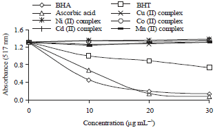
Antioxidant properties: Antioxidant properties of the mixed ligand Mn(II), Co(II), Ni(II), Cu(II) and Cd(II) complexes were determined by four different in vitro methods. Radical scavenging and reducing power properties of the complexes were determined by comparing with standard antioxidants. The antioxidant potentials of the complexes are considered lower when compared to standard molecules such as ascorbic acid, BHT and BHA.
Radical scavenging properties of the complexes were determined by ABTS and DPPH assays, separately. The DPPH assay is very common method, to determine the radical scavenging ability of various samples. This method is based on the reduction of DPPH in ethanolic solution in the presence of a hydrogen-donating antioxidant due to the formation of the non-radical form DPPH-H in the reaction (Bursal and Gulcin, 2011). According to the result of this study, complexes of the ligand did not show significant DPPH free radical scavenging activity. The DPPH• radical scavenging activity of standard antioxidants (ascorbic acid, BHT and BHA) and metal complexes were demonstrated in Fig. 5.
The ABTS assay is based on measuring the reduction of ABTS•+ (cation radicals) caused by antioxidant substances. By presenting an antioxidant, the dark blue color of ABTS radical cation become lighter. Decreasing absorbance at 734 nm indicates the amount of ABTS radical cation scavenging. According to the result of this study, complexes of the ligand did not show significant ABTS•+ radical scavenging activity. Low activity of ABTS radical scavenging of complexes is dependent on the molecule’s structure that has low hydrogen donation potential.
Antioxidant compounds are able to give electrons or hydrogen atoms to the reactive radicals and reduce them into more stable and unreactive species (Koksal et al., 2011). In this study, reducing power of the complexes were determined by FRAP and CUPRAC methods.
The FRAP method based on reduction of Fe3+ to Fe2+ which was related to donating an electron to a radical by an antioxidant compound. Absorbance increase for the samples indicates stronger reducing power of the samples. Compared to the standard antioxidants (BHA, BHT and ascorbic acid), complexes displayed weak reducing activities of Fe3+ to Fe2+. The results were demonstrated in Fig. 6.
 | |
| Fig. 5: | DPPH free radical scavenging activities of the complexes and standard antioxidants (ascorbic acid, BHT and BHA) |
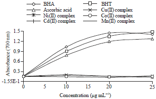 | |
| Fig. 6: | Reducing powers of the complexes and standard antioxidants (ascorbic acid, BHT and BHA) by FRAP assay |
 | |
| Fig. 7: | Reducing powers of the complexes and standard antioxidants (ascorbic acid, BHT and BHA) by CUPRAC assay |
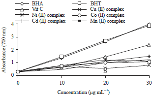
Antioxidant reducing powers of the complexes were also determined by CUPRAC, a method analyzing the reduction of Cu2+ to Cu+. In this assay, a higher absorbance indicates higher cupric ion (Cu2+) reducing ability. According to the results, complexes displayed effective cupric ion (Cu2+) reducing ability (Fig. 7). However, these values are lower than those of standard antioxidants (BHA, BHT and ascorbic acid).
CONCLUSION
The present investigation was carried out for the synthesis and characterization of the novel mixed ligand-metal complexes obtained from saccharinate complexes and neutral ligand (mnppa) and investigated their antioxidant properties. The data obtained by DPPH free radical scavenging, ABTS cation radical scavenging, FRAP and CUPRAC assays showed that studied chemical complex materials had very low antioxidant properties, yet some of them performed better cupric ion reducing power.
ACKNOWLEDGMENT
The authors are grateful to Research Foundation of Tunceli University (TUNIBAP-MFTUB013-05) supporting this study.
REFERENCES
- Alaghaz, A.N.M.A., H.A. Bayoumi, Y.A. Ammar and S.A. Aldhlmani, 2013. Synthesis, characterization and antipathogenic studies of some transition metal complexes with N,O-chelating Schiff's base ligand incorporating azo and sulfonamide Moieties. J. Mol. Struct., 1035: 383-399.
CrossRefDirect Link - Allen, M.J., E. Boyland, C.E. Dukes, E.S. Horning and J.G. Watson, 1957. Cancer of the urinary bladder induced in mice with metabolites of aromatic amines and tryptophan. Br. J. Cancer, 11: 212-228.
Direct Link - Apak, R., K. Güçlü, M. Özyürek and S.E. Karademir, 2004. Novel total antioxidant capacity index for dietary polyphenols and vitamins C and E, using their cupric ion reducing capability in the presence of neocuproine: CUPRAC method. J. Agric. Food Chem., 52: 7970-7981.
CrossRefDirect Link - Baran, E.J., 2005. The saccharinate anion: A versatile and fascinating ligand in coordination chemistry. Quimica Nova, 28: 326-328.
CrossRefDirect Link - Baran, E.J. and V.T. Yilmaz, 2006. Metal complexes of saccharin. Coord. Chem. Rev., 250: 1980-1999.
CrossRefDirect Link - Bursal, E. and I. Gulcin, 2011. Polyphenol contents and in vitro antioxidant activities of lyophilised aqueous extract of kiwifruit (Actinidia deliciosa). Food Res. Int., 44: 1482-1489.
CrossRefDirect Link - Tsai, P.C. and I.J. Wang, 2007. A facile synthesis of some new pyrazolo[1,5-a]pyrimidine heterocyclic disazo dyes and an evaluation of their solvatochromic behaviour. Dyes Pigm., 74: 578-584.
CrossRefDirect Link - Chohan, Z.H., M. Arif, A.M. Akhatar and C.T. Supuran, 2006. Metal-based antibacterial and antifungal agents: Synthesis, characterization and in vitro biological evaluation of Co(II), Cu(II), Ni(II) and Zn(II) complexes with amino acid-derived compounds. Bioinorganic Chem. Appl., 2006: 1-13.
CrossRefDirect Link - Cohen-Addad, N., M. Chatterjee, I. Bekersky and H.P. Blumenthal, 1986. In utero-exposure to saccharin: A threat? Cancer Lett., 32: 151-154.
CrossRefDirect Link - Elnagdi, M.H., M.M. Sallam, H.M. Fahmy, S.A.M. Ibrahim and M.A.M. Elias, 1976. Reactions with the arylhydrazones of α-cyanoketones: The structure of 2-Arylhydrazono-3-ketimino-nitriles. Helvetica Chim. Acta, 59: 551-557.
CrossRefDirect Link - El-Sonbati, A.Z., R.M. Issa and A.M. Abd El-Gawad, 2007. Supramolecular structures and stereochemical versatility of azoquinoline containing novel rare earth metal complexes. Spectrochim. Acta Part A: Mol. Biomol. Spectrosc., 68: 134-138.
CrossRefDirect Link - Esener, H., R. Adiguzel, Z. Ergin, E. Aktan, N. Turan and M. Sekerci, 2011. Synthesis and characterization of novel Mn(II), Co(III), Ni(II) and Cd(II) complexes from 4-(2-Nitrophenylazo)-1H-pyrazole-3,5-diamine. Adv. Sci. Lett., 4: 3669-3675.
CrossRefDirect Link - Guney, S., V.T. Yilmaz and W.T.A. Harrison, 2006. Synthesis, crystal structure and properties of trans-[Cu(sac)2(en)2] (sac=saccharinato and en=ethylenediamine). J. Coord. Chem., 59: 1123-1130.
CrossRefDirect Link - Hamamci, S., V.T. Yilmaz, S. Gumus and O. Buyukgungor, 2008. A one-dimensional silver(I) coordination polymer with saccharin and pyrazine: Synthesis, crystal structure, thermal analysis, FTIR spectra and DFT studies. Struct. Chem., 19: 123-129.
CrossRefDirect Link - Hu, F., X. Yin, J. Lu, Y. Mi, J. Zhuang and W. Luo, 2010. Crystal structure, bioactivities and electrochemistry properties of four diverse complexes with a new pyrazole ligand. J. Coord. Chem., 63: 263-272.
CrossRefDirect Link - Isik, M., M. Korkmaz, E. Bursal, I. Gulcin and E. Koksa et al., 2015. Determination of antioxidant properties of Gypsophila bitlisensis bark. Int. J. Pharmacol., 11: 366-371.
CrossRefDirect Link - Karabocek, S. and N. Karabocek, 1997. Mono- and dinuclear copper(II) complexes of a schiff base ligand, 4',5'- bis(salicylideneimino)benzo-15-crown-5. Polyhedron, 16: 1771-1774.
CrossRefDirect Link - Karci, F., N. Sener, M. Yamac, I. Sener and A. Demircali, 2008. The synthesis, antimicrobial activity and absorption characteristics of some novel heterocyclic disazo dyes. Dyes Pigm., 80: 47-52.
CrossRefDirect Link - Masoud, M.S., M.F. Amira, A.M. Ramadan and G.M. El-Ashry, 2008. Synthesis and characterization of some pyrimidine, purine, amino acid and mixed ligand complexes. Spectrochim. Acta Part A: Mol. Biomol. Spectrosc., 69: 230-238.
CrossRefDirect Link - Munro, J.C., C.A. Moodie, D. Krewski and H.C. Grice, 1975. A carcinogenicity study of commercial saccharin in the rat. Toxicol. Applied Pharmacol., 32: 513-526.
CrossRefDirect Link - Pons, J., A. Chadghan, J. Casabo, A. Alvarez-Larena, J.F. Piniella and J. Ros, 2001. Cu(II) complexes with pyrazole-derived ligands. Crystal structure of {[diaquanitrato(3-phenyl-5-(2-pyridyl)pyrazole)]copper(II)} nitrate. Polyhedron, 20: 2531-2536.
CrossRefDirect Link - Sharaby, C.M., 2005. Preparation, characterization and biological activity of Fe(III), Fe(II), Co(II), Ni(II), Cu(II), Zn(II), Cd(II) and UO2(II) complexes of new cyclodiphosph(V)azane of sulfaguanidine. Spectrochimica Acta Part A: Mol. Biomol. Spectroscopy, 62: 326-334.
CrossRefDirect Link - Sonmez, M., A. Levent and M. Sekerci, 2004. Synthesis, characterization and thermal investigation of some metal complexes containing polydentate ONO-donor heterocyclic schiff base ligand. Russian J. Coord. Chem., 30: 655-659.
CrossRefDirect Link - Suzuki, N. and H. Suzuki, 1995. Suppression of saccharin-induced mutagenicity by interferon-α in human RSa cells. Cancer Res., 55: 4253-4256.
Direct Link - Tayim, H.A. and A.S. Salameh, 1983. Reactions of 2-thiazolin-2-ylthiophene with some metal ions. Polyhedron, 2: 1091-1094.
CrossRefDirect Link - Turan, N., B. Gunduz, H. Korkoca, R. Adiguzel, N. Colak and K. Buldurun, 2014. Study of structure and spectral characteristics of the zinc(II) and copper(ıı) complexes with 5,5-dimethyl-2-(2-(3-nitrophenyl) hydrazono)cyclohexane-1,3-dione and their effects on optical properties and the developing of the energy band gap and ınvestigation of antibacterial activity. J. Mex. Chem. Soc., 58: 65-75.
Direct Link - Turan, N., H. Korkoca, R. Adiguzel, N. Colak and K. Buldurun, 2015. Synthesis, structural characterization and biological activity of novel cyclohexane-1, 3-dione ligands and their metal complexes. Molecules, 20: 9309-9325.
CrossRefDirect Link - West, R.W., W.G. Sheldon, D.W. Gaylor, M.G. Haskin, R.R. Delongchamp and F.F. Kadlubar, 1986. The effects of saccharin on the development of neoplastic lesions initiated with N-methyl-N-nitrosourea in the rat urothelium. Toxicol. Sci., 7: 585-600.
CrossRefDirect Link - Widlicka, D.W., E.H. Wong, G.R. Weisman, K.C. Lam, R.D. Sommer, C.D. Incarvito and A.L. Rheingold, 2000. A preconstrained tricyclic biimidazoline ligand adaptable to diverse coordination modes. Inorg. Chem. Commun., 3: 648-652.
CrossRefDirect Link - Yilmaz, V.T., Y. Topcu and A. Karadag, 2002. Thermal decomposition of triethanolamine and monoethanolethylenediamine complexes of some transition metal saccharinates. Thermochim. Acta, 383: 129-135.
CrossRefDirect Link - Yilmaz, V.T., S. Hamamci and O. Buyukgungor, 2006. Synthesis and characterization of silver(I) saccharinate complexes with pyrazole and imidazole ligands: [Ag(sac)(pz)(H2O)]n and [Ag(sac)(im)] • 2H2O. Zeitschrift fur Naturforschung B, 61: 189-193.
CrossRefDirect Link - Yilmaz, V.T., S. Hamamci and O. Buyukgungor, 2008. One-dimensional silver coordination polymers generated from saccharinate, piperazine and N,N′-bis(2-hydroxyethyl)piperazine. Polyhedron, 27: 1761-1766.
CrossRefDirect Link - Yousef, T.A., G.M. Abu El-Reash, O.A. El-Gammal and R.A. Bedier, 2013. Synthesis, characterization, optical band gap, in vitro antimicrobial activity and DNA cleavage studies of some metal complexes of pyridyl thiosemicarbazone. J. Mol. Struct., 1035: 307-317.
CrossRefDirect Link - Jovanovski, G., 2000. Metal saccharinates and their complexes with N-donor ligands. Croat. Chem. Acta, 73: 843-868.
Direct Link