
Sana Ullah
Laboratory of Fisheries and Aquaculture, Department of Animal Sciences, Quaid-i-Azam University, Islamabad 45320, Pakistan
Maryam Begum
Department of Zoology, University of Malakand, District Lower Dir, Khyber Pakhtunkhwa, Pakistan
Saeed Ahmad
Department of Zoology, University of Malakand, District Lower Dir, Khyber Pakhtunkhwa, Pakistan
Kuldeep Dhama
Division of Pathology, ICAR-Indian Veterinary Research Institute (IVRI), Izatnagar 243122, Bareilly, Uttar Pradesh, India
LiveDNA: 91.4710
International Journal of Pharmacology
Year: 2016 | Volume: 12 | Issue: 3 | Page No.: 169-176


ABSTRACT
The current study was designed to determine the genotoxic effect of endosulfan at sublethal concentrations (66, 50 and 33% of LC50, 1.5 μg L–1) in peripheral blood erythrocytes of an economically important indigenous Indian major carp Mori, Cirrhinus mrigala . A total of 180 fish were divided into four groups, each group received 45 individuals. Group 1st served as control (received no endosulfan), while group 2nd (0.5 μg L–1), 3rd (0.75 μg L–1) and 4th (1 μg L–1) were exposed to endosulfan. For investigating the induced DNA damage, the blood samples were collected from the caudal veins of the fingerlings in all the groups after 7, 14, 21 and 28 days of endosulfan exposure. Endosulfan induced DNA damage in all the treated groups at all concentrations, in terms of percentage of damaged cell (% damage cell) and Genetic Damage Index (GDI) based on visual classification of the extent of damage (Class 0-4) and cumulative tail length (μm). A concentration and time dependent increase was observed in DNA damage in the exposed groups, the highest damage was observed in group 4th (1 μg L–1) followed by group 3rd (0.75 μg L–1). Similarly, the highest level of DNA damage was observed in peripheral blood erythrocytes sampled after 28 days, followed by 21 days after exposure. The current study displayed the severe genotoxic potential of endosulfan in Cirrhinus mrigala , even at sublethal concentrations. Therefore, the indiscriminate and injudicious use of endosulfan should be strictly monitored and banned or at least controlled by the responsible governmental authorities.
PDF Abstract XML References Citation
How to cite this article
Sana Ullah, Maryam Begum, Saeed Ahmad and Kuldeep Dhama, 2016. Genotoxic Effect of Endosulfan at Sublethal Concentrations in
Mori (Cirrhinus mrigala) Fish Using Single Cell Gel Electrophoresis
(Comet) Assay. International Journal of Pharmacology, 12: 169-176.
DOI: 10.3923/ijp.2016.169.176
URL: https://scialert.net/abstract/?doi=ijp.2016.169.176
DOI: 10.3923/ijp.2016.169.176
URL: https://scialert.net/abstract/?doi=ijp.2016.169.176
INTRODUCTION
The enormous use of pesticides in modern world is as evident as bright day light. Pesticides are used to deter, control, mitigate and repel pests in both commercial and household agricultural activities, causing environmental pollution (Ullah, 2015). These are also used for controlling vectors of different diseases (Ullah et al., 2014). Chemical pollution poses a potential threat to humans as well as livestock specifically mammals, birds and fish. Among the various classes of pesticides, pyrethroids and organochlorine are widely employed (Ullah et al., 2015, 2016a). Organochlorine insecticides are majorly used for controlling pests of vegetables, fruits, tea and some non-food crops including cotton and tobacco.
Indiscriminate and injudicious use of pesticides has been a matter of concern for fisheries toxicologist since very long. Studies have shown that less than 0.1% of the applied pesticides reach the target while 99% lead to ecosystems, contaminating land, air and water (Yekeen and Fawole, 2011). Pesticides are one of the major contributors to water pollution. More than 200 types of pesticides are being used in thousand different products, containing different heavy metals including manganese, zinc, lead, copper, cadmium, nickel, iron and chromium (Latif et al., 2013). These heavy metals threaten the survival of different economically important aquatic organisms at lethal concentrations while adversely affect the biological systems of these organisms at sublethal concentrations (Ullah and Zorriehzahra, 2015).
Organochlorine insecticides are widely consumed on large scale due to their strong insecticidal properties and broader applications. However, higher persistence in the environment and being toxic to non-target organisms, some organochlorine insecticides are banned (Sharma et al., 2011). Endosulfan, an organochlorine insecticide is identified as one of the extensively used pesticides in Pakistan. Although, endosulfan is less persistent than other organochlorine insecticides, yet induces different form of toxicities in non-target animals (Lee et al., 2013). Despite being a higher agricultural yield due to endosulfan use, it has also been reported to be highly toxic to different non-target organisms including fish (Suneetha et al., 2010). It alters physiology, behaviour, metabolism, endocrine and defence systems of the fish and ultimately affects their survival (Ullah and Zorriehzahra, 2015; Ullah et al., 2016a).
The mutagenic and genotoxic effects of endosulfan have been reported in various fish species including Mystus vittatus (Sharma et al., 2007), Clarias gariepinus (Yekeen and Fawole, 2011), Carassius carassius (Dar et al., 2014, 2015) and Labeo rohita (Ullah et al., 2016b) but literature regarding sublethal genotoxic effects of endosulfan in mori (Cirrhinus mrigala) is still scanty. Therefore the current study was aimed to investigate endosulfan induced DNA damage in mori, an economically important indigenous Indian major carp.
MATERIALS AND METHODS
Test animal acclimatization: A total of 180 fingerlings of Indian major carp mori (Cirrhinus mrigala) (weight: 9.21±1.12 g; length: 10.45±1.32 cm) were acclimatized for fifteen days and were fed (35% basal protein diet) twice daily at the rate of 5% b.wt., prior starting the experiment. Feed remains and excretory wastes were siphoned off daily to avoid stress. Water quality parameters were checked daily.
Experimental design: After acclimatization, the fish were grouped (45 fingerlings in each group, 15 individuals per aquarium) and exposed to sublethal concentrations (0.5, 0.75 and 1 μg L–1) of endosulfan in triplicates. Group 1st (control group) was not exposed to endosulfan while group 2nd, 3rd and 4th were exposed to 0.5, 0.75 and 1 μg L–1 of endosulfan, respectively. Blood was collected of caudal veins after 7, 14, 21 and 28 days for assessing DNA damage.
Comet assay: The DNA damage was assessed through comet assay by following Singh et al. (1988). The slides, gently neutralized (0.4 M tris buffer, pH 7.5) after electrophoresis were stained (Acridine orange stain, 3-4 mL of 0.2 mg mL–1 of distilled water) and analyzed through epifluorescent microscopy, 400X (Nikon AFX-1 Optihot).
Cells having comet like appearance were having damaged DNA while intact nuclei was having no DNA damage. The DNA damage was observed as DNA migration length in the tails of the comets (Grover et al., 2003). While comet scoring, cells having dispersed heads or no heads were excluded, considering them as apoptotic cell. The captured digital images were analyzed by following Collins (2004) through comets’ visual inspection (Class 0-4, as given in Fig. 1).
The DNA damage was assessed in terms of Genetic Damage Index (GDI), percent damage cells and cumulative tail length of the comets, as this comet scoring method gives sufficient calculable and quantifiable resolution, rational for multipurpose (Liao et al., 2009).
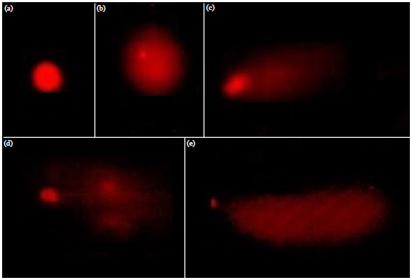 | |
| Fig. 1(a-e): | DNA damage types/classes (comets), (a) Type 0, (b) Type 1, (c) Type 2, (d) Type 3 and (e) Type 4, induced in erythrocytes of Cirrhinus mrigala |
Statistical analysis: Data expressed as Mean±SD was analysed through ANOVA followed by LSD test in Statistix Version X. Value of p<0.05 was considered as significant statistically.
RESULTS
The DNA damage was observed in all endosulfan treated groups. A time and concentration dependent increase was observed in DNA damage induced in peripheral blood erythrocytes of mori. Table 1-4 are showing DNA damage classes (damage type 0-4) induced after exposure to endosulfan at all three sublethal concentrations, while Table 5-8 are showing DNA damage, in terms of genetic damage index, percentage of damaged cell and cumulative tail length, observed after 7-28 days, respectively.
Water physico-chemical parameters: The experiment was carried out in ambient water, having physico-chemical parameters within permissible limits. During the experiment water temperature ranged between 22 and 25°C, pH <7.7, hardness <295 mg L–1, ammonia <0.24 ppm while DO ranged between 6.9-7.5 mg L–1.
Percentage of damaged cell (%): Percent damaged cell, in control group (not exposed to endosulfan) ranged from 3.50±0.1 to 4.60±1.0, in group 2nd (0.5 μg L–1) 23.1±2.1 to 35.0±3.4, in group 3rd (0.75 μg L–1) 34.0±1.2 to 43.7±1.3 while in group 4th (1 μg L–1) percent damage cell ranged from 38.4±2.3 to 48.2±2.21 after 7-28 days of exposure to sublethal concentration of endosulfan.
Genetic Damaged Index (GDI): An increasing trend was observed in GDI with concentration, as the highest level of GDI was observed in Group 4th (1 μg L–1) followed by Group 3rd (0.75 μg L–1). A similar trend was observed with exposure time, as the highest GDI was observed after 28 days followed by 21 days of exposure. The GDI observed after 7 days was 0.1202±0.01, 0.9468±0.17, 1.3156±0.61 and 1.6350±0.71 in group 1st, 2nd, 3rd and 4th while 0.0848±0.00, 1.3262±0.06, 1.7088±0.02 and 1.8770±0.01 after 28 days of exposure, respectively.
Cumulative tail length of comets (μm): Cumulative tail length was highest in group 4th, followed by group 3rd after 28 days while least in group 1st after 7 days, followed by the same group after 14 days. Cumulative tail length ranged between 4.210±0.071 and 5.610±2.120 in group 1st, 113.87±5.91 and 152.51±12.6 in group 2nd, 155.56±9.87 and 198.42±10.6 in group 3rd and between 186.31±11.7 and 234.63±13.3 in group 4th.
| Table 1: | Endosulfan induced DNA damage in Cirrhinus mrigala after 7 days |
 | |
| Data are represented as Mean±SD (n = 6). Means followed by different letters within the column are significantly different (p<0.05), ANOVA followed by LSD test | |
| Table 2: | Endosulfan induced DNA damage in Cirrhinus mrigala after 14 days |
 | |
| Data are represented as Mean±SD (n = 6). Means followed by different letters within the column are significantly different (p<0.05), ANOVA followed by LSD test | |
| Table 3: | Endosulfan induced DNA damage in Cirrhinus mrigala after 21 days |
 | |
| Data are represented as Mean±SD (n = 6). Means followed by different letters within the column are significantly different (p<0.05)., ANOVA followed by LSD test | |
| Table 4: | Endosulfan induced DNA damage in Cirrhinus mrigala after 28 days |
 | |
| Data are represented as Mean±SD (n = 6). Means followed by different letters within the column are significantly different (p<0.05), ANOVA followed by LSD test | |
| Table 5: | Endosulfan induced geno-toxicity in Cirrhinus mrigala after 7 days |
 | |
Data are represented as Mean±SD (n = 6). Means followed by different letters within the column are significantly different (p<0.05), ANOVA followed by LSD test, Damaged cell (%)* = Type II+type III+type IV, **Genetic damage index = Type I+2(type II)+3(type III)+4(type IV)/type 0+type I+type II+type III+type IV | |
| Table 6: | Endosulfan induced genotoxic damage in Cirrhinus mrigala after 14 days |
 | |
| Data are represented as Mean±SD (n = 6). Means followed by different letters within the column are significantly different (p<0.05), ANOVA followed by LSD test | |
| Table 7: | Endosulfan induced genotoxicity in Cirrhinus mrigala after 21 days |
 | |
| Data are represented as Mean±SD (n = 6). Means followed by different letters within the column are significantly different (p<0.05), ANOVA followed by LSD test | |
| Table 8: | Endosulfan induced genotoxic damage in Cirrhinus mrigala after 28 days |
 | |
| Data are represented as Mean±SD (n = 6). Means followed by different letters within the column are significantly different (p<0.05), ANOVA followed by LSD test | |
DISCUSSION
Currently, over thousand chemicals are classified as pesticides. Some of these pesticides have been studied against different animal models and humans for their genotoxic potentials (Zeljezic and Garaj-Vrhovac, 2002; Bhalli et al., 2006, 2009). Animals including different species of mammals, birds, amphibians and fish have shown different extent of DNA damage after exposure to these pesticides. Different aquatic organism, specifically fish, have been employed at most of the occasions due to their prominent role in trophic web, economic worth and capability of accumulating toxic pollutants as well as responsiveness to different carcinogenic, genotoxic and mutagenic toxicants even at very low concentration (Osman et al., 2007; Banu et al., 2001; Ali et al., 2008; De Andrade et al., 2004; Jha, 2008).
Genotoxicological studies is a significant approach to achieve a greater intuition regarding the induced DNA damage, the capability of DNA repair of an organism as well as its protective mechanisms against different pollutants. Moreover, tissues specific response to specific mutagens can also be assessed. Such as, in case of fish, employing erythrocytes for evaluating DNA damage using single cell gel electrophoresis might be beneficial on account of being an easy tissues to collect and easy processing through SCGE, specifically in case of tiny fish species (Sumathi et al., 2001; Ullah et al., 2016c). Previous studies regarding investigations of DNA damage revealed SCGE as a significant, versatile and useful assay (Frenzilli et al., 2009; Galindo et al., 2010). The SCGE has been employed for measuring DNA damage in different organisms including, mollusks (Cotelle and Ferard, 1999; Canty et al., 2009), reptiles (Bronikowski, 2008), amphibians (Cotelle and Ferard, 1999; Yin et al., 2009), mammals (Park et al., 2007; Garaj-Vrhovac et al., 2009) and birds (Baos et al., 2006).
In the current study a significant (p<0.05) genotoxic effect of endosulfan was observed, from time of exposure as well as concentrations, which clearly indicated the genotoxic potential of endosulfan. The current findings were in agreement with the previous studies conducted on different fish species including Ameiurus nebulosus (Pandrangi et al., 1995), Tilapia mossambica (Banu et al., 2001), Dreissena polymorpha (Pavlica et al., 2001), Channa punctatus (Kushwaha et al., 2000; Pandey et al., 2006; Ali et al., 2008), Mugil sp. and Netuma sp., (De Andrade et al., 2004), Carassius auratus (Masuda et al., 2004), Cyprinus carpio (Buschini et al., 2004; Gustavino et al., 2005) and Labeo rohita (Ullah, 2015; Ullah et al., 2016b, c).
The DNA damage observed in the current study could possibly be initiated from DNA single or double strand breaks, formations of DNA adducts and DNA-Protein or DNA-DNA cross links, which might resulted due to the interaction of DNA and pesticide or its metabolites (Fairbairn et al., 1995; Mitchelmore and Chipman, 1998). However, further study is required in order to know the precise mechanism of endosulfan induced genotoxicity as well as to know regarding, which metabolite of endosulfan (alpha and beta) is responsible for DNA strand breaks or either both are responsible (Lu et al., 2000). Yet clastogenic activity, of either of the isomers or their metabolites may exist (Dzwonkowska and Hubner, 1986; Khan and Sinha, 1993). Genotoxicity in fish can be correlated with clastogenicity of endosulfan, as an elevation was observed in DNA migration in the current study as well as in previous studies after use of different environmental mutagens (Russo et al., 2004).
Endosulfan is highly toxic to different species of mammals and fish, which might be attributed to its estrogenic activity. It may possibly bio-accumulate in various edible aquatic organisms and can probably cause human fatalities (Naqvi and Newton, 1990; Belpaeme et al., 1998). However, the genotoxic potential of endosulfan is still to be explored on different fish species. The observed DNA damage in the current study might be linked to Reactive Oxygen Species (ROS) formation during biotransformation of endosulfan, as ROS is highly toxic to fish (Ullah et al., 2016a). The ROS directly break DNA via hydrogen peroxide or hydroxyl ions, subsequently result in oxidized bases of DNA (Akcha et al., 2003). Antioxidant defense system of fish neutralizes ROS but when ROS production exceeds antioxidant enzymes production, it leads to cellular lesions which result in DNA damage (Cadet et al., 2003; Cavalcante et al., 2008; Jha, 2008). Hence, DNA damage is much attributed by oxidative DNA damage due to higher production of ROS (Wilson et al., 1998; Pavlica et al., 2001).
CONCLUSION
Endosulfan was found as highly genotoxic to mori, even at lower sublethal concentration. The results of the current study revealed apprehension regarding the potential dangers of endosulfan to fish, as the similar concentration of endosulfan has been reported in river systems of some African countries. The current findings indicated SCGE/comet assay, as a reliable sensitive assay for investigating genotoxicity induced in fish after pesticides or other toxicants exposure. Moreover, four weeks period of experimentation appears to be satisfactory for determining DNA damage induced by sublethal concentrations of endosulfan in fish using SCGE/comet assay.
REFERENCES
- Dar, S.A., A.R. Yousuf, M.H. Balkhi, F.A. Ganai and F.A. Bhat, 2014. Investigation of the genotoxicity of endosulfan to freshwater Cyprinid fish Crucian carp (Carassius carassius L.) using the micronucleus and chromosomal aberration as biomarkers. Nucleus, 57: 87-98.
CrossRefDirect Link - Dar, S.A., A.R. Yousuf, M.H. Balkhi, F.A. Ganai and F.A. Bhat, 2015. Assessment of endosulfan induced genotoxicity and mutagenicity manifested by oxidative stress pathways in freshwater cyprinid fish crucian carp (Carassius carassius L.). Chemosphere, 120: 273-283.
CrossRefDirect Link - Dzwonkowska, A. and H. Hubner, 1986. Induction of chromosmal aberrations in the Syrian hamster by insecticides tested in vivo. Arch. Toxicol., 58: 152-156.
CrossRefDirect Link - Fairbairn, D.W., P.C. Olive and K.L. O'Neill, 1995. The comet assay: A comprehensive review. Mut. Res./Rev. Genet. Toxicol., 339: 37-59.
CrossRefPubMedDirect Link - Grover, P., K. Danadevi, M. Mahboob, R. Rozati, B.S. Banu and M.F. Rahman, 2003. Evaluation of genetic damage in workers employed in pesticide production utilizing the Comet assay. Mutagenesis, 18: 201-205.
CrossRefDirect Link - Lee, S.E., C. Young-Woong, H.H. Mo, J. Son, K. Park and K. Cho, 2013. Endosulfan-induced biomarkers in Japanese rice fish (Oryzias latipes) analyzed by SELDI-TOF-MS. Int. J. Biol. Sci., 9: 343-349.
CrossRefDirect Link - Naqvi, S.M. and D.J. Newton, 1990. Bioaccumulation of endosulfan (ThiodanR insecticide) in the tissues of Louisiana crayfish, Procambarus clarkii. J. Environ. Sci. Health Part B: Pestic. Food Contam. Agric. Wastes, 25: 511-526.
CrossRefDirect Link - Park, E., M. Glei, Y. Knobel and B.L. Pool-Zobel, 2007. Blood mononucleocytes are sensitive to the DNA damaging effects of iron overload-in vitro and ex vivo results with human and rat cells. Mutation Res./Fund. Mol. Mechan. Mutagen., 619: 59-67.
CrossRefDirect Link - Sharma, A., M. Mishra, K.R. Ram, R. Kumar, M.Z. Abdin and D.K. Chowdhuri, 2011. Transcriptome analysis provides insights for understanding the adverse effects of endosulfan in Drosophila melanogaster. Chemosphere, 82: 370-376.
CrossRefDirect Link - Sharma, S., N.S. Nagpure, R. Kumar, S. Pandey, S.K. Srivastava, P.J. Singh and P.K. Mathur, 2007. Studies on the genotoxicity of endosulfan in different tissues of fresh water fish Mystus vittatus using the comet assay. Arch. Environ. Contaminat. Toxicol., 53: 617-623.
CrossRefDirect Link - Sumathi, M., K. Kalaiselvi, M. Palanivel and P. Rajaguru, 2001. Genotoxicity of textile dye effluent on fish (Cyprinus carpio) measured using the comet assay. Bull. Environ. Contaminat. Toxicol., 66: 407-414.
PubMed - Suneetha, K., K.G. Kumar and K. Veeraiah, 2010. Changes in protein subunits induced by endosulfan and fenvalerate in fresh water fish Labeo rohita through SDS-PAGE. J. Exp. Biol., 31: 759-763.
Direct Link - Rafiq Ullah, A. Zuberi, M. Naeem and Sana Ullah, 2015. Toxicity to hematology and morphology of liver, brain and gills during acute exposure of Mahseer (Tor putitora) to cypermethrin. Int. J. Agric. Biol., 17: 199-204.
Direct Link - Ullah, R., A. Zuberi, S. Ullah, I. Ullah and F.U. Dawar, 2014. Cypermethrin induced behavioral and biochemical changes in mahseer, Tor putitora. J. Toxicol. Sci., 39: 829-836.
CrossRefDirect Link - Ullah, S., M. Begum, K. Dhama, S. Ahmad, S. Hassan and I. Alam, 2016. Malathion induced DNA damage in freshwater fish, Labeo rohita (Hamilton, 1822) using alkaline single cell gel electrophoresis. Asian J. Anim. Vet. Adv., 11: 98-105.
CrossRefDirect Link - Ullah, S., Z. Hasan and K. Dhama, 2016. Toxic effects of endosulfan on behaviour, protein contents and antioxidant enzyme system in gills, brain, liver and muscle tissues of rohu, Labeo rohita. Int. J. Pharmacol., 12: 1-10.
CrossRefDirect Link - Yekeen, T.A. and O.O. Fawole, 2011. Toxic effects of endosulfan on haematological and biochemical indices of Clarias gariepinus. Afr. J. Biotechnol., 10: 14090-14096.
Direct Link - Zeljezic, D. and V. Garaj-Vrhovac, 2002. Sister chromatid exchange and proliferative rate index in the longitudinal risk assessment of occupational exposure to pesticides. Chemosphere, 46: 295-303.
CrossRefDirect Link - Akcha, F., F.V. Hubert and A. Pfhol-Leszkowicz, 2003. Potential value of the comet assay and DNA adduct measurement in dab (Limanda limanda) for assessment of in situ exposure to genotoxic compounds. Mutat. Res./Genet. Toxicol. Environ. Mutagen., 534: 21-32.
CrossRefDirect Link - Ali, D., N.S. Nagpure, S. Kumar, R. Kumar and B. Kushwaha, 2008. Genotoxicity assessment of acute exposure of chlorpyrifos to freshwater fish Channa punctatus (bloch) using micronucleus assay and alkaline single-cell gel electrophoresis. Chemosphere, 71: 1823-1831.
CrossRefDirect Link - Banu, B.S., K. Danadevi, M.F. Rahman, Y.R. Ahuja and J. Kaiser, 2001. Genotoxic effect of monocrotophos to sentinel species using comet assay. Food Chem. Toxicol., 39: 361-366.
CrossRefDirect Link - Baos, R., R. Jovani, N. Pastor, J.L. Tella and B. Jimenez et al., 2006. Evaluation of genotoxic effects of heavy metals and arsenic in wild nestling white storks (Ciconia ciconia) and black kites (Milvus migrans) from southwestern spain after a mining accident. Environ. Toxicol. Chem., 25: 2794-2803.
CrossRefPubMedDirect Link - Belpaeme, K., K. Cooreman and M. Kirsch-Voiders, 1998. Development and validation of the in vivo alkaline comet assay for detecting genomic damage in marine flatfish. Mutat Res., 15: 167-184.
Direct Link - Bhalli, J.A., Q.M. Khan and A. Nasim, 2006. DNA damage in pakistani pesticide-manufacturing workers assayed using the comet assay. Environ. Mol. Mutagen., 47: 587-593.
CrossRefDirect Link - Bhalli, J.A., T. Ali, M.R. Asi, Z.M. Khalid, M. Ceppi and Q.M. Khan, 2009. DNA damage in Pakistani agricultural workers exposed to mixture of pesticides. Environ. Mol. Mutagen., 50: 37-45.
CrossRefPubMedDirect Link - Bronikowski, A.M., 2008. The evolution of aging phenotypes in snakes: A review and synthesis with new data. Age, 30: 169-176.
CrossRefDirect Link - Buschini, A., A. Martino, B. Gustavino, M. Monfrinotti and P. Poli et al., 2004. Comet assay and micronucleus test in circulating erythrocytes of Cyprinus carpio specimens exposed in situ to lake waters treated with disinfectants for potabilization. Mutat. Res./Genet. Toxicol. Environ. Mutagen., 557: 119-129.
CrossRefDirect Link - Cadet, J., T. Douki, D. Gasparutto and J.L. Ravanat, 2003. Oxidative damage to DNA: Formation, measurement and biochemical features. Mutation Res./Fundam. Mol. Mech. Mutagen., 531: 5-23.
CrossRefPubMedDirect Link - Canty, M.N., T.H. Hutchinson, R.J. Brown, M.B. Jones and A.N. Jha, 2009. Linking genotoxic responses with cytotoxic and behavioural or physiological consequences: Differential sensitivity of echinoderms (Asterias rubens) and marine molluscs (Mytilus edulis). Aquatic Toxicol., 94: 68-76.
CrossRefDirect Link - Cavalcante, D.G.S.M., C.B.R. Martinez and S.H. Sofia, 2008. Genotoxic effects of Roundup® on the fish Prochilodus lineatus. Mutat. Res./Genet. Toxicol. Environ. Mutagenesis, 655: 41-46.
CrossRefDirect Link - Collins, A.R., 2004. The comet assay for DNA damage and repair: Principles, applications and limitations. Mol. Biotechnol., 26: 249-261.
CrossRefPubMedDirect Link - Cotelle, S. and J.F. Ferard, 1999. Comet assay in genetic ecotoxicology: A review. Environ. Mol. Mutagen., 34: 246-255.
CrossRefPubMed3.0.CO;2-V/abstract? target='_blank' class='btn btn-sm btn-outline-primary mr-3 mt-3'>Direct Link - De Andrade, V.M., T.R.O. de Freitas and J. da Silva, 2004. Comet assay using mullet (Mugil sp.) and sea catfish (Netuma sp.) erythrocytes for the detection of genotoxic pollutants in aquatic environment. Mutat. Res./Genet. Toxicol. Environ. Mutagen., 560: 57-67.
CrossRefDirect Link - Frenzilli, G., M. Nigro and B.P. Lyons, 2009. The comet assay for the evaluation of genotoxic impact in aquatic environments. Mutat. Res./Rev. Mutat. Res., 681: 80-92.
CrossRefDirect Link - Galindo, B.A., G. Troilo, I.M.S. Colus, C.B.R. Martinez and S.H. Sofia, 2010. Genotoxic effects of aluminum on the neotropical fish Prochilodus lineatus. Water Air Soil Pollut., 212: 419-428.
CrossRefDirect Link - Garaj-Vrhovac, V., G. Gajski, I. Trosic and I. Pavicic, 2009. Retracted: Evaluation of basal DNA damage and oxidative stress in wistar rat leukocytes after exposure to microwave radiation. Toxicology, 259: 107-112.
CrossRefDirect Link - Gustavino, B., A. Buschini, M. Monfrinotti, M. Rizzoni, L. Tancioni, P. Poli and C. Rossi, 2005. Modulating effects of humic acids on genotoxicity induced by water disinfectants in Cyprinus carpio. Mutat. Res./Genet. Toxicol. Environ. Mutagen., 587: 103-113.
CrossRefDirect Link - Jha, A.N., 2008. Ecotoxicological applications and significance of the comet assay. Mutagenesis, 23: 207-221.
CrossRefDirect Link - Khan, P.K. and S.P. Sinha, 1993. Antimutagenic efficacy of higher doses of vitamin C. Mutat. Res./Genet. Toxicol., 298: 157-161.
CrossRefPubMedDirect Link - Kushwaha, B., S.K. Srivastava, B. Singh, N.S. Nagpure and A.G. Ponniah, 2000. Evaluation of comet assay and micronuclei test as genotoxic assays in channa punctatus. Natl. Acad. Sci. Lett., 23: 177-179.
Direct Link - Liao, W., M.A. McNutt and W.G. Zhu, 2009. The comet assay: A sensitive method for detecting DNA damage in individual cells. Methods, 48: 46-53.
CrossRefDirect Link - Masuda, S., Y. Deguchi, Y. Masuda, T. Watanabe and H. Nukaya et al., 2004. Genotoxicity of 2-[2-(acetylamino)-4-[bis(2-hydroxyethyl)amino]-5-methoxyphenyl]-5-amino-7-bromo-4-chloro-2H-benzotriazole (PBTA-6) and 4-amino-3,3'-dichloro-5,4'-dinitro-biphenyl (ADDB) in goldfish (Carassius auratus) using the micronucleus test and the comet assay. Mutat. Res./Genet. Toxicol. Environ. Mutagen., 560: 33-40.
CrossRefPubMedDirect Link - Mitchelmore, C.L. and J.K. Chipman, 1998. DNA strand breakage in aquatic organisms and the potential value of the comet assay in environmental monitoring. Mutat. Res./Fund. Mol. Mechan. Mutagen., 399: 135-147.
CrossRefDirect Link - Osman, A.G.M., S. Wuertz, I.A. Mekkawy, H.J. Exner and F. Kirschbaum, 2007. Lead induced malformations in embryos of the African catfish Clarias gariepinus (Burchell, 1822). Environ. Toxicol., 22: 375-389.
CrossRefPubMedDirect Link - Pandey, S., N.S. Nagpure, R. Kumar, S. Sharma, S.K. Srivastava and M.S. Verma, 2006. Genotoxicity evaluation of acute doses of endosulfan to freshwater teleost Channa punctatus (Bloch) by alkaline single-cell gel electrophoresis. Ecotoxicol. Environ. Saf., 65: 56-61.
CrossRefDirect Link - Pandrangi, R., M. Petras, S. Ralph and M. Vrzoc, 1995. Alkaline single cell gel (comet) assay and genotoxicity monitoring using bullheads and carp. Environ. Mol. Mutagen., 26: 345-356.
CrossRefDirect Link - Pavlica, M., G.I.V. Klobucar, N. Mojas, R. Erben and D. Papes, 2001. Detection of DNA damage in haemocytes of zebra mussel using comet assay. Mutat. Res./Genet. Toxicol. Environ. Mutagen., 490: 209-214.
CrossRefDirect Link - Russo, C., L. Rocco, M.A. Morescalchi and V. Stingo, 2004. Assessment of environmental stress by the micronucleus test and the comet assay on the genome of teleost populations from two natural environments. Ecotoxicol. Environ. Saf., 57: 168-174.
CrossRefDirect Link - Singh, N.P., M.T. McCoy, R.R. Tice and E.L. Schneider, 1988. A simple technique for quantitation of low levels of DNA damage in individual cells. Exp. Cell Res., 175: 184-191.
CrossRefPubMedDirect Link - Ullah, S. and M.J. Zorriehzahra, 2015. Ecotoxicology: A review of pesticides induced toxicity in fish. Adv. Anim. Vet. Sci., 3: 40-57.
CrossRefDirect Link - Wilson, J.T., P.L. Pascoe, J.M. Parry and D.R. Dixon, 1998. Evaluation of the comet assay as a method for the detection of DNA damage in the cells of a marine invertebrate, Mytilus edulis L. (Mollusca: Pelecypoda). Mutat. Res./Fundam. Mol. Mechan. Mutagen., 399: 87-95.
CrossRefPubMedDirect Link - Yin, X., G. Zhu, X.B. Li and S. Liu, 2009. Genotoxicity evaluation of chlorpyrifos to amphibian Chinese toad (Amphibian: Anura) by Comet assay and Micronucleus test. Mutat. Res./Genet. Toxicol. Environ. Mutagen., 680: 2-6.
CrossRefPubMedDirect Link - Lu, Y., K. Morimoto, T. Takeshita, T. Takeuchi and T. Saito, 2000. Genotoxic effects of alpha-endosulfan and beta-endosulfan on human HepG2 cells. Environ. Health Perspect., 108: 559-561.
PubMedDirect Link - Latif, A., M. Ali, A.H. Sayyed, F. Iqbal, K. Usman, M. Rauf and R. Kaoser, 2013. Effect of copper sulphate and lead nitrate, administered alone or in combination, on the histology of liver and kidney of Labeo rohita. Pak. J. Zool., 45: 913-920.
Direct Link