
Wang Lei
Che Zhan Nan Road No. 47, Nanyang, Hennan, China
Wang Chuan
Che Zhan Nan Road No. 47, Nanyang, Hennan, China
International Journal of Pharmacology
Year: 2015 | Volume: 11 | Issue: 2 | Page No.: 148-151


ABSTRACT
The objective of this study was to evaluate the effectiveness of L-carnitine on osteoporosis in men. This was an unbalanced (2:1), double-blind, randomized placebo-controlled trial. Participants were 172 chinese men with primary osteoporosis. L-carnitine at 4 g day‾1 (n = 113) or placebo (n = 59) was administered. Lumbar spine (L2-L4), femoral neck and total hip Bone Mineral Density (BMD), Appendicular Skeletal Muscle (ASM) and biochemical bone markers were measured. Baseline characteristics were similar in both groups in the whole population (age, 63.61±23.79 years; lumbar spine BMD T-score, -3.0±0.9; femoral neck BMD T-score, -3.0±1.3). Men who received L-carnitine over 2 years had gained more substantially in lumbar spine BMD than those received placebo. The relative changes in femoral neck BMD total hip BMD and ASM were significantly different between 2 groups on 24 months which were 25.30±31.09, 10.67±21.35 and 21.58±19.47%. At the end of treatment, mean levels of serum cross-linked telopeptides of type I collagen, a marker of bone resorption, fell in both the L-carnitine group (12.24±36.18%; p = 0.004) and the placebo group (28.06±57.90%, p = 0.02). The corresponding mean changes of bone alkaline phosphatase, a marker of bone formation, were significantly different between L-carnitine group and placebo group on 12 months or 24 months. L-carnitine is efficacious on osteoporotic men, supporting its use in the treatment of osteoporosis in men.
PDF Abstract XML References Citation
Received: November 11, 2014;
Accepted: January 20, 2015;
Published: February 16, 2015
How to cite this article
Wang Lei and Wang Chuan, 2015. Efficacy of L-Carnitine in the Treatment of Osteoporosis in Men. International Journal of Pharmacology, 11: 148-151.
DOI: 10.3923/ijp.2015.148.151
URL: https://scialert.net/abstract/?doi=ijp.2015.148.151
DOI: 10.3923/ijp.2015.148.151
URL: https://scialert.net/abstract/?doi=ijp.2015.148.151
INTRODUCTION
Osteoporosis in men is a great challenge in public health. Mortality after osteoporotic vertebral, nonvertebral and hip fractures is higher in men than that in women (Leboime et al., 2010). A variety of pharmacological and nonpharmacological options were used to administrated osteoporosis in men. Current pharmacological treatments for osteoporosis-alendronate (Orwoll et al., 2000), risedronate (Boonen et al., 2009) zoledronate (Orwoll et al., 2010) and teriparatide (Orwoll et al., 2003).
L-carnitine, a necessary cofactors of fatty acid metabolism, plays a vital role in energy metabolism in bone and muscle. L-carnitine levels in tissues have been found to decline with age (Maccari et al., 1990). L-carnitine may improve age-related bone loss in the elderly patients because of efficient improving total muscle mass in elderly patients (Colucci et al., 2005). Furthermore, it has been shown that the osteoblast generate 40-80% of their energy demands through fatty acid oxidation (Adamek et al., 1987). A study suggests that modulation of fatty acid oxidation may regulate the availability of energy for protein synthesis in osteoblasts (Patano et al., 2008). L-carnitine can influence bone density and slow the rate of bone turnover (Hooshmand et al., 2008). One study shows that subcutaneous injections of L-carnitine reduced bone loss in ovariectomised rats (Orsal et al., 2013). For these reason L-carnitine maybe act as one of effective medicines in the treatment of osteoporosis. We proposed a hypothesis that L-carnitine is efficient in treatment for osteoporosis in men.
MATERIALS AND METHODS
Patients: All patients enrolled in voluntarily were informed the side effect of medicine taken, then signed the informed consent. Criteria for inclusion at the selection visit included:
(1) Chinese men aged ≥65 years, (2) Low Bone Minal Density (BMD): Low lumbar spine (L2-L4) BMD (≤0.840 g cm‾2) with a dual energy x-ray absorptiometry (DEXA) device and/or low femoral neck BMD (≤0.600 g cm‾2 T-score ≤-2.5).
Criteria for exclusion at the selection visit included: (1) A history of or increased risk for venous thromboembolism, severe hypogonadism, (2) Skeletal diseases (such as secondary osteoporosis, Paget disease, osteomalacia, hyperparathyroidism and hypoparathyroidism), (3) Previous treatment acting on bone metabolism (including long-term oral or inhaled glucocorticoid treatment in the previous year, bisphosphonate injection in the previous year or tablets in the previous 18 months, calcitriol and 1 α-vitamin D in the previous 6 months and parathyroid hormone or derivatives, i.e., teriparatide), (4) Severe osteoporosis (T-score ≤-4.0 at any site), (5) Prevalent osteoporotic vertebral fractures and (6) Those with a history of stroke, coronary artery disease, thyroid disease, lung disease, thyroids disease, liver disease and renal disease. Patients visit was on December 11, 2009 and recruitment ended on March 12, 2010. The last patient visit was on March 24, 2013.
Treatment: Patients were allocated to L-carnitine (4 g day‾1) or placebo orally for 2 years. Patients and investigators were blinded to treatment allocation and the study treatments were identically packaged and labeled. All patients received calcium and Vitamin D supplementation (1 g 800 IU daily) for 2 years from the selection visit.
Measurement of bone density and appendicular skeletal muscle mass: All participants underwent the DXA (Hologic Inc., MA, USA) for assessment of BMD and body composition. The BMD (g cm‾2 or T-score) of the femoral neck and lumbar spine (L1-4) were detected at both 1 year and 2 years. The diagnosis of osteoporosis was made using the WHO T-score criteria (T-score <-2.5). Appendicular Skeletal Muscle (ASM) mass was calculated as the sum of muscle mass in arms and legs at both 1 year and 2 years, assuming that all non-fat and non-bone tissue is skeletal muscle (16). These outcomes were used adjusted by muscle mass indexes (ASM/Ht2, kg m‾2) (5, 7, 17). Biochemical bone markers (bone alkaline phosphatase [b-ALP], serum cross-linked telopeptides of type I collagen [s-CTX]) and Quality Of Life (QOL), were investigated at both 1 year and 2 years.
Statistical methods: Descriptive statistics of baseline characteristics are presented with numbers and percentage, qualitative data are presented with Means±SD. Baseline characteristics were studied by independent Student’s-t test. Intergroup differences in the relative change from baseline to end were analyzed by a general linear model with age as covariate to produce an estimate (E) of the treatment group difference, SE of the estimate with the associated 95% Confidence Interval (CI) and p-value. Intergroup differences the treatment effect on the BMD of the femoral neck and lumbar spine, ASM, b-ALP and s-CTX was studied using a general linearmode. That of QOF was were studied by a nonparametric approach. Statistical analyses were performed with SPSS 19.0 software.
RESULTS
One hundred and seventy two patients of the 384 patients selected were included and randomly assigned (113 L-carnitine and 59 placebo). One hundred and sixty two patients completed the entire follow up period and premature drop-out was due to nausea (n = 4), excessive demand (n = 2), diarrhea (n = 2) or miscellaneous symptoms (n = 2). Drop out rates and reasons were not different between both 2 groups. Oral supplementation of L-carnitine substantially increased L-carnitine serum plasma levels up to 60% of the basic value after 6 months (p<0.009) in the L-carnitine group, while a constant decline of L-carnitine plasma levels was evident during the observation period in the placebo group.
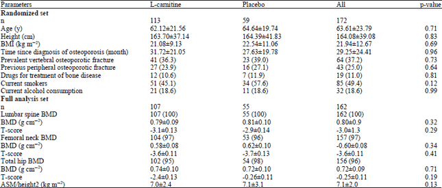
The baseline characteristics of the two groups were similar (Table 1).
| Table 1: | Baseline characteristics |
 | |
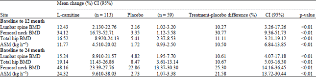
| Table 2: | Mean percentage changes in BMD from baseline to 12 months, 24 months in men receiving L-carnitine or placebo |
 | |
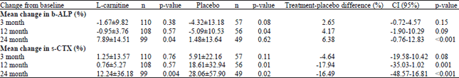
| Table 3: | Mean percentage changes in b-ALP and s-CTX from baseline to each visit in men receiving L-carnitine or placebo |
 | |
The 29.5% vs. 31.7% patients in 2 group had at least one previous treatment, including calcium (31.13% vs. 29.32%), vitamins vitamin Dor analogs (27.47% vs. 23.75%), calcitonin salmon (13.51% vs. 16.84%) and bisphosphonates (9.7% vs. 8.3%). Levels of testosterone were also similar in the two groups (20.3±7.1 vs. 19.6±6.6 ng L‾1, levels of bone markers were also comparable; mean levels of b-ALP were 14.5±5.3 ng mL‾1 vs. 12.9±6.8 ng mL‾1 and that of s-CXT were 0.78±0.39 ng mL‾1 vs. 0.6±0.4 ng mL‾1.
Over 1 year, the analysis showed that lumbar spine (L2-L4) BMD in 2 groups was 0.90±0.12 g cm‾2 (L-carnitine group) and 0.84±0.17 g cm‾2 (placebo group); The relative changes for the two groups (Table 2) were 12.43±6.61% in L-carnitine groups vs. 2.16±3.16% in placebo group. Femoral neck BMD was 0.78±0.13 g cm‾2 (L-carnitine group) and 0.66±0.16 g cm‾2 (placebo group). The relative changes for the two groups were 34.12±11.24% in L-carnitine groups vs. 3.35±4.46% in placebo group. The total hip BMD in 2 groups was 0.86±0.16 g cm‾2 (L-carnitine group) and 0.76±0.13 g cm‾2 (placebo group). The relative changes for the two groups were 16.52±7.64% in L-carnitine groups vs. 5.41±3.59% in placebo group. ASM in 2 groups were 7.76±4.87 kg m‾2 vs. 7.21±6.19 kg m‾2, meanwhile, the relative changes for the two groups were 11.77±6.29% with L-carnitine vs. 1.72±5.48% with placebo.
Over 2 years, the analysis showed that lumbar spine (L2-L4) BMD in 2 groups was 0.91±0.13 g cm‾2 (L-carnitine group) and 0.85±0.15 g cm‾2 (placebo group). The relative changes for the two groups (Table 2) were 15.24±6.76% in L-carnitine groups vs. 4.82±3.44% in placebo group. Femoral neck BMD was 0.88±0.11 g cm‾2 (L-carnitine group) and 0.76±0.13 g cm‾2 (placebo group). The relative changes for the two groups were 48.16±12.03% in L-carnitine groups vs. 22.86±9.42% in placebo group. The total hip BMD in 2 groups was 0.88±0.13 g cm‾2 (L-carnitine group) and 0.78±0.16 g cm‾2 (placebo group). The relative changes for the two groups were 19.14±7.32% in L-carnitine groups vs. 8.47±6.40% in placebo group. ASM in 2 groups were 8.71±4.87 kg m‾2 vs. 7.35±3.24 kg m‾2, meanwhile, the relative changes for the two groups were 24.32±9.17% with L-carnitine vs. 2.73±5.74% with placebo.
Mean levels of s-CTX fell more greatly in the L-carnitine group (Table 3) than in the placebo group from 3 months onward (p<0.001). The relative change from baseline to end was 10.7±58.0% (p<0.022) in the L-carnitine group vs. 34.9±65.8% (p<0.001) in the placebo group. Meanwhile, the relative changes from baseline to end of b-ALP were 6.4±28.5% (p<0.005) in the L-carnitine group vs. 1.9±25.4% (p = 0.51) in the placebo group.
Patients in L-carnitine group had a better QOL (i.e., a decrease in score) from baseline to study end. Moreover, patients in L-carnitine group improved in QOL from baseline to 24 months (p = 0.009). More patients receiving L-carnitine had eased pain in the middle/upper part of back (32% vs. 23%; p = 0.28), pain interfering with sleep (23% vs. 15%; p = 0.39) and discomfort in the same position (35% vs. 27%; p = 0.36) than patients receiving placebo; this improvement was significant for pain when walking/climbing stairs (17% vs. 4%; p = 0.019).
DISCUSSION
Orsal et al. (2013) first carried out a study to assess the protective bone-sparing effect of L-carnitine on chronic inflammation-induced bone loss in ovariectomised rats. The result showed that L-carnitine administration was able to restore BMD in ovariectomised rats. Another study showed that muscle mass was positively correlated with bone density in men (Kim et al., 2014). However, the effect of improving muscle mass on bone density in men was not evaluated up to now. Our study population has the typical features of a population of men with osteoporosis in terms of age and T-score at baseline. These changes in BMD in our study shows that L-carnitine supplement can improve bone density, muscle, marker of bone turnover, QOL in men with osteoporosis. This is in keeping with previously published studies.
Treatment with L-carnitine in this population was associated with significant increases in BMD at the lumbar spine, femoral neck, total hip and ASM throughout the study compared with placebo. BMD can predict osteoporotic fracture in men, independent of age, body weight, or prevalent fracture. Study identified that sarcopenia can increase the risk of osteoporosis and may lead to increased risk of bone fracture. With regard to L-carnitine, changes in femoral neck BMD and ASM have been associated with a reduction in vertebral and hip fracture risk. Our findings suggest that L-carnitine may have antifracture efficacy in men with osteoporosis. Present study also confirms that treatment with L-carnitine increased BMD after 2 year. Meanwhile, the markers of bone turnover were dropped down in men with osteoporosis after L-carnitine supplementing. QOL results indicate an improvement in QOL in patients treated with L-carnitine compared with placebo. The positive trend was confirmed over 24 months, particularly with regard to pain when walking/climbing stairs.
Our study was a single-center study, therefore, a multi-center, large scale, double-blinded study is imperative to examining efficacy of L-carnitine in osteoporosis men.
The study could suggest that the clinical benefit of an inexpensive oral L-carnitine supplementation may reach the clinical benefit for osteoporosis in men.
REFERENCES
- Leboime, A., C.B. Confavreux, N. Mehsen, J. Paccou, C. David and C. Roux, 2010. Osteoporosis and mortality. Joint Bone Spine, 77: S107-S112.
CrossRefDirect Link - Orwoll, E., M.D.M. Ettinger, M.D.S. Weiss, M.D.P. Miller and M.D.D. Kendler et al., 2000. Alendronate for the treatment of osteoporosis in men. N. Engl. J. Med., 343: 604-610.
CrossRefDirect Link - Boonen, S., E.S. Orwoll, D. Wenderoth, K.J. Stoner, R. Eusebio and P.D. Delmas, 2009. Once-weekly risedronate in men with osteoporosis: Results of a 2-year, placebo-controlled, double-blind, multicenter study. J. Bone Miner. Res., 24: 719-725.
CrossRefDirect Link - Orwoll, E.S., P.D. Miller, J.D. Adachi, J. Brown and R.A. Adler et al., 2010. Efficacy and safety of a once-yearly i.v. Infusion of zoledronic acid 5 mg versus a once-weekly 70-mg oral alendronate in the treatment of male osteoporosis: A randomized, multicenter, double-blind, active-controlled study. J. Bone Miner. Res., 25: 2239-2250.
CrossRefDirect Link - Orwoll, E.S., W.H. Scheele, S. Paul, S. Adami and U. Syversen et al., 2003. The effect of teriparatide [human parathyroid hormone (1-34)] therapy on bone density in men with osteoporosis. J. Bone Miner. Res., 18: 9-17.
CrossRefDirect Link - Orsal, E., Z. Halici, Y. Bayir, E. Cadirci and H. Bilen et al., 2013. The role of carnitine on ovariectomy and inflammation-induced osteoporosis in rats. Exp. Biol. Med., 238: 1406-1412.
PubMed - Colucci, S., G. Mori, S. Vaira, G. Brunetti and G. Greco et al., 2005. L-carnitine and isovaleryl L-carnitine fumarate positively affect human osteoblast proliferation and differentiation in vitro. Calcified Tissue Int., 76: 458-465.
CrossRefDirect Link - Adamek, G., R. Felix, H.L. Guenther and H. Fleisch, 1987. Fatty acid oxidation in bone tissue and bone cells in culture. Characterization and hormonal influences. Biochem. J., 248: 129-137.
Direct Link - Patano, N., L. Mancini, M.P. Settanni, M. Strippoli and G. Brunetti et al., 2008. L:-carnitine fumarate and isovaleryl-L:-carnitine fumarate accelerate the recovery of bone volume/total volume ratio after experimetally induced osteoporosis in pregnant mice. Calcified Tissue Int., 82: 221-228.
CrossRefDirect Link - Hooshmand, S., A. Balakrishnan, R.M. Clark, K.Q. Owen, S.I. Koo and B.H. Arjmandi, 2008. Dietary l-carnitine supplementation improves bone mineral density by suppressing bone turnover in aged ovariectomized rats. Phytomedicine, 15: 595-601.
CrossRefDirect Link - Maccari, F., A. Arseni, P. Chiodi, M.T. Ramacci and L. Angelucci, 1990. Levels of carnitines in brain and other tissues of rats of different ages: Effect of acetyl-L-carnitine administration. Exp. Gerontol., 25: 127-134.
CrossRefDirect Link - Kim, S., C.W. Won, B.S. Kim, H.R. Choi and M.Y. Moon, 2014. The association between the low muscle mass and osteoporosis in elderly Korean people. Korean Med. Sci., 29: 995-1000.
CrossRefPubMedDirect Link