
E.R. Yuslianti
Departement of Biochemistry and Molecular Biology, Faculty of Medicine, Jenderal Achmad Yani University, Jl. Ters. Jenderal Sudirman, Cimahi, Indonesia
I.P.R. Sabirin
Oral Biology, Dentistry Programme, Faculty of Medicine, Jenderal Achmad Yani University, Jl. Ters. Jenderal Sudirman, Cimahi, Indonesia
E. Sovia
Departement of Pharmacology, Faculty of Medicine, Jenderal Achmad Yani University, Jl. Ters. Jenderal Sudirman, Cimahi, Indonesia
International Journal of Pharmacology
Year: 2013 | Volume: 9 | Issue: 5 | Page No.: 318-321


ABSTRACT
This study was proposed to find the beneficence of noni (Morinda citrifolia L.) leaves ethanol extract as a treatment for wound healing in rat skin. This experimental study was done by making excisional wound on the back of Wistar rats then divided them into four groups which were given topical treatments of noni extract; control group treated by base gel and three treatment groups were given 10% (w/w) povidone iodine, 10% ethanol extract gel of noni leaves and 20% (w/w) ethanol extract gel of noni leaves, recpectively. The result of this study showed that 10% topical morinda ethanol extract gel (10 mg mL-1) had a significant effect on rat skin excisional wound healing compared to 10% povidone iodine (p<0.05). The level of plasma MDA of rats in every group was also examined, however there was no significant difference between treatment and control groups. This research showed that noni extract could improved excisional woung healing.
PDF Abstract XML References Citation
Received: September 27, 2013;
Accepted: January 20, 2014;
Published: February 04, 2014
How to cite this article
E.R. Yuslianti, I.P.R. Sabirin and E. Sovia, 2013. Effect of Topical Ethanol Extracts of Morinda citrofilia L. Leaves on Excisional Wound Healing. International Journal of Pharmacology, 9: 318-321.
DOI: 10.3923/ijp.2013.318.321
URL: https://scialert.net/abstract/?doi=ijp.2013.318.321
DOI: 10.3923/ijp.2013.318.321
URL: https://scialert.net/abstract/?doi=ijp.2013.318.321
INTRODUCTION
Wound healing is a natural process to get the skin or mucosa back to normal from previous injury. This process consists of three phases: inflammatory, proliferation and remodeling. Wound healing can be failed if the natural ability of the tissue healing is decreased, such as systemic disease condition or if the wound not properly treated. An optimal wound healing is achieved if there is minimal or no complication related to the healing process such as either deficiency or excess of skin component like epithelial and connective tissue or unnecessary wound contraction (Peterson, 2004; Nayak et al., 2009).
The inflammatory phase which occurs along with hemostasis indicated with infiltration of inflammatory cells in the blood clot and release of mediators from the leukocyte. The proliferation phase of wound healing consists of neovascularization, formation of granulation tissue, primary wound contraction and the beginning of epithelialization. On the granulation tissue, Platelet-Derived Growth Factor (PDGF) produced by platelet degranulation was responsible to fibroblast proliferation, eventually secreted collagen to fill the previously wounded area and provide the space for new blood capillaries. The fibroblast migrated from the edge of the wound to the center along the formed fibrin. During the remodeling phase, epithelial cells proliferated along side the previously wounded area, determined by Epidermal Growth Factor (EGF) released by fibroblast (Peterson, 2004; Kumar et al., 2004).
Morinda citrifolia L. also known as noni, is a tropical plant distributed in South East Asia, Pacific, South America and Central America. The fruit, leaves, seed and flower could be used as traditional medicine. Empirically, it used topically for wound, sprained ankle and reducing pain. The active substances that found in the leaves of noni are saponins, triterpens, tannin, alkaloids, iridoid glycosides and flavonoids. In the role of wound healing the subtances work as antibacterial agent, haemostatic and astringent, analgesics, anti-inflammatory and antioxidant (Suwiti, 2010; Mills and Bone, 2000; Rasal et al., 2008). Prevoius study showed that ethanol extract of 10 mg mL-1 of noni leaves have a potent antimicrobial activity against E. coli, Bacillus, Staphylococcus aereus and subtilis (Kumar et al., 2010). Lipid peroxidation is a continuous oxidative chain reaction which provides free radicals to trigger a subsequent peroxidation. This reaction will continue to biological cell membranes respons (e.g., Mitochondrion and nucleus) and eventually break the lipid chain into many substances including Malondialdehyde (MDA). The lipid peroxidation process that occurs continuously can be assessed by detected the MDA level in the blood plasma, as an indicator for the damage of cell membranes by oxidants (Hamid et al., 2010).
In vivo model, such as rats, can be used to analyze cellular or molecular mechanism of wound healing. This model is able to investigation complex tissue movement during the healing process like bleeding and hemostasis, formation of granulation tissue and neovascularization and re-epithelialization. It is also able to help choosing any agents or substances that will stimulate or demote the healing process and eventually creating a satisfactory result of the new-formed skin, both functional and aesthetic (DiPietro and Burns, 2003).
In this research, excisional wound were made on the back of the rats and followed by application of 10% and 20% ethanol extract of noni leaves to discover the ideal dose of the topical extract. The blood plasma of MDA level also measured to examine the influence of injury and its treatment to the rate of oxidative stress.
MATERIALS AND METHODS
The research conducted at Biochemistry and Molecular Biology Laboratory of Medical Faculty General Achmad Yani University and Pharmacy Laboratory of Science Faculty General Achmad Yani University, Cimahi, West Java for the period of 16-30 December 2012. Wistar rats were utilized in this research after an ethical clearance was received from Ethical Committee of Hasan Sadikin Hospital, Bandung, West Java.
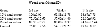
Twenty-four Wistar rats are used in this experiment, which devided into four groups. One control group and three treatment group treated with 10% of noni extract, 20% of noni extract and 10% povidone iodine ointment, respectively. Local anesthetic (Ketamin) was given before excisional wounding. The wound area was about 10x10x2 mm. Treatment was given immediately after the wounds were created, with 1 mL of the proper formulations and repeated once a day. The clinical appearance of the wound such as wound closure and hair growth on all groups were also examined and compared on 3, 7 and 14th day to represent inflammatory, proliferation and remodeling phase of wound healing. The wound closure was measured at three different times (3, 7 and 14th day) on every group and calculated with a equation:
A (c+p) = (Nc+0,2xNp) x 0,02 |
Details: A(c+p): Area complete (c) dan partial (p)
Examination of MDA with TBARs method: The plasma MDA level was assessed at the 14th day after excisional wounding. Blood lipid peroxidation was measured by TBARs method (Thiobarbituric Acid Reactive Like substance) which could observed oxidative stress according to thiobarbituric acid reaction with malondialdehyde (MDA). Rats’ blood was centrifuged and the plasma was acquired. To prevent lipid oxidation, during the examination Butylated Hidroxytoluene (BHT) and Ethylenedinitrilo Tetraacetic Acid Disodium Salt Dihydrate (EDTA) were added to the sample before protein precipitation with acetic acid. Thiobarbituric acid were also added and the formulation was heated in 45 min at 100°C. The result was a light reddish substance which can be read at 532 nm wavelength. The final result was multiplied with a standard level of malondialdehyde bis-dimetilasetal (Repetto et al., 2012).
Data analysis of this research using Mann-Whitney test for two or more independent groups.
RESULTS
On the 3rd day after wounding, the clinical appearance of the wound which was treated topically by 10% ethanol extract of noni leaves showed a significant healing compared to other groups (Fig. 1). The wound appeared drier than others groups. The 10% povidone iodine group was not healed properly.
The observation of the wound on 7th day showed that the wound area of all groups were smaller, with the better healing could be seen on the group that treated with 10% noni leaves ethanol extract (Fig. 2). On the 14th day, the wound of 10% noni leaves ethanol extract group showed the best healing, wound closure and hair growth compared to the others groups (Fig. 3).
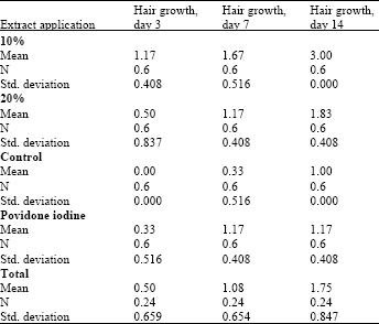
Malondialdehyde level was evaluated by obtaining the blood from orbital vein on the 14th day after wounding. The MDA level of 10% noni leaves extract, 20% noni leaves extract, povidone iodine and control group were 0,07; 0,19; 0,24 and 0,09 nm mL-1, respectively (Table 1-4).
The statistical analysis showed statistically significant wound closure occurred only in the 3rd day after wounding on the group treated by 10% of morinda leaves extract.
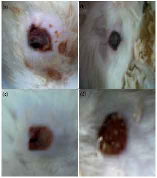 | |
| Fig. 1(a-d): | Excisional wound on 3rd day, (a) Povidon iodine group, (b) 10% noni extract group, (c) 20% noni extract group and (d) Control group |
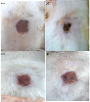 | |
| Fig. 2(a-d): | Excisional wound on 7th day, (a) Povidon iodine group, (b) 10% noni extract group, (c) 20% noni extract group and (d) Control group |
The hair growth as a sign of good wound healing was statistically significant on all observation day in the group treated by 10% morinda extract.
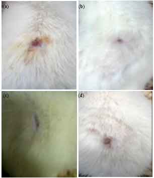 | |
| Fig. 3(a-d): | Excisional wound on 14th day, (a) Povidon iodine group, (b) 10% noni extract group, (c) 20% noni extract group and (d) control group |
| Table 1: | Wound area mean value (mm2) |
 | |
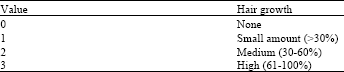
| Table 2: | Semi-quantitative parameters for hair growth |
 | |
However, the MDA level on all groups was not statistically significant.
DISCUSSION AND CONCLUSION
The management of the wound playing important role in both skin and mucosa wound healing process. It can help accelerating the healing process and optimize the final result. This research showed that 10% topical morinda leaves ethanol extract was more effective for wound healing compared to 20% morinda leaves extract, 10% povidone iodine and control group. Wound closure was decent by administration of 10% of morinda leaves extract, but the result was only statistically significant on the 3rd day of the observation.
| Table 3: | Mean value of hair growth |
 | |
| Table 4: | Statisical analysis on all groups compared to the control group |
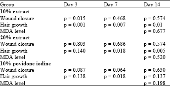 | |
The hair growth rate was also better with the application of 10% morinda leaves extract compared to that of control group. The observation on post-wound hair growth was also showed that 10% ethanol extract treated group had the best hair regrowth in 14 days. This research find out that topical application of 10% morinda leaves extract was better than 20% of the same extract, 10% of povidone iodine and untreated wound on wound healing process, although it is not statistically significant on every parameter. This result supported previous research that showed antimicrobial activity of morinda leaves E. coli, Bacillus sp. Staphilococcus aureus and S. subtilis (Kumar et al., 2010). If the dose was administered correctly, the ethanol extract of morinda leaves can accelerate wound healing by preventing infection, invasion of bacteria and its toxin and release of excess mediator from leukocyte which can prolong the wound healing. There was a possibility that if the extract dose was higher that therapeutic dose, it can prolong the wound healing process, but didn’t have a dangerous effect.
The MDA level of treated group did not showed a significant decrease compared to control group. This result indicated that oxidative stress generated after skin wounding was not influenced by topical application of medicine on the wound. The result was different with previous research which stated that blood MDA level was lower on the post-wounded subject treated by oral administration of ethanol extract of morinda leaves (Nayak et al., 2009).
REFERENCES
- Hamid, A.A., O.O. Aiyelaagbe, L.A. Usman, O.M. Ameen and A. Lawal, 2010. Antioxidants: Its medicinal and pharmacological applications. Afr. J. Pure Applied Chem., 4: 142-151.
Direct Link - Kumar, K.T., D.S. Panda, U.N. Nanda and S. Khuntia, 2010. Evaluation of antibacterial, antifungal and anthelmintic activity of Morinda citrifolia L. (Noni). Int. J. PharmTech Res., 2: 1030-1032.
Direct Link - Nayak, B.S., S. Sandiford and A. Maxwell, 2009. Evaluation of the wound-healing activity of ethanolic extract of Morinda citrifolia L. leaf. Evidence-Based Complement. Altern. Med., 6: 351-356.
CrossRefDirect Link - Rasal, V.P., A. Sinnathambi, P. Ashok and S. Yeshmaina, 2008. Wound healing and antioxidant activities of Morinda citrifolia leaf extract in rats. Iran. J. Pharmacol. Therapeut., 7: 49-52.
Direct Link - Suwiti, N.K., 2010. Deteksi histologik kesembuhan luka pada kulit pasca pemberian daun mengkudu (Morinda citrofilia Linn) [The histological detection of skin wound after treatment with mengkudu leaves (Morinda citrifolia Linn.)]. Buletin Veteriner Udayana, 2: 1-9.
Direct Link