
S. O. Oyedemi
School of Biological Sciences, University of Fort Hare, Alice 5700, South Africa
A. J. Afolayan
School of Biological Sciences, University of Fort Hare, Alice 5700, South Africa
International Journal of Pharmacology
Year: 2011 | Volume: 7 | Issue: 2 | Page No.: 248-256


ABSTRACT
The present study investigated antioxidant activities of Leonotis leonurus extract both in vivo and in vitro to provide scientific basis to traditional usage of this plant. The in vitro antioxidants activity was evaluated by determining ferric reducing power, total flavonoids, phenolics, flavonols and proanthocyanidins contents using standard assay methods. The ability of the extract to scavenge 2, 2 diphenyl-2-picrylhydrazyl (DPPH), Nitric Oxide (NO), 2, 2-azinobis [3-ethylbenzothiazoline-6-sulfonic acid] diammonium salt were also assessed using spectroscopic method. Oral administration of L. leonurus extract at the doses of 125, 250 and 500 mg kg-1 body weight was evaluated in Wistar rats induced with carbon tetrachloride (CCl4) for 7 days. The extract effectively increased the percentage inhibition of reduced glutathione (GSH), superoxide dismutase (SOD) and catalase (CAT). However, lipid peroxidation was significantly decreased in CCl4 treated rats with the extract when compared with the diabetic control rats. The plant extract (0.8 mg mL-1) scavenge DPPH.+ ABTS.+ and NO.+ by inhibiting 72.48, 78.02 and 70.43% of the radicals, respectively. The reducing power of the extract was found to be concentration dependent. In addition, the extracts yielded higher phenolics content followed by flavonoids, proanthocyanidins and flavonols. A positive linear correlation was established between these polyphenols and the free radical scavenging activities. The results obtained from this study indicate that L. leonurus is a potential source of antioxidants and thus could prevent many radical related diseases.
PDF Abstract XML References Citation
Received: July 31, 2010;
Accepted: December 18, 2010;
Published: March 16, 2011
How to cite this article
S. O. Oyedemi and A. J. Afolayan, 2011. In vitro and in vivo Antioxidant Activity of Aqueous Leaves Extract of Leonotis leonurus (L.) R. Br. International Journal of Pharmacology, 7: 248-256.
DOI: 10.3923/ijp.2011.248.256
URL: https://scialert.net/abstract/?doi=ijp.2011.248.256
DOI: 10.3923/ijp.2011.248.256
URL: https://scialert.net/abstract/?doi=ijp.2011.248.256
INTRODUCTION
Free radicals are chemically unstable atoms or molecules that cause extensive damage to cells as a result of imbalance between the generation of Reactive Oxygen Species (ROS) and the antioxidant enzymes. Reactive oxygen species are a group of highly reactive molecules due to the presence of unpaired electron. The example includes superoxide anions, hydroxyl and hydrogen peroxide radicals. They are often generated as byproducts of oxidative damage to the DNA molecules, lipids and proteins (Farber, 1994). This damage could leads to several human diseases such as diabetes mellitus, cancer, atherosclerosis, arthritis, anemia, asthma, inflammation and neurodegenerative (Polterat, 1997). These diseases can be delayed or prevented by inhibiting the initiation or propagation of oxidative chain reaction (Gerber et al., 2002). The intake of natural and synthetic antioxidant products have been reported to reconcile their upshot by quenching their catalytic metal ions (Robak and Marcinkiewicz, 1995). Recently, there is an increasing interest in finding natural antioxidants especially from plant that can protect human body from free radical related diseases (Kinsella et al., 1993). This upsurge development could be attributed to the multiple biological activities exerted by the plants as a result of broad range of phytochemical compounds with antioxidant activities (Alan and Miller, 1996).
Leonotis leonurus (L.) R. Br. (Lamiaceae) known as Wild dagga in English, is commonly found along forest margins, rocky hillsides and riverbanks. It is also found in the grassland area of Eastern and Western Cape, Kwazulu-Natal as well as Mpumalanga provinces of South Africa (Van Wyk et al., 2000). The bright yellow-green leaves of the plant which are rough in texture have a characteristic aromatic-pungent odour. The leaves are commonly used in South Africa traditional medicine for the treatment of various diseases such as cough, cold, influenza, chest infections, diabetes mellitus, eczema, epilepsy, delayed menstruation, intestinal worms, constipation, scorpion stings, spider and snake bite (Jager et al., 1996; Van Wyk et al., 2000; Ososki et al., 2002). It is also used for the management of haemorrhoids, eczema, skin rashes, influenza, tuberculosis, hypertension, jaundice and boils (Bienvenu et al., 2002; Noumi et al., 1999). Previous studied on this plant evaluated the possible toxicological effect in male and female Wistar rats at different doses (Maphosa et al., 2008; Oyedemi et al., 2010b). Considering the pharmacological importance of this plant, it is therefore necessary to investigate its antioxidant and free radical scavenging properties.To the best of our knowledge, there is no or little information on the in vitro and in vivo antioxidant activities of aqueous leaves extract of L. leonurus. Therefore, this study was aimed to provide information on the antioxidant properties of this plant to justify its folkloric usage.
MATERIALS AND METHODS
Plant material: The leaves of Leonotis leonurus were collected from rockhill field near Ntselamanzi location in Nkokonbe Municipality (Eastern Cape, South Africa) between May and June, 2008. It was authenticated by Prof. DS. Grierson of the Department of Botany, University of Fort Hare. Voucher specimen (Sun MED 2) was deposited at the Giffen herbarium of the University.
Sample preparation: The leaves of the plant after washing with distilled water were air-dried at room temperature for 7 days and pulverized using an electric blender (Waring Products Division, Torrington, USA). One hundred gram (100.00 g) of the sample powder was extracted in 3 L of distilled water (4°C) maintained on a mechanical shaker (Stuart Scientific Orbital Shaker SO1, Essex, UK) for 48 h. The extract was thereafter filtered using Buchner funnel and Whatman No.1 filter paper. The filtrate was freeze-dried with Savant Refrigerated Vapor Trap (RV T41404, California, USA) to give a yield of 23.48 g and later reconstituted in distilled water to give the required concentrations needed in this study.
Animals: Male rats (Rattus norvegicus) of Wistar strain weighing 220.05 g±12.20 were obtained from the animal house of the Agricultural and Rural Development Research Institute, University of Fort Hare. They were kept in aluminum cages placed in a well ventilated house conditions (temperature 23±1°C; photoperiod: 12 h light and 12 h dark cycle; humidity: 40-45%). The animals were allowed free access to rat pellets (Balanced Trusty Chunks (Pioneer Foods (Pty) Ltd., Huguenot, South Africa) and water. The experiment was carried out after approval by the Ethics Committee on the Use and Care of Experimental Animals of the University of Fort Hare.
Animal grouping and extract administration: Thirty male rats were randomized into five groups of six animals each. Group 1 received distilled water only (0.5 mL) repeatedly on daily basis for 7 days. Group 2 animals served as hepatotoxic control, treated with 0.5 mL of 50% CCl4 in olive oil. Group 3-5 animals were treated daily with 0.5 mL of 125, 250 and 500 mg kg-1 body weight of the extract, respectively. Animals in group 3-5 were given 0.5 mL of CCl4 after 6 h of the last treatment. On day 8 the rats were sacrificed by ether anesthesia and liver was excised, rinsed in ice cold of 0.25 M sucrose solution and 10% w/v homogenate was prepared in 0.05 M phosphate buffer (pH 7) centrifuged at 800 g for 10 min at 4°C. The supernatant obtained was used for the estimation of catalase, superoxide dismutase, reduced glutathione and lipid peroxidation.
In vitro antioxidant assay
Total phenolics assay: The amount of phenolics compound in the aqueous leaves extract of L. leonurus was determined with Folin Ciocalteu reagent using the method of Spanos and Wrolstad (1990) modified by Lister and Wilson (2001). To 0.5 mL of plant extract solution (1 mg mL-1) was added 2.5 mL of 10% Folin-Ciocalteu reagent and 2 mL of Na2CO3 (2% w/v). The resulting mixture was incubated at 45°C with shaking for 15 min. The absorbance of the samples was measured at 765 nm using UV/visible light. Total phenolics content was expressed as mg g-1 tannic acid equivalent using the following equation from the calibration curve: Y = 0.1216x, R2 = 0.936512, where x was the absorbance and Y was the tannic acid equivalent (mg g-1). The experiment was conducted in triplicate and the results are reported as mean±SD.
Total flavonoids assay: Total flavonoids content in the extracts was determined by aluminum colorimetric assay (Zhishen et al., 1999) with some modifications. One milliliter of sample was mixed with 3 mL of methanol, 0.2 mL of 10% aluminium chloride, 0.2 mL of 1 M potassium acetate and 5.6 mL of distilled water incubated at room temperature for 30 min. The absorbance of the reaction mixture was measured at 420 nm with UV/visible spectrophotometer. All determinations were done in triplicate and values were calculated from calibration curve obtained from quercetin using the following equations: Y = 0.0255x, R2 = 0.9812, where x was the absorbance and Y the quercetin equivalent (mg g-1).
Total flavonols assay: Total flavonols content was determined following the procedure described by Kumaran and Karunakaran (2007). The reacting mixture consists of 2.0 mL of sample/standard and 2.0 mL of AlCl3 prepared in ethanol with 3.0 mL of (50 g L-1) sodium acetate solution. The absorption was immediately measured at 440 nm after 2.5 h at 20°C. Total flavonoids content was calculated as quercetin (mg g-1) equivalent using the following equation based on the calibration curve: Y = 0.0255x, R2 = 0.9812, where, x was the absorbance and Y the quercetin equivalent (mg g-1).
Total proanthocyanidins assay: Total proanthocyanidins was determined based on the procedure of Sun et al. (1998). The mixture of 3 mL of vanillin-methanol (4% v/v), 1.5 mL of hydrochloric acid was added to 0.5 mL (1 mg mL-1) of aqueous extract and vortexed. The resulting mixture was allowed to stand for 15 min at room temperature followed by the measurement of the absorbance at 500 nm. Total proanthocyanidin content was expressed as catechin equivalents (mg g-1) using the equation obtained from the calibration curve: Y = 0.5825x, R2 = 0.9277, where, x was the absorbance and Y is the catechin equivalent (mg g-1).
Determination of reducing power: The reducing power of the extract was evaluated according to the method of Yen and Chen (1995) with some modifications. One milliliter of the extract or BHT or Ascorbic acid and alpha tocopherol (Vitamin E) prepared in distilled water (0-5.0 mg mL-1) was mixed individually to the mixture containing 2.5 mL of 0.2 M phosphate buffer (pH 6.6) and 2.5 mL of K3Fe(CN)6 (1% w/v). The resulting mixture was incubated at 50°C for 20 min, followed by the addition of 2.5 mL of TCA (10% w/v). About 2.5 mL of the resulting solution after vigorous shaking was mixed with 2.5 mL of distilled water and 0.5 mL of FeCl3 (0.1 %, w/v). The absorbance was measured at 700 nm against blank sample.
DPPH radical scavenging activity: The method described by Liyana-Pathiranan and Shahidi (2005) was used to determine DPPH scavenging activity of the plant extract. A solution of 0.135 mM DPPH was prepared in methanol. One milliliter of the solution was mixed with 1.0 mL of the extract prepared in methanol (0.2-0.8 mg mL-1) or BHT or rutin. The reaction mixture was vortexed thoroughly and left in the dark at room temperature for 30 min. The absorbance of the mixture was measured at 517 nm. BHT and rutin was used as the reference drugs. The ability of the plant extract to scavenge DPPH radical was calculated by the following equation:
where, Abscontrol is the absorbance of DPPH radical + methanol; Abssample is the absorbance of DPPH radical + sample extract/standard.
ABTS radical scavenging activity: The ABTS scavenging activity of the extract was assessed using the method of Re et al. (1999). The working solution was prepared by mixing two stock solutions of 7 mM ABTS solution and 2.4 mM potassium persulphate solution in equal amount and allowed to react for 12 h at room temperature in the dark. The resulting solution was later diluted by mixing 1 mL of freshly prepared ABTS solution to obtain an absorbance of 0.706±0.001 units at 734 nm after 7 min. The percentage inhibition of ABTS.+ by the extract was calculated and compared with that of BHT and rutin using the following equation:
where, Abscontrol is the absorbance of ABTS radical + methanol; Abssample is the absorbance of ABTS radical + sample extract/standard.
Nitric oxide scavenging activity: The method of Garrat (1964) was adopted to determine the nitric oxide radical scavenging activity of aqueous leaves extract of L. leonurus. A volume of 2 mL of 10 mM sodium nitroprusside prepared in 0.5 mM phosphate buffer saline (pH 7.4) was mixed with 0.5 mL of plant extract or BHT or rutin at various concentrations (0.2-0.8 mg mL-1). The mixture was incubated at 25°C for 150 min. After incubation 0.5 mL of the solution was withdrawn and mixed with 0.5 mL of Griess reagent [(1.0 mL sulfanilic acid reagent (0.33% prepared in 20% glacial acetic acid at room temperature for 5 min with 1 mL of naphthylethylenediamine dichloride (0.1% w/v)]. The mixture was furthered incubated at room temperature for 30 min, followed by measuring the absorbance at 540 nm. The amount of nitric oxide radical inhibited by the extract was calculated using the Eq describe below:
where, Abscontrol is the absorbance of Nitric oxide radical+ methanol; Abssample is the absorbance of Nitric oxide radical + sample extract/standard.
Catalase activity: Catalase activity was assayed according to the method of Sinha (1972). To 0.4 mL of hydrogen peroxide (0.2 M) was added 1 mL of 0.01 M phosphate buffer (pH 7) followed by the addition of 0.1 mL clear supernatant of liver homogenate (10% w/v) and gently swirled at room temperature. The reaction of the mixture was stopped by adding 2 mL of dichromate-
acetic acid reagent (5% K2Cr2O7 prepared in glacial acetic acid). The changes in the absorbance was measured at 620 nm and recorded after 3 min interval. Percentage inhibition was calculated using the equation:
Superoxide dismutase activity: Superoxide activity was estimated by the method of Beauchamp and Fridovich (1971). The reaction mixture consisted of 0.5 mL of hepatic PMS, 1 mL of 50 mM Na2CO3, 0.4 mL of 25 μm NBT and 0.2 mL of 0.1 mM hydroxylamine-hydrochloride, followed by the addition of 0.1 mL of liver homogenate (10% w/v). The change in absorbance was recorded at 560 nm over 3 min interval. The inhibitory effect of superoxide dismutase was calculated as:
Determination of GSH activity: Reduced glutathione was determined by the modified method of Ellman (1959). An aliquot of 1.0 mL of liver tissue supernatant was treated with 0.5 mL of Ellman’s reagent (19.8 mg of DTNB in 100 mL of 0.1% sodium nitrate) and 3.0 mL of phosphate buffer (0.2 M, pH 8.0). The absorbance was measured at 412 nm. The percentage inhibition was calculated using the equation:
where, Ao is the absorbance of the control without extract and A1 is the absorbance of the sample extract.
Estimation of lipid peroxidation: Lipid peroxidation in the liver was estimated colorimetrically by Thiobarbituric Acid Reactive Substances (TBARS) followed the modification method of Niehius and Samuelson (1968). One-tenth millimeter (0.1 mL) of tissue homogenate (10%) was treated with 2 mL of (1:1:1 ratio) TBA-TCA-HCl reagent (thiobarbituric acid 0.37%, 0.25 N HCl). The reaction mixture was stopped by the addition of 15% TCA. All the tubes were placed in a boiling water bath for 30 min and cooled. The amount of malondialdehyde formed in each of the samples was assessed by measuring the absorbance of clear supernatant at 535 nm against reference blank. Percentage inhibition was calculated using this Eq:
where, Ao is the absorbance of the control and A1 is the absorbance of the sample extract.
RESULTS
The result of total phenolics, flavonoids, flavonols and proanthocyanidins contents in aqueous leaves extract of Leonotis leonurus is shown in Fig. 1. The plant extract depicted high phenolics contents (220 mg g-1 tannic acid equivalent) followed by flavonoids (34.16 mg g-1 quercetin equivalent), proanthocyandins (25 mg g-1 quercetin equivalent) and flavonols (7.75 mg g-1 catechin equivalent). These compounds have been reported to possess strong antioxidant potentials and may contribute significantly to the radical scavenging activities and reducing power of the extract as observed in this study.
Figure 2 illustrates the reducing capacity of the extract and reference drugs at various concentrations. The transformation of Fe3+ to Fe2+ in the presence of plant extract was used to measure its antioxidant activity. The reducing power of the extract, BHT, Vitamin C and alpha tocopherol (Vitamin E) was concentration dependent. The reductive capability is shown in the following order: Vitamin C> Vitamin E> BHT> aqueous extract.
DPPH scavenging potential of the extract at the concentrations investigated in this study was determined together with the reference drugs (BHT and rutin) (Fig. 3). The plant extract showed concentration-response curves comparable with that of BHT and rutin.
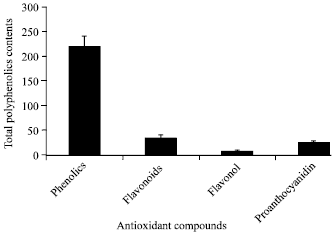 | |
| Fig. 1: | Total polyphenol contents of aqueous leaves extract of L. leonurus |
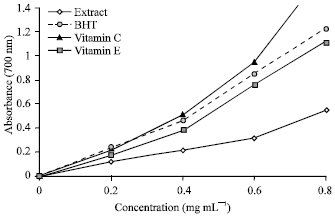 | |
| Fig. 2: | The reductive abilities of equeous leaves extract of L. Leonurus, BHT, Vitamin C and Vitamin E |
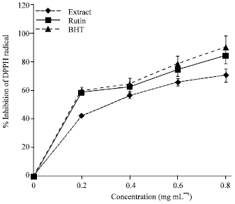 | |
| Fig. 3: | The DPPH radical scavenging activity of L. leonurus extract and the standard rutin and BHT. The data represent the percentage DPPH inhibition. Each value represents mean±S.D (n = 3) |
This activity was increased with increasing concentrations of both extract and reference drugs.
Leonotis leonorus extract was an effective scavenger of ABTS radicals as shown in Fig. 4. The ABTS radical scavenging activity of the extract was comparable to that of BHT and rutin in a concentration dependent manner. At 0.8 mg mL-1 plant extract, BHT and rutin scavenge ABTS radical by 78.02, 89.86 and 92.1%, respectively.
The result of nitric oxide antiradical activity of the extract was increased with increasing concentration (Fig. 5). BHT and rutin were used as reference compounds. The extract showed high concentration-dependent inhibition of nitric oxide. At 0.8 mg mL-1, the percentage inhibitions of nitric oxide were 72.33, 75.86 and 82.73% for rutin, extract and BHT, respectively.
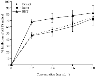 | |
| Fig. 4: | The ABTS radical scavenging activity of L. leonurus extract and the standard rutin and BHT. The data represent the percentage ABTS inhibition. Each value represents mean±S.D (n = 3) |
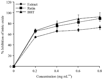 | |
| Fig. 5: | The nitric oxide radical scavenging activity of L. leonurus extract and the standard rutin and BHT. The data represent the percentage nitric oxide inhibition. Each value represents mean±S.D (n = 3) |
The activity of BHT was more pronounced than the plant extract but stronger than rutin. The results of SOD, CAT, GSH and lipid peroxidation in the liver of control, hepatotoxic and experimental animals are shown in Table 1. SOD, CAT and GSH activities were significantly lowered in CCl4 induced rats (Group 2) than the control rats (Group1) but were significantly increased when compared with hepatotoxic treated groups with plant extract at the doses of 125, 250 and 500 mg kg-1 body weight. The lipid peroxidation levels indicated by TBARS was significantly higher in the animals induced with CCl4 than the normal rats.
| Table 1: | Antioxidant property of aqueous leaves extract of L. leonurus on liver superoxide dismutase, catalase, reduced glutathione and lipid peroxidation |
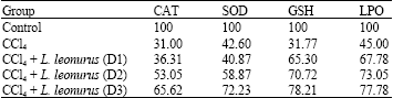 | |
| D1 = 125 mg kg-1; D2 = 250 mg kg-1 and D3 = 500 mg kg-1 body weight. Results are expressed as percentage inhibition of the control. Values are means of six replicates | |
Following plant extract administration at the doses investigated, the lipid peroxidation level was considerably lowered than the hepatotoxic group.
DISCUSSION
Leonotis leonurus is commonly used as a traditional herbal medicine for the treatment of various human diseases in South Africa. The decoction of the leaves of this plant is frequently used while the extract is taken orally for longer period of time depending on the severity of the ailments (Oyedemi et al., 2009). Meanwhile, the mechanism of actions is yet to be known. It has been reported that antioxidant activities depicted by medicinal plants are derived from phenolics compounds (Bravo, 1998; Cai et al., 2004). The results obtained from this study confirmed that aqueous leaves extract of L. leonurus contains high amount of phenolics and flavonoids compounds than some other plants reported in the literature (Sofidiya et al., 2008; Adedapo et al., 2009; Hazra et al., 2009). The significance of these compounds in plant extract has been recognized to exhibit various biological activities such as anti-inflammatory, anticancerogenic, antiviral, vasodilatory and antimicrobial effect. The presence of these compounds could be responsible for the antiradicals activities observed in this study which may due to their ability to adsorb, neutralize and quench free radicals (Duh et al., 1999).
The antioxidant activity of the plant extract was investigated by measuring the transformation of Fe3+ to Fe2+ (Meir et al., 1995). The reductive capability was monitored by the formation of Perl’s blue at 700 nm. The data obtained from this study showed that the extract and the standard drugs increased their reducing ability with increasing concentration. The antioxidants activities of BHT, Vitamin C and Vitamin E were significantly higher than the plant extract. This result corroborates with that of Afolayan et al. (2008) on methanol extract of Malva parviflora. It could be inferred from this study that the reference drugs and L. leonurus extract are electron donors and thus could possibly reduce the oxidized intermediates of lipid peroxidation (Ordonez et al., 2006). This observation might be attributed to the presence of reductones which are known terminators of free radical chain reactors by donating hydrogen atom (Gordon, 1990).
The scavenging properties of plant extract against DPPH a stable free radical is based on its ability to decolorize the visible deep purple color which was measured from the changes in absorbance at wavelength of 517 nm. Both extract and reference drugs demonstrated strong DPPH scavenging properties in a concentration related manner (Fig. 3). Although, the DPPH radical scavenging of the extract was lower than those of standard drugs but showed proton donating ability thus could serve as free radical inhibitors. This observation is in agreement with our previous finding on aqueous stem bark extract of Strychnos henningsii (Oyedemi et al., 2010a).
The ability of plant extract to scavenge DPPH could also reflect its ability to inhibit the formation of ABTS+. However, the results obtained in this study was contrary to the report of Wang et al. (1998) who reported that compounds which exhibit ABTS+ scavenging activity may not possess DPPH scavenging activity. Factor such as stereoselectivity of the radicals or the solubility of the extract in different testing system have been implicated to affect antiradicals activity of the extract. This implies that the plant extract may be useful for treating radical-related pathological damage.
ABTS radical is a blue chromophore produced by the reaction of ABTS and potassium persulphate. The addition of plant extract to this pre-formed radical cation reduced it to ABTS with increasing concentrations. The result obtained from this assay revealed the high level of proton radical scavenging ability of the plant extract (Fig. 4). The ABTS scavenging activity of the extract at the highest concentration (78.02%) was significantly different with that of BHT (92.10%) and rutin (89.86%) used as reference drugs in this study. The presence of polyphenolics compounds could be responsible for this observation which has been reported of oxidizing the proton radical generated within the system (Mathew and Abraham, 2006). This result showed similar trend with that of Adedapo et al. (2008) on methanol stem extracts of Acokanthera oppositifolia and Adenia gummifera.
Nitric oxide is well known to play a crucial role in the pathogenesis of various diseases caused by inflammation especially when combined with superoxide radical to form peroxynitrite anion (Moncada et al., 1991). It is generated from sodium nitroprusside in aqueous solution and reacts with oxygen to form nitrite. The scavenging activity of the extract against nitric oxide was detected by its ability to inhibit the formation of nitrite through direct competition with oxygen and oxides of nitrogen in the reacting mixture (Marcocci et al., 1994). The significant decrease in the concentration of nitric oxide radical was comparable with the standard drug which is due to the scavenging ability of the plant extract. This observation corroborated with the report of Londonkar and Kamble (2009) on Pandanus odoratissimus but contradicted with our previous work on Strychnos henningsii. The percentage inhibition displayed by the extract showed a potent scavenger of nitric oxide.
The administration of carbon tetrachloride (CCl4) to the experimental animals causes changes in the liver marker and antioxidant enzymes (Singh, 1980). These hepatotoxic effects are largely based on membrane lipid peroxidation and induction of trichloromethyl radical (Johntson and Kroening, 1998). In this study, the level of lipid peroxidation was significantly increased in carbon tetrachloride treated rats. This observation suggests the inability of antioxidant defense mechanism to prevent formation of excessive free radicals. The treatment with aqueous extract of L. leonotis was significantly reduces the level of lipid peroxides generated in all the doses investigated than the untreated hepatotoxic rats.
Superoxide dismutase has been reported as one of the most important antioxidant defense enzymes that scavenge superoxide anion by converting to hydrogen peroxide thus diminish the toxic effect caused by this radical (Curtis et al., 1972). The decreased percentage inhibition of superoxide dismutase as shown in carbon tetrachloride treated rats might be due to the damage caused by this chemical. Meanwhile, following plant extract administration at the doses investigated the percentage inhibition of the enzyme was remarkably increased in a dose dependent manner. This observation suggests the protective mechanism of L. leonurus extract in scavenging superoxide anion which may be attributed to the high contents of phenolics and flavonoids compounds (Robak and Glyglewski, 1988).
Catalase is an antioxidant enzyme widely distributed in all animal tissues. The enzyme is known to protect the system from highly reactive hydroxyl radicals through hydrogen peroxide decomposition (Chance and Greenstein, 1952). Depletion of this enzyme may enhance the cellular damage caused by assimilation of superoxide and hydrogen peroxide. However, oral administration of the extract to the hepatotoxic rats resulted to significant improvement on hepatic catalase levels in a dose dependent manner. It can be conclude from the present study that L. leonurus extract possess hepatoprotective ability against liver damage.
GSH is a non enzymatic biological antioxidant that plays a vital role in coordinating the antioxidant defensive system of the body. Deficiency of this enzyme in the body system has been reported to induce oxidative stress that could lead to tissue disorder and injury (Mansour et al., 2001). The decreased level of GSH observed in this study after administration of CCl4 to the rats is associated with an increased in the level of lipid peroxidation (Table 1). However, it has been demonstrated that the level of GSH could be enhanced in rats pretreated with Nigella sativa (Mansour et al., 2001). Correspondingly, the percentage inhibition of this enzyme was reduced in rats treated with CCl4 as a result of low enzyme activities. Meanwhile, it was significantly increased after plant extract treatment in a dose dependent manner. This consideration is in accordance with the level of lipid peroxidation in extract treated groups as observed in this study. Furthermore, this observation corroborates with the report of Bhandarkar and Khan (2004) on antihepatotoxic effect of Nymphaea stellata. A similar effect was also observed in our previous studies on the in vivo antioxidant properties of Strychnos henningsii in Wistar rats induced with carbon tetrachloride (Oyedemi et al., 2010a). Therefore, it clearly obvious that aqueous leaves extract of L. leonurus have protective role against oxidative damage in the hepatic tissue. The liver cells innate ability was awaken and maintain defense against oxidants by secreting more antioxidants.
In conclusion, there is enough evidence to support that the aqueous leaves extract of L. leonurus exhibited strong antioxidant activity and free radical scavenging activity both in vitro and in vivo which may due to the high content of phenolics, flavonoids and proanthocyanidins antioxidant compounds. The result obtained from this study has provided scientific credence to the ethnotherapeutic usage of this plant traditionally.
ACKNOWLEDGMENT
The authors are grateful to the Govan Mbeki Research and Development Center, University of Fort Hare, Alice, South Africa for their financial support.
REFERENCES
- Adedapo, A.A., F.O. Jimoh, A.J. Afolayan and P.J. Masika, 2009. Antioxidant properties of the methanol extracts of the leaves and stems of Celtis africana. Rec. Nat. Prod., 3: 23-31.
Direct Link - Beauchamp, C. and I. Fridovich, 1971. Superoxide dismutase: Improved assays and an assay applicable to acrylamide gels. Anal. Biochem., 44: 276-287.
CrossRefPubMedDirect Link - Bhandarkar, M.R. and A. Khan, 2004. Antihepatotoxic effect of Nymphaea stellata willd., against carbon tetrachloride-induced hepatic damage in albino rats. J. Ethnopharmacol., 91: 61-64.
CrossRefDirect Link - Bienvenu, E., G.J. Amabeoku, P.K. Eagles, G. Scott and E.P. Springfield, 2002. Anticonvulsant activity of aqueous extract of Leonotis leonurus. Phytomedicine, 9: 217-223.
Direct Link - Bravo, L., 1998. Polyphenols: Chemistry, dietary sources, metabolism and nutritional significance. Nutr. Rev., 56: 317-333.
CrossRefPubMedDirect Link - Cai, Y., Q. Luo, M. Sun and H. Corke, 2004. Antioxidant activity and phenolic compounds of 112 traditional Chinese medicinal plants associated with anticancer. Life Sci., 74: 2157-2184.
CrossRefPubMedDirect Link - Chance, B., D.S. Greenstein and F.J.W. Roughton, 1952. The mechanism of catalase action. I. Steady-state analysis. Arch. Biochem. Biophys., 37: 301-321.
CrossRefPubMedDirect Link - Curtis, S.J., M. Moritz and P.J. Snodgrass, 1972. Serum enzymes derived from liver cell fractions I. The response to carbon tetrachloride intoxication in rats. Gastroenterology, 62: 84-92.
PubMedDirect Link - Duh, P.D., Y.Y. Tu and G.C. Yen, 1999. Antioxidant activity of water extract of Harng Jyur (Chrysanthemum morifolium Ramat). LWT-Food Sci. Technol., 32: 269-277.
CrossRefDirect Link - Ellman, G.L., 1959. Tissue sulfhydryl groups. Arch. Biochem. Biophys., 82: 70-77.
CrossRefPubMedDirect Link - Farber, J.L., 1994. Mechanisms of cell injury by activated oxygen species. Environ. Health Perspect., 102: 17-24.
PubMed - Gerber, M., M.C.B. Rault, S. Herberg, E. Riboli, A. Scalbert and M.H. Siess, 2002. Food and cancer state of the art about the protective effect of fruits and vegetables. Bull. Cancer, 89: 293-312.
PubMed - Hazra, B., R. Sarkar, S. Mandal, S. Biswas and N. Mandal, 2009. Studies on antioxidant and antiradical activities of Dolichos biflorus seed extract. Afr. J. Biotechnol., 8: 3927-3933.
Direct Link - Jager, A.K., A. Hutchings and J. Staden, 1996. Screening of Zulu medicinal plants for prostaglandin-synthesis inhibitors. J. Ethnopharmacol., 52: 95-100.
PubMedDirect Link - Johnston, D.E. and C. Kroening, 1998. Mechanism of early carbon tetrachloride toxicity in cultured rat hepatocytes. Pharmacol. Toxicol., 83: 231-239.
PubMed - Kumaran, A. and R.J. Karunakaran, 2007. In vitro antioxidant activities of methanol extracts of five Phyllanthus species from India. LWT-Food Sci. Technol., 40: 344-352.
CrossRefDirect Link - Liyana-Pathirana, C.M. and F. Shahidi, 2005. Antioxidant activity of commercial soft and hard wheat (Triticum aestivum L.) as affected by gastric pH conditions. J. Agric. Food Chem., 53: 2433-2440.
CrossRefDirect Link - Mansour, M.A., O.T. Ginawi, T. El-Hudiyah, A.S. El-Khatib, O.A. Al-Shabanah and H.A. Al-Sawaf, 2001. Effects of volatile oil constituents of Nigella sativa on carbon tetrachloride induced hepatotoxicity in mice. Res. Commun. Mol. Pathol. Pharmacol., 110: 239-251.
Direct Link - Marcocci, L., L. Packer, M.T. Droy-Lefaix, A. Sekaki and M. Gardes-Albert, 1994. Antioxidant action of Ginkgo biloba extract EGb 761. Methods Enzymol., 234: 462-475.
CrossRefPubMedDirect Link - Mathew, S. and T.E. Abraham, 2006. In vitro antioxidant activity and scavenging effects of Cinnamomum verum leaf extract assayed by different methodologies. Food. Chem. Toxicol., 44: 198-206.
CrossRefDirect Link - Maphosa, V., P.J. Masika and A.A. Adedapo, 2008. Safety evaluation of the aqueous extract of Leonotis leonurus shoots in rats. Hum. Exp. Toxicol., 27: 837-843.
CrossRefPubMedDirect Link - Meir, S., J. Kanner, B. Akiri and S. Philosoph-Hadas, 1995. Determination and involvement of aqueous reducing compounds in oxidative defense systems of various senescing leaves. J. Agric. Food Chem., 43: 1813-1819.
CrossRefDirect Link - Miller, A.L., 1996. Antioxidant flavonoids: Structure, function and clinical usage. Altern. Med. Rev., 1: 103-111.
Direct Link - Moncada, S., R.M. Palmer, E.A. Higgs, 1991. Nitric oxide: Pathology, pathophysiology and pharmacology. Pharmacol. Rev., 43: 109-142.
PubMedDirect Link - Niehaus, Jr. W.G. and B. Samuelsson, 1968. Formation of malonaldehyde from phospholipid arachidonate during microsomal lipid peroxidation. Eur. J. Biochem., 6: 126-130.
CrossRefPubMedDirect Link - Noumi, E., F. Houngue and D. Lontsi, 1999. Traditional medicines in primary health care: plants used for the treatment of hypertension in Bafia Cameroon. Fitoterapia, 70: 134-139.
CrossRef - Ososki, A.L., P. Lohr, M. Reiff, M.J. Balick, J. Kronenberg, A. Fugh-Berman and B. O`Connor, 2002. Ethnobotanical literature survey of medicinal plants in the Dominican Republic used for women`s health conditions. J. Ethnopharmacol., 79: 285-298.
PubMed - Oyedemi, S.O., G. Bradley and A.J. Afolayan, 2010. In-vitro and -vivo antioxidant activities of aqueous extract of Strychnos henningsii Gilg. Afr. J. Pharm. Pharmacol., 4: 70-78.
Direct Link - Oyedemi, S.O., G. Bradley and A.J. Afolayan, 2010. Effect of aqueous extract of Leonotis leonurus (L.) R. Br. leaves in male Wistar rats. Hum. Exp. Toxicol., 29: 377-384.
CrossRefDirect Link - Re, R., N. Pellegrini, A. Proteggente, A. Pannala, M. Yang and C. Rice-Evans, 1999. Antioxidant activity applying an improved ABTS radical cation decolorization assay. Free Radical Biol. Med., 26: 1231-1237.
CrossRefDirect Link - Robak, J. and E. Marcinkiewiez, 1995. Scavenging of reactive oxygen species as the mechanism of drug action. Pol. J. Pharmacol., 47: 89-98.
Direct Link - Sinha, A.K., 1972. Colorimetric assay of catalase. Anal. Biochem., 47: 389-394.
CrossRefPubMedDirect Link - Sofidiya, M.O., F.O. Jimoh, A.A. Aliero, A.J. Afolayan, O.A. Odukoya and O.B. Familoni, 2008. Antioxidant and antibacterial properties of Lecaniodiscus cupanioides. Res. J. Microbiol., 3: 91-98.
CrossRefDirect Link - Spanos, G.A. and R.E. Wrolstad, 1990. Influence of processing and storage on the phenolic composition of Thompson Seedless grape juice. J. Agric. Food Chem., 38: 1565-1571.
CrossRefDirect Link - Sun, J.S., Y.W. Tsuang, J.J. Chen, W.C. Huang, Y.S. Hang and F.J. Lu, 1998. An ultra-weakchemiluminescence study on oxidative stress in rabbits following acute thermal injury. Burns, 24: 225-231.
PubMed - Wang, M., J. Li, M. Rangarajan, Y. Shao, E.J. LaVoie, T.C. Huang and C.T. Ho, 1998. Antioxidative phenolic compounds from sage (Salvia officinalis). J. Agric. Food Chem., 46: 4869-4873.
CrossRefDirect Link - Yen, G.C. and H.Y. Chen, 1995. Antioxidant activity of various tea extracts in relation to their antimutagenicity. J. Agric. Food Chem., 43: 27-32.
CrossRefDirect Link - Zhishen, J., T. Mengcheng and W. Jianming, 1999. The determination of flavonoid contents in mulberry and their scavenging effects on superoxide radicals. Food Chem., 64: 555-559.
CrossRefDirect Link - Oyedemi, S.O., G. Bradley and A.J. Afolayan, 2009. Ethnobotanical survey of medicinal plants used for the management of diabetes mellitus in the Nkonkobe municipality of South Africa. J. Med. Plants Res., 3: 1040-1044.
Direct Link - Londonkar, R. and A. Kamble, 2009. Evaluation of free radical scavenging activity of Pandanus odoratissimus. Int. J. Pharmacol., 5: 377-380.
CrossRefDirect Link - Afolayan, A.J., O.M. Aboyade and M.O. Sofidiya, 2008. Total phenolic content and free radical scavenging activity of Malva parviflora L. (Malvaceae). J. Biol. Sci., 8: 945-949.
CrossRefDirect Link - Ordonez, A.A.L., J.D. Gomez, M.A. Vattuone and M.I. Lsla, 2006. Antioxidant activities of Sechium edule (Jacq.) Swartz extracts. Food Chem., 97: 452-458.
CrossRefDirect Link - Adedapo, A.A., F.O. Jimoh, A.J. Afolayan and P.J. Masika, 2008. Antioxidant activities and phenolic contents of the methanol extracts of the stems of Acokanthera oppositifolia and Adenia gummifera. BMC Complement. Altern. Med., Vol. 8.
CrossRefDirect Link - Robak, J. and R.J. Gryglewski, 1988. Flavonoids are scavengers of superoxide anions. Biochem. Pharmacol., 37: 837-841.
CrossRefDirect Link