
E. Woode
Department of Pharmacology, Faculty of Pharmacy and Pharmaceutical Sciences, College of Health Sciences, Kwame Nkrumah University of Science and Technology, Kumasi, Ghana
E. Boakye-Gyasi
Department of Pharmacology, Faculty of Pharmacy and Pharmaceutical Sciences, College of Health Sciences, Kwame Nkrumah University of Science and Technology, Kumasi, Ghana
C.A. Danquah
Department of Pharmacology, Faculty of Pharmacy and Pharmaceutical Sciences, College of Health Sciences, Kwame Nkrumah University of Science and Technology, Kumasi, Ghana
C. Ansah
Department of Pharmacology, Faculty of Pharmacy and Pharmaceutical Sciences, College of Health Sciences, Kwame Nkrumah University of Science and Technology, Kumasi, Ghana
M. Duwiejua
Department of Pharmacology, Faculty of Pharmacy and Pharmaceutical Sciences, College of Health Sciences, Kwame Nkrumah University of Science and Technology, Kumasi, Ghana
International Journal of Pharmacology
Year: 2009 | Volume: 5 | Issue: 3 | Page No.: 181-190


ABSTRACT
The anti-arthritic effect of an ethanolic leaf extract of Palisota hirsuta, a plant used locally in Ghana for various painful inflammatory conditions was assessed, using the Freund’s adjuvant induced-arthritis model in rats. Palisota hirsuta Extract (PHE) as well as dexamethasone and methotrexate, used as positive controls, showed significant dose-dependent anti-arthritic properties prophylactically, curatively and also in combination therapy. PHE (30-300 mg k-1) significantly reduced the arthritic edema in the ipsilateral paw with the highest dose used giving a maximum inhibition of 13.02±8.77%. PHE 300 mg k-1 also significantly prevented the spread of the edema from the ipsilateral to the contralateral paw indicating inhibition of systemic spread. Dexamethasone (0.3-3 mg k-1) and methotrexate (0.1-1.0 mg k-1) significantly and in a dose dependent manner also inhibited polyarthritis edema as well as completely preventing the spread of the arthritis from the ipsilateral to the contralateral paws of the treated animals. PHE in combination with methotrexate did not show any significant effect, however, there was a significant inhibition of arthritis in both the acute and the polyarthritis phases when PHE was combined with dexamethasone. Dexamethasone in combination with methotrexate gave the greatest inhibition of both phases with an extreme level of significance as expected. Overall, the present results demonstrate that PHE has anti-arthritic effect which could be similar to that exhibited by methotrexate.
PDF Abstract XML References Citation
How to cite this article
E. Woode, E. Boakye-Gyasi, C.A. Danquah, C. Ansah and M. Duwiejua, 2009. Anti-Arthritic Effects of Palisota hirsuta K. Schum. Leaf Extract in Freund’s Adjuvant-Induced Arthritis in Rats. International Journal of Pharmacology, 5: 181-190.
DOI: 10.3923/ijp.2009.181.190
URL: https://scialert.net/abstract/?doi=ijp.2009.181.190
DOI: 10.3923/ijp.2009.181.190
URL: https://scialert.net/abstract/?doi=ijp.2009.181.190
INTRODUCTION
Adjuvant-Induced Arthritis (AIA) in rats, a chronic inflammatory disease characterized by infiltration of the synovial membrane and associated with destruction of the joints, resembles rheumatoid arthritis in humans in terms of immunological and biochemical features (Behar and Porcelli, 1995; Kumar et al., 2002). The inflammatory response has received a great deal of interest in the field of medical research because inflammation and pain underlie almost every disease process (Kapoor et al., 2005).
Palisota hirsuta K. Schum. (Family: Commelinaceae), known locally in Ghana as somenini or mpentemi (Twi), sombenyin (Fante) and sumbe (Ewe) is a robust herb found in forest regrowths and is about 2-4 m high. In Ghana, this plant and several others are used either alone or as combination therapy with orthodox medicine in the treatment of various painful inflammatory conditions (Burkill, 1985; Dokosi, 1998).
The whole plant and various parts are used extensively in West African traditional medicine for various painful inflammatory conditions. In Ghana, the whole plant is used for stomach pains and the sap from the roasted leaves is instilled in the ear for earache whilst the heated leaves are applied over the lumbar region for kidney pains (Burkill, 1985). Also, a leaf infusion or poultice is taken orally or applied locally for piles (Dokosi, 1998; Burkill, 1985). The dried leaves are smoked for toothache (Burkill, 1985; Dokosi, 1998). The Igbos of Obompa in Nigeria prepares an ointment made from this plant for gunshot wounds and swelling (Burkill, 1985).
Apart from the anti-viral study done by Anani et al. (2000) and Hudson et al. (2000) not much has been reported on this plant. The anti-inflammatory and antipyretic effect of an ethanolic root extract of this plant has also been recently reported by Boakye-Gyasi et al. (2008). The present study reports on the anti-arthritic effects of the ethanolic extract of the leaves in Freund’s adjuvant-induced arthritis in rats using a curative protocol (administered after onset of arthritis), prophylactic protocol (administered before onset of arthritis) and combination therapy. This would help provide scientific basis for the use of this plant locally for treating arthritic conditions as well as provide an alternative for the drugs currently being used for controlling arthritis. In this study dexamethasone a steroidal anti-inflammatory agent and methotrexate, a Disease Modifying Anti-Rheumatoid Drug (DMARD) were used as reference anti-arthritic drugs.
MATERIALS AND METHODS
Plant material: Leaves of the plant Palisota hirsuta were collected from the Botanic Gardens of Kwame Nkrumah University of Science and Technology, Kumasi, Ghana, between January and February, 2007. The leaves were authenticated by Mr. Amissah, the curator of the garden and a voucher specimen (No. FP 10081) has been kept in the Faculty of Pharmacy Herbarium, KNUST, Kumasi.
Preparation of extract: Leaves were air-dried indoor for a week and pulverized with a hammer-mill. This was extracted by cold maceration using 70% (v/v) ethanol over a period of 72 h. The resultant extract was concentrated under low temperature (60°C) and pressure to a syrupy mass in a rotary evaporator which was then dried to a dark brown semi-solid mass using water bath and kept in a desiccator till it was ready to be used. Final yield was 10.5%. This is subsequently referred to as PHE or extract.
Drugs: Dexamethasone sodium phosphate was purchased from Pharm-Inter, Brussels, Belgium and methotrexate sodium from Dabur Pharma, India.
Animals: Sprague-Dawley rats of both sexes (150-200 g) were purchased from Noguchi Memorial Institute for Medical Research, University of Ghana, Legon, Ghana and housed in the animal facility of the Department of Pharmacology, Kwame Nkrumah University of Science and Technology (KNUST). The animals were housed in groups of six in stainless steel cages (34x47x18 cm3) with soft wood shavings as bedding, fed with normal commercial pellet diet (GAFCO, Tema) and given water ad libitum and maintained under laboratory conditions (temperature 24-28°C, relative humidity 60-70% and 12 h light-dark cycle). All procedures and techniques used in these studies were in accordance with the National Institute of Health Guidelines for the Care and Use of Laboratory Animals (NIH, Department of Health and Human Services Publication No. 85 -23, revised 1985). All protocols used were approved by the Departmental Ethics Committee.
Phytochemistry: The presence of saponins, alkaloids, triterpenes, flavonoids, glycosides, reducing sugars and tannins were tested by simple qualitative and quantitative methods of Trease and Evans (1989) and Sofowora (1993).
Induction of arthritis: Adjuvant arthritis was induced as previously described by Pearson (1956) as modified by Woode et al. (2008). Briefly, animals were injected intraplantar with 0.1 mL Complete Freund’s Adjuvant (CFA) into the right hind paw of each rat. CFA was prepared by triturating heat-killed Mycobacterium tuberculosis [strains C, DT and PN (mixed) obtained from the Ministry of Agriculture, Fisheries and Food, UK] in paraffin oil to make a 3 mg mL-1 suspension. Animals were randomly assigned to groups of six animals. Arthritic control group received only intraplantar injection of CFA, whilst non-arthritic control/IFA group received only intraplantar injection of 0.1 mL Incomplete Freund’s Adjuvant (IFA) (sterile paraffin oil). P. hirsuta extract (30, 100 and 300 mg k-1), dexamethasone (0.3, 1.0 and 3 mg k-1) and methotrexate (0.1, 0.3 and 1 mg k-1) were administered to rats in the various groups, respectively.
Effect of drugs on adjuvant-induced arthritis: Three sets of experiments were performed.
In the first set of experiments, effect of the drugs on established arthritis (curative protocol), was studied. Arthritis was induced by intraplantar injection of 0.1 mL CFA. Drugs were administered on day 9 with the onset of arthritis.
The animals were randomly grouped as follows:
| Group I | : | Arthritic control/CFA (intraplantar injection of 0.1 mL CFA) |
| Group II | : | Non-arthritic control/IFA (intraplantar injection of 0.1 mL of IFA) |
| Group III-V | : | Treated with dexamethasone 0.3 to 3 mg k-1 i.p., 9 days after intraplantar injection of CFA |
| Group VI-VIII | : | Treated with methotrexate 0.1 to 1 mg k-1 i.p., 9 days after intraplantar injection of CFA |
| Group IX-XI | : | Treated with extract 30-300 mg k-1 p.o., 9 days after intraplantar injection of CFA |
In the second set of experiments (prophylactic protocol) the effect of drugs were investigated when given before induction of arthritis. Drugs were administered on day 0 and CFA was injected intraplantar 24 h later. The animals were grouped as follows for this study:
| Group I | : | Arthritic control/CFA (intraplantar injection of 0.1 mL CFA) |
| Group II | : | Non-arthritic control/IFA (intraplantar injection of 0.1 mL of IFA) |
| Group III | : | Pre-treated with dexamethasone 1.0 mg k-1 i.p., from day 0 |
| Group IV | : | Pre-treated with methotrexate 0.3 mg k-1 i.p., from day 0 |
| Group V | : | Pre-treated with extract 100 mg k-1 p.o., from day 0 |
For the last set of experiments (combination therapy), the extract and standard drugs were combined and the rats were grouped as follows:
| Group VI | : | Pre-treated with dexamethasone 1 mg k-1 + extract 100 mg k-1 |
| Group VII | : | Pre-treated with methotrexate 0.3 mg k-1+ extract 100 mg k-1 |
| Group VIII | : | Pre-treated with dexamethasone 1 mg k-1+ methotrexate 0.3 mg k-1 |
In all experiments, foot volume was measured by water displacement plethysmography (Fereidoni et al., 2000) for both the ipsilateral (injected paw) and the contralateral paw (non-injected paw) before intraplantar injection of CFA (day 0) and every other day (day 2, 4, 6, 8,…., 28). The edema component of inflammation was quantified by measuring the difference in foot volume between day 0 and the various time points. The extract was administered every day whilst dexamethasone and methotrexate were administered every other day and every 4 days, respectively. Doses of drugs used for this study were selected based on earlier study data and preliminary studies done in the laboratory (Woode et al., 2008).
The extract was suspended in 2% tragacanth mucilage, whilst the reference drugs (methotrexate and dexamethasone) were dissolved in normal saline. Test drugs were prepared such that doses were administered in volumes not exceeding 10 mL k-1. All drugs were freshly prepared. Inflamed control animals were given 2% tragacanth mucilage (100 mL k-1, p.o.).
Raw scores for ipsilateral and contralateral paw volumes were individually normalized as percentage of change from their values at day 0, then averaged for each treatment group.
Arthritis score: The arthritis score was evaluated blindly by the same person in all the rats on day 29 using photographs and radiographs of the affected hind limbs. Photographs were taken with a digital camera (Sony digital camera DCR-DVD 705E). Whilst radiographs were taken with a UMB type-2 X-ray unit (Softex Ltd., Tokyo, Japan) and industrial X-ray film (Fuji Photo Film, Tokyo, Japan) after animals were anaesthetized with chloroform. The X-ray apparatus was operated at 30 kV peak and 10 sec exposure with a 45 cm tube-to-film distance for lateral projections.
Using the photographs, the severity of arthritis of each paw was scored as described by Kinne et al. (1995) according to the extent of erythema and edema of the periarticular tissues, using a scale of 0-4. The arthritis score of each rat on day 0 was determined to be 0. The radiographs from the X-rays were also used to score the severity of bone and joint destruction blindly by the same person for each hind limb, according to the extent of osteoporosis, osteophytes, joint spaces and joint structure, as described by Pohlers et al. (2007). The severity of bone destruction of each paw was scored using a scale of 0-4. The radiological score for normal control rats was determined to be 0.
The hind paw volume and arthritis score were used as the measurement parameters of inflammation and arthritis.
Data analysis: In all experiments, a sample size of five (n = 5) was utilized. Data was presented as the effect of drugs on the time course and the total edema response of adjuvant-induced arthritis for the 28 days period.
The time-course curves for paw volume were subjected to two-way (treatmentxtime) repeated measures analysis of variance with Bonferroni’s post hoc t-test.
Total foot volume for each treatment was calculated in arbitrary unit as the Area Under the Curve (AUC) and to determine the percentage inhibition for each treatment, the following equation was used.
Differences in AUCs were analyzed by ANOVA followed by Student-Newman-Keuls’ post hoc test. Doses for 50% of the maximal effect (ED50) for each drug were determined by using an iterative computer least squares method, with the following nonlinear regression (three-parameter logistic) equation:
where, X is the logarithm of dose and Y is the response. Y starts at a (the bottom) and goes to b (the top) with a sigmoid shape.
The fitted midpoints (ED50s) of the curves were compared statistically using F-test (Miller, 2003; Motulsky and Christopoulos, 2003). GraphPad Prism for Windows version 4.03 (GraphPad Software, San Diego, CA, USA) was used for all statistical analysis and ED50 determinations. p<0.05 was considered statistically significant.
RESULTS
Phytochemical analysis: The phytochemical analysis of P. hirsuta showed it contains alkaloids, flavonoids, tannins and terpenoids with tannins and flavonoids being the most dominant chemical constituent.
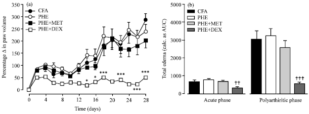
Effect of drugs on adjuvant-induced arthritis: The phases of the adjuvant arthritis investigated in the present study were the acute and polyarthritic/chronic phases corresponding to day 0-10 and days 10-28 post adjuvant inoculation, respectively. Two-way ANOVA (treatment x time) revealed a significant (F3,144 = 3.65, p = 0.0355) effect of drug treatment. Total edema produced by each treatment is expressed in arbitrary units as AUC of the time-course curves.
All arthritic control animals showed acute inflammatory edema at the ipsilateral (injected paw) around days 4-6 followed by subsequent chronic polyarthritic phase which begins around day 10-12 as earlier described by Weichman (1989). The progress of inflammatory edema in the contralateral (non-injected) paw was evident on day 12 indicative of systemic inflammation. Throughout the 28 day experiment, there was no significant change in the paw volume of the non-inflamed control groups injected IFA.
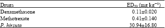
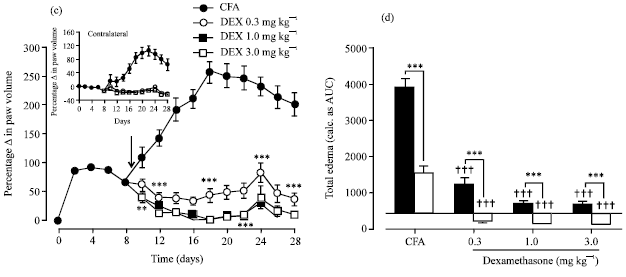
PHE (30-300 mg k-1) modified the time course curve significantly (F3,16 = 3.59, p = 0.0149) and reduced the acute edema in the ipsilateral paw with the highest dose used (300 mg k-1) significantly (F3,16 = 4.13, p = 0.0240) reducing the edema with a percentage inhibition of 13.02±8.77% (Fig. 1a). PHE 300 mg k-1 also significantly (F3,16 = 3.84, p = 0.0302) prevented the spread of the edema form the ipsilateral to the contralateral paw indicating inhibition of systemic spread (Fig. 1b). Dexamethasone (0.3-3 mg k-1) a steroidal anti-inflammatory agent profoundly and significantly (F3,16 = 96.91, p<0.0001) in a dose dependent manner inhibited polyarthritis edema with a maximal inhibition of 91.59±2.06% (Fig. 1c). Dexamethasone also completely prevented the spread of the arthritis (F3,16 = 36.74, p<0.0001) from the ipsilateral to the contralateral paws of the treated animals (Fig. 1d). Methotrexate (0.1-1.0 mg k-1) also dose dependently reduced the edema in the ipsilateral paw but this effect was not so significant (F3,16 = 13.76, p = 0.0001). However, methotrexate completely prevented the spread of the arthritis with a high level of significance (F3,16 = 21.57, p<0.0001) as shown in Fig. 1f.
Figure 2 also shows the dose-response curves of the effects of the drugs under test. The steroidal anti-inflammatory drug, dexamethasone showed the greatest potency (Table 1). PHE as well after oral administration exhibited significant potency when it was administered 10 days after the onset of arthritis.
| Table 1: | ED50 values for Adjuvant-induced arthritis |
 | |
Similarly, the DMARD, methotrexate also exhibited a significantly greater potency (Table 1).
PHE was found to be approximately 75.46x less potent than methotrexate (F1,28 = 61.55, p<0.0001) and 281.27x less potent than dexamethasone (F1,28 = 5.28, p = 0.0299). Dexamethasone was also found to be 3.73x more potent than methotrexate (F1,28 = 9.73, p = 0.0042).
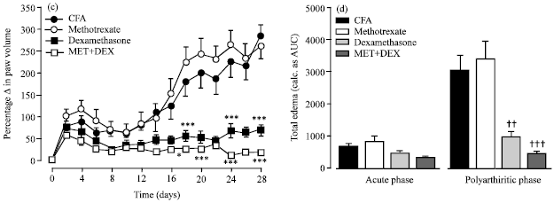
Figure 4 shows the effect of PHE (100 mg k-1), dexamethasone (1 mg k-1) and methotrexate (0.3 mg k-1) alone (prophylactic protocol) and in various combinations with each other. Two-way ANOVA (treatmentxtime) revealed a significant (F3,224 = 13.06, p = 0.0001) effect of drug treatment. Total edema produced by each treatment is expressed in arbitrary units as AUC of the time-course curves.
Effect of PHE when given alone and when combined with methotrexate did not show any significant effect in both the acute (p>0.05) and polyarthritis (p>0.05) phases. PHE in combination with dexamethasone however showed a significant inhibition of arthritis in both the acute (F3,16 = 8.20, p = 0.0016) and the polyarthritis (F3,16 = 10.28, p = 0.0005) phases. Dexamethasone in combination with methotrexate gave the greatest inhibition of both phases with an extreme level of significance (phase 1 F3,16 = 4.27, p = 0.0214; phase 2 F3,16 = 14.38, p<0.0001).
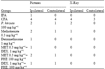
From the arthritic score of the photographs (Fig. 3), the CFA group showed widespread erythema and swelling in both the ipsilateral and contralateral paws. PHE and methotrexate treated animals however exhibited slight erythema and swelling whilst the dexamethasone treated and the IFA groups showed no sign of erythema or swelling.
Measurement of paw or joint swelling only gives an indication of edematous changes in these regions; however, the most obvious damage takes place in the tibiotarsals joint itself. Hence, radiographs of the hind paws for each group were scored for the extent of bone damage at the joints as shown in Table 2 and 3. CFA/Arthritic control group gave the highest score (Fig. 3) demonstrating severe bone enlargement with active osteophytosis in the bone metaphysis, reduced bone density, focal areas of excessive bone resorption and no visible joint spaces, whilst the bones were intact in the IFA/non arthritic control which recorded the lowest score.
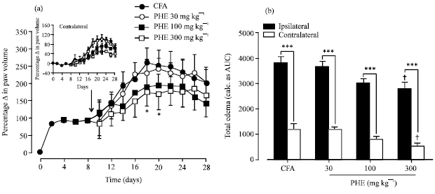 | |
| Fig. 1: | Effect of PHE (30-300 mg k-1 p.o.), dexamethasone (0.3-3 mg k-1 i.p.) and methotrexate (0.1-1 mg k-1 i.p.) on time course curve (a, c and e, respectively) and the total edema response (b, d and f, respectively) in adjuvant induced arthritis in rats. Values are expressed as Means±SEM (n = 5). ***p<0.001; **p<0.01; *p<0.05 compared to vehicle-treated group (Two-way ANOVA followed by Bonferroni’s post hoc test). †††p<0.001; ††p<0.01; †p<0.05 compared to vehicle-treated group (One-way ANOVA followed by Newman-Keul’s post hoc test |
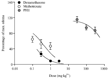 | |
| Fig. 2: | Dose response curves for dexamethasone (0.3-3.0 mg k-1 i.p.), methotrexate (0.1-1 mg k-1 i.p.) and PHE (30-300 mg k-1 p.o.) on adjuvant induced arthritis in rats |
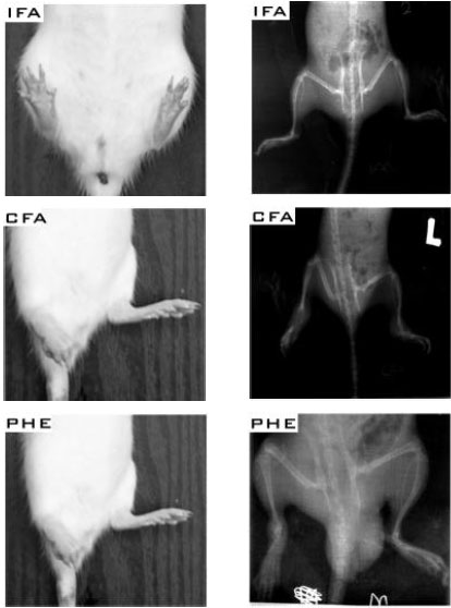 | |
| Fig. 3: | Photographs and radiographs of rat pre treated with IFA/non arthritic, CFA/arthritic control and PHE in the rat adjuvant induced arthritis |
 | |
| Fig. 4: | Time course effects of P. hirsuta 100 mg k-1, P. hirsuta 100 mg k-1+methotrexate 0.3 mg k-1 and P. hirsuta 100 mg k-1+dexamethasone 1.0 mg k-1, (a) methotrexate 0.3 mg k-1, dexamethasone 1.0 mg k-1 and methotrexate 0.3 mg k-1 +dexamethasone 1.0 mg k-1 (b) on CFA induced increase in the ipsilateral paw volume and (b and d) the AUC (total edema) for 28 days in the acute and polyarthritic phase. Each point in (a and c) and column in (b and d) represents the Mean±SEM (n = 5). ***p<0.001, **p<0.01, *p<0.05 compared to vehicle-treated group (Two-way ANOVA followed by Bonferroni’s post hoc test). †††p<0.001; †p<0.05 compared to vehicle-treated group (One-way ANOVA followed by Neuman-Keul’s post hoc test PHE, dexamethasone and metothrexate all suppressed the pathological changes seen in adjuvant induced-arthritis with dexamethasone and methotrexate exhibiting dose-dependency as shown in the radiological scores (Table 2, 3). |
| Table 2: | Arthritic scores for representative animals for groups treated curatively with PHE, methotrexate, dexamethasone, or vehicle |
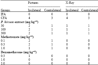 | |
| Table 3: | Arthritic scores for representative animals for groups treated with PHE, methotrexate, dexamethasone, or vehicle prophylactically and in combination therapy |
 | |
DISCUSSION
This present study has demonstrated that the oral treatment of rodents with ethanolic extract from leaves of the plant Palisota hirsuta exhibited potent anti-arthritic properties in the rat adjuvant-induced arthritis when administered prophylactically, curatively or in combination with standard anti-rheumatic drugs.
Rat adjuvant-induced arthritis is a commonly used animal model for preclinical studies of non-steroidal anti-inflammatory drugs and disease-modifying anti-rheumatic drugs and it is suggested as the most convenient model for studying drugs affecting human arthritis (Pearson, 1956; Whitehouse, 2007) and has often been used to study the mechanisms of action and preventive effects of a number of disease-modifying anti-rheumatic drugs (Hoffmann et al., 1997). Development of rat adjuvant-induced arthritis can be divided into three phases just like human rheumatoid arthritis. These phases start with the induction phase without evidence of synovitis, followed by early synovitis and finally late synovitis with progressive joint destruction (Hoffmann et al., 1997). A good anti-rheumatic agent should be able to block one or more of these phases.
In this study, P. hirsuta extract was able to suppress the joint inflammation and synovitis. It also proved very effective in preventing the systemic spread and ultimately reducing the destruction of joints as seen in the scores for the pictures and the radiographs. Radiographs are necessary to determine true remission of disease and for accurate evaluation of disease status (Kitamura et al., 2007). Reduced bone formation and increased resorption are the causes of bone loss in adjuvant-induced arthritis in rats (Aota et al., 1996; Makinen et al., 2007; Findlay and Haynes, 2005). The X-rays scores clearly show increased bone loss in arthritic groups and decreased bone loss in drug treated groups. It has been suggested that, new therapeutic strategies for chronic forms of arthritis have to aim at both, suppression of inflammation and bone protection and as such, joint protection plus suppression of synovitis are known to be the ultimate goals of a better RA treatment (Hoffmann et al., 1997; Sharma et al., 2004; Atzeni and Sarzi-Puttini, 2007) which is indicative that PHE can have a role to play in the management of human rheumatoid arthritis.
Phytochemical analysis of the leaf extract showed it contains flavonoids, tannins, terpenoids and alkaloids. A lot of these secondary plant metabolites identified have also been shown to exhibit anti-arthritic properties (Kupeli and Yesilada, 2007; Clavin et al., 2007) and as such, even though the exact mechanism involved in the anti-arthritic effect of PHE is not known, it can be speculated that one or more of these plant metabolites could probably be responsible for this particular pharmacological effect.
As a number of disease-modifying anti-rheumatic drugs in monotherapy often have unexpected side effects, combined treatment at lower doses may be necessary in order to expand the margin between efficacy and toxicity (Hisadome et al., 2004; Makinen et al., 2007). Based on this premise, the effect of combined lower doses of PHE and methotrexate or dexamethasone on the progression of hind paw inflammation and joint destruction in rats was studied. PHE in combination with dexamethasone had strong inhibitory effect on arthritis in rats; showing a synergistic suppression of both the increase in hind paw volume and also joint destruction thus producing a better remission of adjuvant-induced arthritis than PHE or dexamethasone alone. It is therefore possible that PHE and dexamethasone suppress a completely different stage of the inflammatory process in arthritic rats thus their combined effect gives a better remission. The effect of PHE was however not potentiated by methotrexate which is indicative that both drugs may be suppressing a similar stage of the inflammatory process in arthritic rats. As expected, dexamethasone combined with methotrexate synergistically suppressed arthritic progression which was extremely significant (Brown et al., 2006).
In respect to joint destruction, there is very little evidence on the role of most of the anti-rheumatic drugs currently being used, such as gold, sulfasalazine and methotrexate, when used alone in the modification of long term disease outcome (Zhao et al., 2006). Combination therapy in rheumatoid arthritis is acknowledged to give a better remission of disease than monotherapy (Capell et al., 2007; Mottonen et al., 2006). It is therefore necessary to develop new agents from natural sources, which when used in combination with other anti rheumatic drugs will be less toxic and at the same time, be affordable and effective for preventing joint destruction, as well as synovial inflammation, in RA, thus increasing efficacy of the treatment for patients with RA (Ronday et al., 1998) and as shown with this study, PHE is a very promising candidate for monotherapy and/or combination therapy in the effective treatment of rheumatoid arthritis.
ACKNOWLEDGMENTS
The authors are grateful for the technical assistance offered by Messrs Thomas Ansah, Gordon Darku and George Ofei of the Department of Pharmacology, Faculty of Pharmacy and Pharmaceutical Sciences, KNUST, Kumasi and also to Paa Kweku Woode for editing the pictures.
REFERENCES
- Anani, K., J.B. Hudson, C. DeSouza, K. Akpagana, G.H.N. Tower, J.T. Arnason and M. Gbeassor, 2000. Investigation of medicinal plants of togo for antiviral and antimicrobial activities. Pharma. Biol., 38: 40-45.
CrossRef - Aota, S., T. Nakamura, K. Suzuki, Y. Tanaka, Y. Okazaki, Y. Segawa, M. Miura and S. Kikuchi, 1996. Effects of indomethacin administration on bone turnover and bone mass in adjuvant-induced arthritis in rats. Calcif. Tissue Int., 59: 385-391.
CrossRefDirect Link - Atzeni, F. and P. Sarzi-Puttini, 2007. Early rheumatoid arthritis. Reumatismo, 59: 100-117.
PubMedDirect Link - Boakye-Gyasi, E., E. Woode, G.K. Ainooson, D.D. Obiri, C. Ansah, M. Duwiejua and A. Donkoh, 2008. Anti-Inflammatory and antipyretic effects of an ethanolic extract of Palisota hirsute K. Schum roots. Afr. J. Pharm. Pharmacol., 2: 191-199.
Direct Link - Brown, A.K., M.A. Quinn, Z. Karim, P.G. Conaghan and C.G. Peterfy et al., 2006. Presence of significant synovitis in rheumatoid arthritis patients with disease-modifying antirheumatic drug-induced clinical remission: evidence from an imaging study may explain structural progression. Arth. Rheum., 54: 3761-3773.
PubMed - Capell, H.A., R. Madhok, D.R. Porter, R.A.L. Munro, I.B. McInnes and J.A. Hunter et al., 2007. Combination therapy with sufasalazine and methotrexate is more effective than either drug alone in patients rheumatoid arthritis with a suboptimal response to sulfasalazine: Results from the double-blind placebo-controlled MASCOT study. Ann. Rheum. Dis., 66: 235-241.
CrossRefDirect Link - Clavin, M., S. Gorzalczany, A. Macho, E. Munoz, G. Ferraro, C. Acevedo and V. Martino, 2007. Anti-inflammatory activity of flavonoids from Eupatorium arnottianum. J. Ethnopharmacol., 112: 585-589.
CrossRef - Fereidoni, M., A. Ahmadiani, S. Semnanian and M. Javan, 2000. An accurate and simple method for measurement of paw edema. J. Pharmacol. Toxicol. Methods, 43: 11-14.
CrossRef - Findlay, D.M. and D.R. Haynes, 2005. Mechanisms of bone loss in rheumatoid arthritis. Mod. Rheumatol., 15: 232-240.
CrossRef - Hisadome, M., T. Fukuda, K. Adachi and H. Komatsu, 2004. Combination benefit of a pyrimidylpiperazine derivative (Y-40138) and methotrexate in arthritic rats. Eur. J. Pharmacol., 497: 351-359.
CrossRef - Kapoor, M., O. Shaw and I. Appleton, 2005. Possible anti-inflammatory role of COX-2-derived prostaglandins: Implications for inflammation research. Curr. Opin. Invest. Drugs, 6: 461-466.
PubMedDirect Link - Kinne, R.W., C.B. Schmidt-Weber, R. Hoppe, E. Buchner, E. Palombo-Kinne, E. Nurnberg and F. Emmrich, 1995. Long-term amelioration of rat adjuvant arthritis following systemic elimination of macrophages by clodronate-containing liposomes. Arthritis Rheum., 38: 1777-1790.
CrossRefPubMedDirect Link - Kitamura, T., J. Hashimoto, T. Murase, T. Tomita, T. Hattori, H. Yoshikawa and K. Sugamoto, 2007. Radiographic study of joint destruction patterns in the rheumatoid elbow. Clin. Rheumatol., 26: 515-519.
CrossRef - Kumar, D.A., P. Manikandan, M. Sumitra, K.V. Raju, C. Gayathri, N. Arutselvan and R. Puvanakrishnan, 2002. A novel peptide derivative exhibits anti inflammatory and antioxidant activity in adjuvant-induced arthritis in rats. Mol. Cell. Biochem., 229: 9-17.
CrossRef - Kupeli, E. and E. Yesilada, 2007. Flavonoids with anti-inflammatory and antinociceptive activity from Cistus laurifolius L. leaves through bioassay-guided procedures. J. Ethnopharmacol., 112: 524-530.
CrossRef - Makinen, H., H. Kautiainen, P. Hannonen, T. Mottonen and M. Leirisalo-Repo et al., 2007. Sustained remission and reduced radiographic progression with combination disease modifying antirheumatic drugs in early rheumatoid arthritis. J. Rheumatol., 34: 316-321.
PubMed - Mottonen, T., P. Hannonen, M. Leirisalo-Repo, M. Korpela, M. Hakala and H. Kautiainen, 2006. Efficacy of combination therapy in rheumatoid arthritis: Comment on the review by Smolen et al. Arth. Rheum., 54: 2032-2034.
CrossRef - Motulsky, H.J. and A. Christopoulos, 2003. Fitting Model to Biological Data using Linear and Nonlinear Regression a Practical Guide to Curve Fitting. 1st Edn., GraphPad Software Inc., San Diego, CA.
Direct Link - Pearson, C.M., 1956. Development of arthritis, periarthritis and periostitis in rats given adjuvants. Exp. Biol. Med., 91: 95-101.
CrossRefPubMedDirect Link - Pohlers, D., A. Beyer, D. Koczan, T. Wilhelm, H.J. Thiesen and R.W. Kinne, 2007. Constitutive upregulation of the transforming growth factor-beta pathway in rheumatoid arthritis synovial fibroblasts. Arth. Res. Ther., 9: R59-R59.
CrossRef - Ronday, H.K., J.M. Te Koppele, R.A. Greenwald, S.A. Moak and J.A. De Roos et al., 1998. Tranexamic acid, an inhibitor of plasminogen activation, reduces urinary collagen cross-link excretion in both experimental and rheumatoid arthritis. Br. J. Rheumatol., 37: 34-38.
CrossRef - Sharma, P.K., D. Hota and P. Pandhi, 2004. Biologics in rheumatoid arthritis. J. Assoc. Physicians India, 52: 231-236.
PubMed - Whitehouse, M.W., 2007. Adjuvant arthritis 50 years on: The impact of the 1956 article by C. M. Pearson, Development of arthritis, periarthritis and periostitis in rats given adjuvants. Inflamm. Res., 56: 133-138.
CrossRef - Woode, E., G.K. Ainooson, E. Boakye-Gyasi, C. Ansah and D.D. Obiri et al., 2008. Anti-arthritic and antioxidant properties of the ethanolic stem bark extract of Newbouldia laevis (P. Beauv.) Seaman ex Bureau (Bignoniaceae). J. Med. Plants Res., 2: 180-188.
Direct Link - Anani, K., J.B. Hudson, C. DeSouza, K. Akpagana, G.H.N. Tower, J.T. Arnason and M. Gbeassor, 2000. Investigation of medicinal plants of togo for antiviral and antimicrobial activities. Pharma. Biol., 38: 40-45.
CrossRef