
S. Fakurazi
Pharmacology Unit, Department of Human Anatomy,Universiti Putra Malaysia, 43400, UPM Serdang, Selangor, Malaysia
U. Nanthini
Department of Pathology, Faculty of Medicine and Health Sciences, Universiti Putra Malaysia, 43400, UPM Serdang, Selangor, Malaysia
I. Hairuszah
Department of Pathology, Faculty of Medicine and Health Sciences, Universiti Putra Malaysia, 43400, UPM Serdang, Selangor, Malaysia
International Journal of Pharmacology
Year: 2008 | Volume: 4 | Issue: 4 | Page No.: 270-275


ABSTRACT
This study is conducted to investigate the possible hepatoprotective action of Moringa oleifera Lam. (MO), a high value medicinal plant against a single high dose of APAP induced hepatotoxicity. Male Sprague Dawley rats were dosed with APAP (3000 mg kg-1 body weight; p.o.) to induce hepatocellular damage. In rats that were pretreated with MO (200 and 800 mg kg-1; p.o.) for 14 days prior to APAP treatment, there was a reduction of liver enzymes (ALT, AST and ALP) and also the restoration of glutathione level. The biochemical results showed parallel finding with the histopathological analysis in which liver sections obtained from rats pretreated with MO, the damage was blocked. Intriguingly, MO alone has significantly elevated the level glutathione compared to the control group. The findings has suggested that Moringa oleifera Lam. is a promising product in protecting the liver against APAP induced liver injury via the restoration and elevation of glutathione level in the liver.
PDF Abstract XML References Citation
How to cite this article
S. Fakurazi, U. Nanthini and I. Hairuszah, 2008. Hepatoprotective and Antioxidant Action of Moringa oleifera Lam.Against Acetaminophen Induced Hepatotoxicity in Rats. International Journal of Pharmacology, 4: 270-275.
DOI: 10.3923/ijp.2008.270.275
URL: https://scialert.net/abstract/?doi=ijp.2008.270.275
DOI: 10.3923/ijp.2008.270.275
URL: https://scialert.net/abstract/?doi=ijp.2008.270.275
INTRODUCTION
Induction of parenchymal hepatocellular damage by acetaminophen (APAP) overdose in human and laboratory animals are well-documented (Jaeschke et al., 2003). Following overdosage, APAP is metabolically broken down via cytochrome P4502E1 mediated pathway leads to the formation of reactive metabolite N-acetyl benzoquinoneimine (NAPQI) (James et al., 2003). Cellular glutathione is depleted and oxidative stress ensues (Michell et al., 1973). Subsequently, oxidative stress leads to the increase in reactive oxygen species (ROS), which aggravate heart disease, diabetes, liver injuries, cancer progression as well as aging process (Lampronti et al., 2005; Giordano, 2005). Maintaining the balance between ROS and antioxidant enzymes is therefore crucial and could serve as a major mechanism in preventing damage by oxidative stress.
Moringa oleifera Lam. (MO) belongs to the genus Moringacea (syns Moringa pterygosperma Gaertn). The plant is native to North India but it is now found throughout the tropics. It is commonly known as horse radish or drumstick tree. It is a multipurpose tree with most of its parts being useful for a number of applications (Anwar et al., 2007).
Studies have reported that the plant extract has antihypertensive (Faizi et al., 1995), a highly potent anti-inflammatory (Cáceres et al., 1992) and also as antitumor (Guevara et al., 1999). The extract has also been found to reduce the level of cholesterol (Mehta et al., 2003) and regulate the thyroid status (Costa et al., 2005). It was also been reported to have a hepatoprotective action against antitubercular drugs such as isoniazid and rifampicin-induced liver injury (Kumar and Pari, 2002). The plant seed powder may be useful in chelation therapy (Kumari et al., 2005; Sharma et al., 2006).
The aim of the present study was to investigate the effects of liver function markers following APAP induced hepatotoxicity and the improvement of liver histopathology following MO administration.
MATERIALS AND METHODS
Preparation of plant extract: The fresh leaves of Moringa oleifera were collected from Bandar Sunggala Farm, Port Dickson, Malaysia and authenticated by a botanist at Institute Bioscience (IBS), Universiti Putra Malaysia. A voucher specimen (M.O.SF001) has been kept at the herbarium in IBS for future reference. The crude extract preparation was according to Hossain et al. (1992). One hundred grams of the powder form that was prepared from the shaded dried substance was then extracted using 1000 mL of 80% hydroalcoholic solvent (80% ethanol: 20% distilled water). The extract was then filtered and then concentrated using a rotary evaporator (Rotavor R-200, Buchi, Switzerland) under reduced pressure at 40°C and then lyophilized (Freeze Dry System, FreeZone 4.5, Labconco, USA; -40°C, 120 bar). A dark green mass was then obtained and stored at -20°C until further use.
Experimental hepatoprotective study: Sprague Dawley rats (200-250 g) were obtained from Faculty of Veterinary Medicine, Universiti Putra Malaysia and were acclimatized under control laboratory conditions for 7 days with free access to standard food and water. This study was conducted, according to the rules and regulations by the Animal Care Unit Committee (ACUC), Universiti Putra Malaysia. The rats were randomly assigned into control and treatment groups of 5 rats per group.
Treatment was divided into pretreatment phase of MO in distilled water and the second phase in which the animals were given 3 g kg-1 APAP in 40% sucrose buffer on day 15. The animals were then euthanized 48 h after APAP administration.
Group 1 was pretreated with distilled water for 14 days prior to a single dose of APAP (3 g kg-1 body weight; p.o.) served as hepatotoxin control group. Group 2 and 3 were administered with aqueous extract of MO (200 and 800 mg kg-1 body weight; p.o.) for 14 days before APAP administration. Group 4 served as positive control group where the animals were pretreated with 200 mg kg-1 silymarin in distilled water prior to APAP administration. Group 5 and 6 were treated with MO (200 and 800 mg kg-1) prior to sucrose buffer treatment on day 15.
Forty eight hours after APAP administration the rats were fasted and then anaesthetized with diethylether. Blood was collected via cardiac puncture and the animals were then sacrificed. Livers were then excised, washed in ice-cold saline to remove blood and weighed. A section from the median lobe was preserved in 10% formal saline. The remaining liver was quickly frozen in dry ice and stored at -80°C for further analysis.
Liver function tests: Blood was collected and kept at 4°C overnight before being centrifuged at 3000 rpm at 4°C for 10 min to isolate the serum. The liver enzymes were assessed including alanine aminotransferase (ALT), aspartate aminotransferase (AST) and alkaline phosphatase (ALP) using diagnostic kits obtained from Roche Diagnostics Ltd. (Germany).
Liver histopathology: A section from the median lobe of the liver from each group was removed and fixed in 10% formalin and embedded in the paraffin. Liver sections of 3-4 μm thickness were prepared according to the standard procedure and stained with haematoxylin and eosin (H and E).
Determination of glutathione level: The liver homogenate was prepared using Ultra Turax where liver tissue was homogenized in 0.01 M Phosphate Buffer (pH 7.4) at 4°C. The liver glutathione (GSH) determination was performed according to the method previously described by Ellman (1959).
Statistical analysis: Data were presented as mean±SEM. Statistical analysis is performed using ANOVA Statistical Package for the Social Sciences (SPSS) version 13.0. p-value less than 0.05 (p<0.05) was considered to be statistically significant.
RESULTS AND DISCUSSION
Increased in the level of activities of ALT, AST and ALP in the blood reflect the damage of liver hepatocytes and indirectly impairment of liver functions following APAP-induced hepatotoxicity. In Table 1, ALT, AST and ALP activities were significantly elevated (p<0.05) after administration of APAP as compared to the control group (40% sucrose). Pretreatment with 200 and 800 mg kg-1 of MO extract significantly reduced the elevation of these enzymes (p<0.05). The reduction of liver enzymes were seen to be to the level of the control group (40% sucrose; p<0.05) and it was also similar to the level of group pretreated with silymarin (positive control).
| Table 1: | The activities of alanine aminotransferase (ALT), aspartate aminotransferase (AST) and alkaline phosphatase (ALP) in rats treated with Moringa oleifera Lam. (MO) and a single dose of acetaminophen (APAP) |
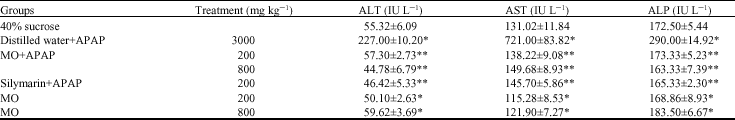 | |
| *p<0.05 significantly different from group treated with 40% sucrose, **p<0.05 significantly different from group treated with distilled water and APAP |
One of the hallmark signs of hepatic injury or damage is apparent leakage of cellular enzymes into plasma. In addition, the extent and type of liver injury or damage can be accessed based on the presence or absence of specific enzymes in the blood stream (Kumar et al., 2004). In general measurement of alanine aminotransferase (ALT), aspartate aminotransferase (AST) and alkaline phosphatase (ALP) are commonly used as marker enzymes in accessing APAP induced hepatotoxicity (Yanpallewar et al., 2003; Asha et al., 2004; Yen et al., 2007). Measurement of ALT is more liver specific to determine hepatocellular damage (Shyamal et al., 2006).
In this study, hepatoprotective effect of MO is evidenced by the improvement ALT, AST and ALP levels. Pretreatment with MO extract suppresses APAP induced AST and ALT elevations. Previous studies have reported elevations of transaminases after APAP treatment (Asha et al., 2004). The increase is time dependent with significant elevation noted after 48 h (p<0.05) suggesting severe hepatocellular damage caused by leakage of these enzymes into circulation that is normally cytoplasmic in location (Chung et al., 2001).
Recovery towards normalization of the enzymes following MO pretreatment suggested that the plant extract have role in preserving structural integrity of hepatocellular membrane, thus prevented enzymes leakage into circulation. Present results are consistent with generally accepted hypothesis that transaminase level return to normal with healing of hepatic parenchyma and the regeneration of hepatocytes (Ahmed and Khater, 2001).
The development of hepatotoxicity induced following APAP treatment was exacerbated following the depletion of glutathione. Therefore, in this study glutathione level was investigated to observe the effect of MO pretreatment to the animals. From Table 2, it was clearly indicated that APAP treatment has significantly reduce the level of glutathione (0.55±0.44 μmol g-1 protein), compared to the control animals (p<0.05).
| Table 2: | The level of glutathione (GSH) in the liver homogenate prepared from rats treated with Moringa oleifera Lam. (MO) and a single dose of acetaminophen (APAP) |
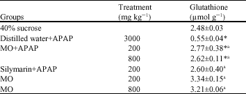 | |
| *p<0.05 significantly different compared to 40% sucrose, ap<0.05 significantly different from group treated with distilled water and APAP |
Pretreatment of the animals with MO (200 and 800 mg kg-1) has clearly restored the level of glutathione significantly (p<0.05). The restoration level of glutathione following MO was similar to that of silymarin. Intriguingly, MO treatment (200 and 800 mg kg-1) has actually increased the level of glutathione in the liver where the level is significantly higher when compared to the control (40% sucrose; p<0.05).
Overdosage of APAP leads to GSH depletion and reactive metabolite formation, NAQPQI. The metabolite subsequently form covalent binding to the macromolecules of cells leading to cellular necrosis which cause damage to the liver. Therefore, GSH, the vital intracellular nonprotein sulfhydryl that maintain cellular macromolecules in functional states, serves as a key determinant of the extent of APAP induced hepatic injury (Yanpallewar et al., 2003). Present results revealed that a high dose of APAP has caused remarkably reduced level of cellular GSH. It has been shown that preservation of GSH from being depleted provides direct protection against APAP induced hepatotoxicity (Ahmed and Khater, 2001). In the current study, GSH depletion was prevented when MO was administered before APAP. This indicates protection against APAP mediated liver toxicity. Similar action of MO in preserving GSH level was reported by Kumar and Pari (2003) following administration of antitubercular drugs such as isoniazid, rifampicin and pyrazinamide.
Interestingly, MO extracts alone increased intracellular GSH levels regardless of the doses. Therefore, the action of MO on enhancing GSH level could support the available GSH pool allowing the cell to detoxify more NAPQI and minimize the damage due to APAP or its metabolite that was evident from histopathological study. Negligible injury of hepatocytes was observed in the close vicinity of centrilobular vein. Present results are consistent with the observation that some hepatoprotective compound has the capability of enhancing GSH level that helps to preserve GSH levels (Hewawasam et al., 2004; Yapar et al., 2007).
The biochemical results obtained was comparable to the histopathological analysis of the liver sections obtained from each group. Figure 1 shows the micrograph of the liver section obtained from groups treated with 40% sucrose, APAP intoxicated and rats that were pretreated with MO. The control group (Fig. 1a) shows normal cellular architecture with distinct hepatocytes structure, sinusoidal spaces and central veins. Meanwhile, severe histopathological changes were clearly observed in APAP-intoxicated rats revealing centrilobular hepatic necrosis, with occasional bridging necrosis. Prominent degree of fatty changes and massive infiltration of lymphocytes and nuetrophils were clearly observed. When MO was treated prior to APAP administration, significant decrease of hepatocellular changes were observed. Normal liver architecture was seen in the liver sections obtained from rats pretreated with both doses (1c and d). Similar observation was seen when either MO alone or silymarin was administered to the animals (1e and f).
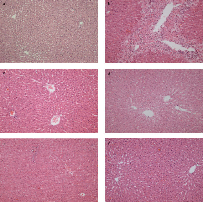 | |
| Fig. 1: | Hepatoprotective action of Moringa oleifera Lam. (MO) against acetaminophen (APAP) induced hepatotoxicity in rats. Liver sections were stained with H and E. (a) 40% sucrose, (b) distilled water and APAP, (c) 200 mg kg-1 MO and APAP, (d) 800 mg kg-1 MO and APAP, (e) 800 mg kg-1 MO alone and (f) 200 mg kg-1 silymarin and APAP Magnification 100x |
CONCLUSION
These results have clearly shown the potential hepatoprotective action of Moringa oleifera and enhancement of the level of glutathione following treatment of Moringa oleifera. Further studies must be conducted such as concurrent treatment of the plant extract in providing the hepatoprotective activities to further elucidate the bioactive component of the plant and assess the mechanism of hepatoprotective action of the plant.
ACKNOWLEDGMENTS
We would like to express our gratitude for tremendous help and contribution of staff in the Department of Nutrition and Dietetic, Department of Human Anatomy, Department of Biomedical Sciences and Laboratory of Molecular Biomedicine, Institute Bioscience, Universiti Putra Malaysia for the technical assistance and advice as well as material provision. A special acknowledgment is owed to Dr. Abdah binti Md. Akim who has generously provided advice and improvements of methodology of enzymatic study.
REFERENCES
- Anwar, F., S. Latif, M. Ashraf and A.H. Gilani, 2007. Moringa oleifera: A food plant with multiple medicinal uses. Phytother. Res., 21: 17-25.
CrossRefPubMedDirect Link - Asha, V.V., S. Akhila, P.J. Wills and A. Subramoniam, 2004. Further studies on the antihepatotoxic activity of Phyllanthus maderaspatensis Linn. J. Ethnopharmacol., 92: 67-70.
CrossRef - Caceres, A., A. Saravia, S. Rizzo, L. Zabala, E. De Leon and F. Nave, 1992. Pharmacologie properties of Moringa oleifera. 2: Screening for antispasmodic, antiinflammatory and diuretic activity. J. Ethnopharmacol., 36: 233-237.
CrossRefPubMedDirect Link - Chung, Y.H., J.A. Kim, B.C. Song Song, I.H. Koh, M.S. Lee, H.C. Eunsil, Y.S. Lee and D.J. Su, 2001. Centrilobular hepatic necrosis; Isocitrate dehydrogenase as a marker of centrilobular model of rats. J. Gastroenterol. Hepatol., 16: 328-332.
CrossRefPubMedDirect Link - Costa-Lotufo, L.V., M.T.H. Khan, A. Ather, D.V. Wilke and P.C. Jimenez et al., 2005. Studies of the anticancer potential of plants used in Bangladeshi folk medicine. J. Ethnopharmacol., 99: 21-30.
CrossRefPubMedDirect Link - Ellman, G.L., 1959. Tissue sulfhydryl groups. Arch. Biochem. Biophys., 82: 70-77.
CrossRefPubMedDirect Link - Faizi, S., B.S. Siddiqui, R. Saleem, S. Siddiqui, K. Aftab and A.H. Gilani, 1995. Fully acetylated carbamate and hypotensive thiocarbamate glycosides from Moringa oleifera. Phytochemistry, 38: 957-963.
CrossRefPubMedDirect Link - Giordano, F.J., 2005. Oxygen, oxidative stress, hypoxia, and heart failure. J. Clin. Invest., 115: 500-508.
CrossRefDirect Link - Hewawasam, R.P., K.A.P.W. Jayatilaka, C. Pathirana and L.K.B. Mudduwa, 2004. Hepatoprotective effect of Epaltes divaricata extract on carbon tetrachloride induced hepatotoxicity in mice. Indian J. Med. Res., 120: 30-34.
PubMed - Hossain, M.Z, B.A. Shibib and R. Rahman, 1992. Hypoglycemic effects of Coccinia indica inhibition of key gluconeogenic enzyme, glucose-6-Phosphatase. Indian. J. Exp. Biol., 30: 418-420.
Direct Link - James, L.P., P.R. Mayeux and J.A. Hinson, 2003. Acetaminophen-induced hepatotoxicity. Drug Metab. Dispos., 31: 1499-1506.
CrossRefPubMedDirect Link - Kumar, N.A. and L. Pari, 2003. Antioxidant action of Moringa oleifera Lam. (Drumstick) against antitubercular drugs induced lipid peroxidation in rats. J. Med. Food, 6: 255-259.
CrossRefPubMedDirect Link - Kumari, P., P. Sharma, S. Srivastava and M.M. Srivastava, 2005. Arsenic removal from the aqueous system using plant biomass: A bioremedial approach. J. Ind. Microbiol. Biotechnol., 32: 521-526.
CrossRefPubMedDirect Link - Lampronti, I., M.T. Khan, N. Bianchi, A. Ather, M. Borgatti, L. Vizziello, E. Fabbri and R. Gambari, 2005. Bangladeshi medicinal plant extracts exhibiting molecular interactions between nuclear factors and target DNA sequences mimicking NF-kappa B binding sites. Med. Chem., 1: 327-333.
PubMedDirect Link - Mehta, L.K., R. Balaraman, A.H. Amin, P.A. Bafna and O.D. Gulati, 2003. Effect of fruits of Moringa oleifera on the lipid profile of normal and hypercholesterolaemic rabbits. J. Ethnopharmacol., 86: 191-195.
CrossRefPubMedDirect Link - Mitchell, J.R., D.J. Jollow, W.Z. Potter, J.R. Gillette and B.B. Brodie, 1973. Acetaminophen-induced hepatic necrosis. IV. Protective role of glutathione. J. Pharmacol. Exp. Ther., 187: 211-217.
PubMedDirect Link - Sharma, P., P. Kumari, M.M. Srivastava and S. Srivastava, 2006. Removal of cadmium from the aqueous system by shelled Moringa oleifera Lam. Seed powder. Bioresour. Technol., 97: 299-305.
CrossRef - Yanpallewar, S.U., S. Sen, S. Tapas, M. Kumar, S.S. Raju and S.B. Acharya, 2003. Effect of Azadirachta indica on paracetamol-induced hepatic damage in albino rats. Phytomedicine, 10: 391-396.
PubMedDirect Link - Yapar, V., A. Kart, M. Karapehlivan, O. Atakisi, R. Tuncac, S. Erginsoy and M. Citil, 2007. Hepatoprotective effect of L-carnitine against acute acetaminophen toxicity in mice. Exp. Toxicol. Pathol., 59: 121-128.
CrossRefPubMedDirect Link - Yen, F.L., T.H. Wu, L.T. Lin and C.C. Lin, 2007. Hepatoprotective and antioxidant effects of Cuscuta chinensis against acetaminophen-induced hepatotoxicity in rats. J. Ethnopharmacol., 111: 123-128.
CrossRefPubMedDirect Link - Guevara, A.P., C. Vargas, H. Sakurai, Y. Fujiwara and K. Hashimoto et al., 1999. An antitumor promoter from Moringa oleifera Lam. Mutat. Res./Genet. Toxicol. Environ. Mutagen., 440: 181-188.
CrossRefDirect Link