
A. Sabdono
Department of Marine Science, FPK, Diponegoro University, Semarang, Indonesia
O.K. Radjasa
Department of Marine Science, FPK, Diponegoro University, Semarang, Indonesia
H.S. Utomo
Rice Research Station, Louisiana State University Ag Center, Rayne, LA, USA
International Journal of Oceanography and Marine Ecological System
Year: 2012 | Volume: 1 | Issue: 1 | Page No.: 11-23


ABSTRACT
Scleractinian corals harbor diverse bacterial communities within their tissue. However, it is still not known the significant role of those bacteria in bioremediation of heavy metal contamination. The present study aimed to investigate the diversity of the bacterial community associated with the corals that have multiple resistance to heavy metals. Sixty-one coral bacteria isolated from three different life-forms of scleractinian coral samples collected from Central Java coastal waters were established by plating on Zobell’s 2214E. Those isolates were screened for their resistance against Pb, Cr, Zn, at 1 mM level using agar diffusion method and 22 isolates were selected. Minimal Inhibitory Concentrations (MIC) of heavy metals were determined and different MIC of these isolates was shown to be highly resistant to Pb, Cr and Zn ions. A rapid grouping by using Repetitive Extragenic Palindromic (REP)-PCR was conducted to estimate the richness of the isolates. Three heavy metal resistant bacterial strains representing three major genetic groups were selected for further studies. Based on analysis of morphological, biochemical and 16S rDNA sequence of these isolates revealed that one strain belongs to γ-proteobacteria division while the other two belong to Firmicutes division. Isolate PL05 was closely related to Pseudoalteromonas sp. while PL12 and PL22 isolates were closely related to Virgibacillus sp. This work provides the first evidence of bacteria possessing multiple resistances against heavy metals can be recovered from corals.
PDF Abstract XML References Citation
Received: July 20, 2011;
Accepted: October 08, 2011;
Published: November 14, 2011
How to cite this article
A. Sabdono, O.K. Radjasa and H.S. Utomo, 2012. Screening of Multi-metal Resistances in a Bacterial Population Isolated from Coral Tissues of Central Java Coastal Waters, Indonesia. International Journal of Oceanography and Marine Ecological System, 1: 11-23.
DOI: 10.3923/ijomes.2012.11.23
URL: https://scialert.net/abstract/?doi=ijomes.2012.11.23
DOI: 10.3923/ijomes.2012.11.23
URL: https://scialert.net/abstract/?doi=ijomes.2012.11.23
INTRODUCTION
Coral reef is one among tropical coastal ecosystems of the world beside mangrove and seagrass (Kathiresan and Alikunhi, 2011). Coral reef ecosystems have a high species diversity that contain hundreds of reef fish, corals, plants and animals (Cesar et al., 2003; Veron, 1986). Due to the abundance of unique chemical properties from certain coral types, coral reefs have been viewed as the medicine cabinets of the sea (Tacio, 2004). Corals have shown a great potential for finding effective chemical agents that provide a large proportion of bioactive compounds with different biological activities (Radjasa and Sabdono, 2009; Shahbudin et al., 2011). Because of its potentials, Bruckner (2002) suggested that coral reefs could be the major source of drugs for the next decade. Corals are host of bacterial life that living on seawater around corals, the surface and tissue of corals and their interactions have been studied in detail (Rohwer et al., 2001, 2002; Lampert et al., 2006; Koren and Rosenberg, 2006; Ibrahim, 2008; Lins-de-Barros et al., 2010). However, little is known about the functional role of bacteria associated with scleractinian corals (Rohwer et al., 2002). Even the understanding of their functional role in coral reef ecosystems is still ignored, Ritchie (2006) reported that coral bacteria play significant role in the antibiotic activity and pigment production. In addition, some investigators stated that coral bacteria were also play important role in cycling of nitrogen, carbon, sulphur and phosphate (Siboni et al., 2008; Sharon and Rosenberg, 2008; Raina et al., 2009). Furthermore, several studies reported that coral bacteria play functional role in phosphatase enzyme activity (Al-Shehri, 2006), anti-pathogenic bacterium Streptococcus equi (Radjasa et al., 2007) and health and disease of corals (Friaz-Lopez et al., 2002). Sabdono and Radjasa (2008) reported that coral bacteria are capable of degrading organophosphate pesticides. Coral bacteria resistant to heavy metal can be used in remediation efforts of various environments, since they can grow under variable salinity and temperature (Koren and Rosenberg, 2006).
Central Java is the third most-populous province in Indonesia and one of the fastest growing provinces in commercial and industrial sectors. Wastes from residential areas, rivers, industries and agricultures have intensified and through migration, runoff and infiltration, they make a way to the coastal waters. Several studies concerning the marine pollutants of Java coastal waters reported the impact of anthropogenic activities on heavy metal contamination (Booij et al., 2001; Takarina et al., 2004; Sabdono, 2009). Because of their toxicity, persistence and bioaccumulation problem, heavy metals are one of the most serious polluting agents in marine environments (Blackmore, 1998; Selvin et al., 2009). Studies showed that marine pollution had severely impacted the microbial ecology (Danovaro et al., 2003; Dell’Anno et al., 2003; Boyd, 2010).
Heavy metals such as lead, chrome and zinc were emphasized in this study due to their extensive use in wood preservation, electroplating, metal-finishing and chemical industries of Central Java. These heavy metals were also detected at high concentration in dead coral tissues (Sabdono, 2009). Heavy metals become toxic to the cell when present at concentration above trace amounts (Nies, 1999). The objective of this study was to investigate the diversity of the bacterial community found within the coral tissues that have multiple resistance to metals.
MATERIALS AND METHODS
Sampling and coral bacterial isolation: Corals representing 3 different life forms (branching, massive, sub-massive) were collected from Central Java coastal waters in July 2008. Specimens of the corals Porites lutea, Galaxea fascicularis and Pocillopora damicornis were collected randomly by scuba diving at depths of 2 to 5 m. Individual specimens were placed separately in plastic bags to avoid contact with air and brought to the surface. The individual samples in the plastic bags containing natural seawater were processed within a few hours after collection. Tissue samples were removed from the skeleton with a sterilized scrapper and the exposed surface tissues were removed with a sterile scalpel blade. The resultant tissues were serially diluted spread on a half-strength ZoBell 2216E marine agar medium and incubated at room temperature for 48 h. On the basis of morphological features, colonies were randomly picked and purified by making streak plates (Madigan et al., 2000).
Screening of metal resistant isolates: A total of 61 bacterial strains isolated from three coral species (P. lutea, G. fascicularis and P. damicornis) were screened for their resistance to three heavy metals according to the Kirby-Bauer disc diffusion method (Bauer et al., 1966). Filter paper disks, 8 mm in diameter (Toyobo, Co, Japan), were soaked in solutions of the appropriate heavy-metal salt (lead, chrome or zinc, supplied as Pb(NO3)2, K2Cr2O7 or ZnCl2) at 1 mM concentrations. The disks were then placed on the surface of plates that previously inoculated with 0.1 mL of isolates. Each plate contained one disk lacking the heavy metal salt and three disks containing each concentration of heavy metal salt. The plates were then incubated at 28°C for approximately 72 h. At the end of incubation period, the zones of inhibition were measured as indicator for resistance. Zone measurements were recorded as the distance from the edge of the zone to the edge of the disk. Isolates that had a zone size (clearance zone) less than 1.00 mm were considered as resistant strain (Rani et al., 2010). Isolates showing resistant to 1 mM metals were further tested at higher concentrations.
Minimum Inhibitory Concentration (MIC) of the heavy metal resistant coral bacteria was determined by gradually increasing 0.5 mM of the heavy metal concentration. The starting concentration used was 1.5 mM. MIC was noted when the isolates formed zone inhibition greater than 1.0 mm in size.
Microscopic and biochemical characterizations: Selected bacterial strains highly resistant to heavy metals were grown in Zobell 2216E medium and underwent further microscopic and biochemical evaluations. Photomicrograph was used to determine the morphology of the isolates. While standard gram staining, motility and biochemical characterizations based on Bergey’s Manual of Determinative Bacteriology (Holt et al., 1994) were used to determine their biochemical properties.
Rep-PCR amplification and grouping of isolates: To determine genetic relatedness among bacterial isolates, DNA fingerprinting were performed according to the method of Sabdono and Radjasa (2008). In the rep-PCR, BOX AIR primer (5’-CTACGGCAAGGCGACGCTG ACG-3’; Versalovic et al., 1994) was used. Genetic grouping analyses of selected isolates was carried out by making matrixes from the positions of bands on the gel which were then analyzed using Free Tree program (Pavlicek et al., 1999). The Tree View ver.1.6.6 program was used in constructing the tree (Page, 1996).
DNA extraction, PCR amplification and sequencing of 16S rDNA: DNA extraction, PCR amplification of partial 16S rDNA of bacterial strain and purification of PCR products were also carried out based on the method of Sabdono and Radjasa (2008). Primers [(forward primer 8-27: 5’-AGAGTTTGATCCTGGCTCAG-3’ (Weisburg et al., 1991) and reverse primer 1510-1492: 5’-GGTTACCTTGTTACGACTT-3’ (Reysenbach et al., 1992) were used to amplify 16S rDNA.
Sequencing and phylogenetic analysis: Based on phylogenetic grouping, the isolates each representing major genetic group were selected and used for DNA sequencing. The sequencing and phylogenetic analysis were conducted according to the method of Sabdono and Radjasa (2008). The PCR product was purified and concentrated with Microcon-100 microconcentrators (Amicon, Beverly, MA, USA) according to manufacturer’s instructions. Sequencing was carried out with a SequiTherm Long-Read Cycle Sequencing Kit (Epicentre Technologies, Madison, WI, USA) and an automated sequencer (the ALF DNA sequences: Pharmacia LKB Biotech, Uppsala, Sweden). The nucleotides sequences obtained from partial sequencing of 16S rDNA were then compared for homology to the BLAST database. A phylogenetic tree was constructed using maximum-likelihood analysis. Clustal X software was used for multiple alignment/pairwise the DNA sequence (Thompson et al., 1997). Phylogenetic analysis was performed with the Phylogenetic Analysis Using Parsimony (PAUP Ver.4) software package (Swofford, 1998). Bootstrap analysis of 1,000 replicates was performed to estimate robustness of the tree.
Nucleotide sequence accession numbers: Nucleotide sequences of the 16S rDNA from three heavy metal bacterial resistant strain obtained in this study have been deposited in the GeneBank database under accession number HQ659245 to HQ659247. The accession numbers of 16S rDNA of other strain cited and used as comparison in this study.
RESULTS AND DISCUSSION
Isolation and screening of coral bacteria resistant to heavy metals: Initial screening using 1 mM metal concentration indicated that out of 61 coral bacterial isolates, 10 isolates (16.4%) were susceptible to any heavy metal tested while 22 (36.1%), 13 (21.3%) and 16 (26.2%) isolates were resistance to three, two and one metal(s), respectively (Table 1). When the three different life forms of corals were compared, the highest percentage of metal resistant bacteria (77.3%) was originated from coral species P. lutea. While, the percentage of metal resistant bacteria isolated from G. fascicularis and P. damicornis were 11.1 and 14.3%, respectively. No literature evidences could be compared to heavy metal resistant bacteria isolated from scleractinian corals. However, there were many studies regarding bacteria associated with other marine invertebrates that resistant to heavy metals. Jeanthon and Prieur (1990) reported that heterotrophic bacteria isolated from two deep-sea hydrothermal vent polychaete Annelids were resistant to high concentration of metal. Most of those isolates (92.3%) displayed multiple resistance to cadmium, zinc, arsenate and silver and tolerated high amounts of copper. Selvin et al. (2009) found out the heavy metal resistance pattern of the bacteria associated with a marine sponge Fasciospongia cavernosa. Similar studies on heavy metal resistance by marine actinomycetes isolated from saltpan soil have been reported (Deepika and Kannabiran, 2010). In addition, heavy metal resistant bacteria were isolated from sediment in the Uppanar Estuary, South East Coast of India (Karthikeyan et al., 2007) and Sunchon Bay, South Korea (Kamala-Kannan and Lee, 2008). The highest percentage metal resistant bacteria found in coral P. lutea indicated that this coral species could be used as material source of bacterial isolation. Al-Rousan et al. (2007) reported that Porites corals (massive) have a high tendency to accumulate heavy metals. Since the bacteria associated with coral Porites were continuously subjected to high levels of metals, this condition could create the emergence of metal resistant bacteria.
| Table 1: | Comparison of the No. isolates of multiple metal resistant bacteria of the three different corals |
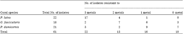 | |
The 22 isolates showing resistance to 3 metals were reassessed further for higher concentrations.
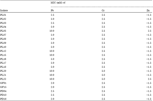
MIC (Minimum Inhibitory Concentration) of 22 isolates: The resistance levels of the 22 coral bacterial isolates to three metal toxicities and their range varied (Table 2). The strains evaluated had a wide resistant range to lead toxicity from 2.0 to 10 mM. They had a narrower range to chromium toxicity from 2.5 to 5 mM and an extremely narrow resistant range to zinc toxicity of <1.5 up to 2.5 mM. Nies, 1999 stated that the bacteria are able to tolerate beyond Cd 0.5 mM, Zn 1.0 mM, Cu 1.0 mM, Pb 5.0 mM and Ni 1.0 mM could be considered as extreme. By using this definition, the results of this study demonstrated that 6, 3 and 2 isolates were extremely resistant to lead, chromium and zinc, respectively. While, the remaining had high and moderate level of resistance. Compared to the previous study, both the highest MIC of lead and chrome in this study were slightly higher than that of Frankia strain (Richards et al., 2002). However, those MIC value was lower for lead and similar for chrome to that of reported by Nieto et al. (1989). The resistance of coral bacteria to heavy metals could be induced due to the industrial and domestic wastewater from coastal regions of Central Java. High concentrations of heavy metals in the coral tissues were found in this polluted coral reef region (Sabdono, 2009). Since bacteria living in the coral tissue were continuously subjected to heavy metal toxicity, this condition could stimulate the emergence of metal resistant coral bacteria. The differences of these results are due to the levels of metal pollution and type of organic structures (Gillan et al., 2005; Bezverbnaya et al., 2005).
| Table 2: | The MICs of the heavy metals tested against coral bacteria determined by zone of inhibition |
 | |
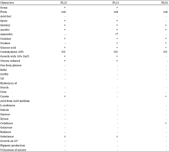
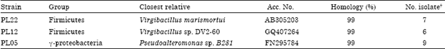
Characterization of selected bacterial isolates: Based on the repetitive-PCR results and constructed dendrogram of the isolates, three groups were created at which similarity level (Fig. 1). The 16S rDNA from strain PL22, PL05 and PL12 representing each of the three groups were characterized and sequenced to obtain information on their identity. Microbiological characteristics and the results of DNA sequencing of those isolates are presented in Table 3 and 4. The two selected isolates (PL22 and PL12) are gram positive while PL05 isolate is gram negative. All three selected isolates were rod-shaped, aerobe, motile, endospore former with positive catalase and oxidase activity. All of them did not produce any kinds of pigments and have no ability to metabolite all the 6 sugars tested. Different from PL05 isolate, the isolate of PL22 and PL12 grew on 10% NaCl concentration.
Analysis of 16S rDNA sequences revealed the presence of two major groups of bacteria: (1) Firmicutes and (2) γ-proteobacteria. BLAST analysis of PL22 and PL12 isolates revealed that these strains were close relative, with 99% similarity, of Virgibacillus marismortui and Virgibacillus sp. DV2-60, respectively. While, BLAST analysis of PL05 isolate revealed that this isolate is a close relative, with 99% similarity, of the strain Pseudoalteromonas sp. B281 (Table 4). The 16S rDNA sequences of these bacteria were submitted to GenBank (Accession No. HQ659245 to HQ659247). Several heavy metal-resistant bacteria isolated from marine environments have been identified. Stuart et al. (2009) reported coastal marine bacteria Synechococcus sp. tolerance to copper. Bacteria Pseudomonas sp. and Delftia sp. resistant to metals isolated from sea water and sediment of Persian Gulf were reported by Zolgharnein et al. (2010). De Souza et al. (2006) found psychotropic bacteria resistant to heavy metal and antibiotic isolated from Antarctic marine water. In addition, Selvin et al. (2009) reported bacteria Streptomyces sp., Salinobacter sp., Roseobacter sp., Pseudomonas sp., Vibrio sp., Micromonospora sp., Saccharomonospora sp. and Alteromonas sp. resistance against heavy metals isolated from marine sponge.
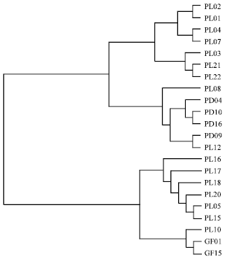 | |
| Fig. 1: | Dendrogram of multi metal resistant isolates (PL22, PL12, PL05 were further selected for DNA sequencing) |
| Table 3: | Microbiological characterization of three selected isolates |
 | |
| Sign+: Positive result; sign -: Negative result; NC: No change; v: No assayed; F: Facultative | |
| Table 4: | Characterization of representative heavy metal-resistant coral bacteria |
 | |
To estimate genetic affiliation of the heavy metal-resistant isolates among coral-associated bacteria, a neighbor-joining tree including identified isolates and representative marine microorganisms is constructed. A phylogenetic analysis of the 16S rRNA data for selected strains belonging to the group of the Firmicutes and Proteobacteria produced the dendrogram shown in Fig. 2. This comparison was made to determine the species to which the three selected isolates are most closely related and how closely the three taxa are related to each other.
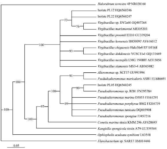 | |
| Fig. 2: | Phylogenetic tree based on the 16S ribosomal DNA sequence data showing the relationships of representative strains with the most closely related bacteria identified in the GenBank database. Halorubrum terrestre was used as out group. Bar indicated 5% dissimilarity of sequences |
The PL12 and PL22 isolates were in the same cluster of phylogenetic tree Virgibacillus sp. while the PL05 isolate was in the cluster of Pseudoalteromonas sp. Many researchers have reported the structure of coral-associated bacterial communities isolated from different coral species. Bourne and Munn (2005) reported that the majority of microbial community obtained from coral tissue P. damicornis was γ-proteobacteria, whereas the coral mucus was dominated by α-proteobacteria. While Koren and Rosenberg (2006) revealed that a large diversity of bacteria associated with coral tissue of Oculina patagonica were Pseudomonas sp., α-proteobacteria and Vibrio species. The results of the present study showed that the bacterial diversity associated with corals was different from the previous reported. The differences in bacterial community structure could be explained as an effect of heavy metal pollution on the coral reef ecosystem. Webster et al. (2001) reported that the total density and counts of microbial communities associated with the sponge Rhopaloeides odorabile were significantly reduced in response to Cu2+ concentrations. Ageret al. (2010) stated that anthropogenic pollution will reduce bacterial species richness, loss of species and change in bacterial community structure. In addition, coastal pollution has an impact on the microbial communities inhabiting healthy coral tissues (Klaus et al., 2007).
It is interesting to note that the PL22, PL12 and PL05 isolates showed high multiple resistant against Pb, Cr, Zn heavy metals. This raises the possibility the use of these bacteria as potential candidates for remediation efforts of heavy metal contaminated coral reef ecosystems. The genus Virgibacillus constitutes a diverse group of gram-positive bacteria, rod-shaped and spore forming (Peng et al., 2009). This members of genus are ubiquitous in different marine environments reflect their wide functional properties. Gupta et al. (2008) reported that Virgibacillus sp. produce extracellular thermostable serine alkaline protease while Kuhlmann et al. (2008) reported that this genus could also produce ectoine as a microbial osmoprotectant. In addition, the genus Virgibacillus sp produce salt-activated extracellular proteinases (Sinsuwan et al., 2007) and possess inhibitory activity against fouling bacteria (Kanagasabhapathy et al., 2005). The member of genus Pseudoalteromonas is a rod-shaped, motile, gram-negative bacteria that usually found in association with marine eukaryotic hosts such as sponges and algae (Bowman, 2007). This genus is well known to produce inhibitory compounds against surface competitors (Thomas et al., 2008). Moreover, Hedlund and Staley (2006) reported that genus Pseudoalteromonas could degrade polycyclic aromatic hydrocarbons while Mimura et al. (2008) reported that this genus could absorb trybutylin.
CONCLUSION
Bacteria living in the coral tissue are complex and diverse. Further studies are needed to investigate their mechanisms of metal resistance that could be useful in the bioremediation of contaminated coral reef ecosystems. This paper reported that coral bacteria Virgibacillus marismortui PL22, Virgibacillus sp. PL12 and Pseudoalteromonas sp. PL05 showed high multiple resistant activity against Pb, Cr, Zn heavy metals.
ACKNOWLEDGMENT
This study was supported by grant from Directorate General of Higher Education (DIKTI), Indonesian Ministry of National Education under competent research grant scheme (HIBAH KOMPETENSI, No. 013/HIKOM/DP2M/2008) and Program Academic Recharging (PAR-C), No. 921/D4.4/PK/2010. The study also was partly supported by Agricultural Research Center-Louisiana State University, Baton Rouge, Louisiana, USA).
REFERENCES
- Ager, D., S. Evans, H. Li, A.K. Lilley and C.J. Van Der Gas, 2010. Anthropogenic disturbance affects the structure of bacterial communities. Environ. Microbiol., 12: 670-678.
CrossRef - Al-Rousan, S.A., R. Al-Shloul, F. Al-Horani and A. Abu-Hilal, 2007. Heavy metal contents in growth bands of Porites corals: Record of anthropogenic and human developments from the Jordanian Gulf of Aqabah. Mar. Pollut. Bull., 54: 1912-1922.
PubMed - Al-Shehri, A.M., 2006. Factors affecting alkaline phosphatase activity of the marine cyanobacterium Lyngbya majuscula. J. Boil. Sci., 6: 931-935.
CrossRefDirect Link - Bauer, A.W., W.M.M. Kirby, J.C. Sherris and M. Turck, 1966. Antibiotic susceptibility testing by a standardized single disk method. Am. J. Clin. Pathol., 45: 493-496.
CrossRefPubMedDirect Link - Bezverbnaya, I.P., L.S. Buzoleva and N.K. Khristoforova, 2005. Metal-resistant heterotrophic bacteria in coastal waters of Primoye. Biologiya Morya, 31: 89-93.
Direct Link - Blackmore, G., 1998. An overview of trace metal pollution in coastal waters of Hong Kong. Sci. Total Environ., 214: 21-48.
CrossRefPubMedDirect Link - Bowman, J.P., 2007. Bioactive compound synthetic capacity and ecological significance of marine bacterial genus Pseudoalteromonas. Mar. Drugs, 5: 220-241.
CrossRefDirect Link - Booij, K., M.T. Hillebrand, R.F. Nolting and J. Van Ooijen, 2001. Nutrients, trace metals and organic contaminants in Banten Bay, Indonesia. Mar. Pollut. Bull., 42: 1187-1190.
Direct Link - Bourne, D.G. and C.B. Munn, 2005. Diversity of bacteria associated with the coral Pocillopora damicornis from the great barrier reef. Environ. Microbiol., 7: 1162-1174.
PubMed - Boyd, R.S., 2010. Heavy metal pollutants and chemical ecology: Exploring new frontier. J. Chem. Ecol., 36: 46-58.
CrossRef - Danovaro, R., M. Armeni, C. Corinaldesi and M.L. Mei, 2003. Viruses and marine pollution. Mar. Pollut. Bull., 46: 301-304.
CrossRef - Dell’Anno, A., M.L. Mei, C. Lanni and R. Danovaro, 2003. Impact of bioavailable heavy metals on bacterial activities in coastal marine sediments. World J. Microbiol. Biotechnol., 19: 93-100.
CrossRef - Friaz-Lopez, J., A.L. Zerkle, G.T. Bonheyo and B.W. Fouke, 2002. Partitioning of bacterial communities between seawater and healty, black band disease and dead coral surfaces. Applied Environ. Microbiol., 68: 2214-2228.
CrossRefPubMedDirect Link - Gillan, D.C., B. Danis, P. Pernet, G. Joly and P. Dubois, 2005. Structure of sediment-associated microbial communities along a heavy-metal contamination gradient in the marine environment. Applied Environ. Microbiol., 71: 678-690.
CrossRefDirect Link - Gupta, A., B. Joseph, A. Mani and G. Thomas, 2008. Biosynthesis and properties of an extracellular thermostable serine alkaline protease from Virgibacillus pantothenticus. World J. Microbiol. Biotechnol., 24: 237-243.
CrossRefDirect Link - Hedlund, B.P. and J.T. Staley, 2006. Isolation and characterization of Pseudoalteromonas strains with divergent polycyclic aromatic hydrocarbon catabolic properties. Environ. Microbiol., 8: 178-182.
CrossRef - Holt, J.G., N.R. Kreig, P.H.A. Sneath, J.T. Staley and S.T. Williams, 1994. Bergey's Manual of Determinative Bacteriology. 9th Edn., Lippincott Williams and Wilkins, Baltimore, USA., ISBN-13: 9780683006032, Pages: 787.
Direct Link - Ibrahim, A.S.S., 2008. Diversity of coral Eunicea fusca associated bacteria using culture dependent techniques. Res. J. Microbiol., 3: 614-621.
CrossRefDirect Link - Jeanthon, C. and D. Prieur, 1990. Susceptibility to heavy metals and characterization of heterotrophic bacteria isolated from two hydrothermal vent polychaete annelids, Alvinella pompejana and Alvinella caudate. Applied Environ. Microbiol., 56: 3308-3314.
Direct Link - Kamala-Kannan, S. and K.J. Lee, 2008. Metal tolerance and antibiotic resistance of bacillus species isolated from sunchon bay sediments, South Korea. Biotechnology, 7: 149-152.
CrossRefDirect Link - Karthikeyan, R., S. Vijayalakshmi and T. Balasubramanian, 2007. Monthly variation of heavy metal and metal resistant bacteria from uppanar estuary (South East Coast of India). Res. J. Microbiol., 2: 50-57.
CrossRefDirect Link - Kathiresan, K. and N.M. Alikunhi, 2011. Tropical coastal ecosystems: Rarely explored for their interaction!. Ecologia, 1: 1-22.
CrossRefDirect Link - Klaus, J.S., I. Janse, J.M. Heikoop, R.A. Sanford and B.W. Fouke, 2007. Coral microbial communities, zooxanthellae and mucus along gradients of seawater depth and coastal pollution. Environ. Microbiol., 9: 1291-1305.
CrossRefDirect Link - Koren, O. and E. Rosenberg, 2006. Bacteria associated with mucus and tissues of the coral Oculina patagonica in Summer and Winter. Applied Environ. Microbiol., 72: 5254-5259.
CrossRefDirect Link - Kuhlmann, A.U., J. Bursy, S. Gimpel, T. Hoffmann and E. Bremer, 2008. Synthesis of the compatible solute ectoine in Virgibacillus pantothenticus is triggered by high salinity and low growth temperature. Applied Environ. Microbiol., 74: 4560-4563.
CrossRef - Lins-de-Barros, M.M., R.P. Vieira, A.M. Cardoso, V.A. Monteiro and A.S. Turque et al., 2010. Archaea, bacteria and algal plastids associated with the reef-building corals Siderastrea stellata and Mussismilia hispida from Buzios, South Atlantic Ocean, Brazil. Microb. Ecol., 59: 523-532.
CrossRefPubMedDirect Link - Mimura, H., R. Sato, Y. Furuyama, A. Taniike, M. Yagi, K. Yoshida and A. Kitamura, 2008. Adsorption of tributyltin by tributyltin resistant marine Pseudoalteromonas sp. cells. Mar. Pollut. Bull., 57: 877-882.
CrossRef - Nies, D.H., 1999. Microbial heavy-metal resistance. Applied Microbiol. Biotechnol., 51: 730-750.
PubMed - Nieto, J.J., A. Ventosa and F. Ruiz-berraquero, 1989. Susceptibility of halobacteria to heavy metals. Applied Environ. Microbiol., 53: 1199-1202.
Direct Link - Page, R.D.M., 1996. TreeView: An application to display phylogenetic trees on personal computers. Comput. Applied Biosci., 12: 357-358.
CrossRefPubMedDirect Link - Pavlicek, A., S. Hrda and J. Flegr, 1999. Free-Tree-freeware program for construction of phylogenetic trees on the basis of distance data and bootstrap/jackknife analysis of the tree robustness. Application in the RAPD analysis of genus Frenkelia. Folia Biol. (Praha), 45: 97-99.
PubMedDirect Link - Peng, Q.Z., J. Chen, Y.Q. Zhang, Q.H. Chen and D.J. Peng et al., 2009. Virgibacillus zhanjiangensis sp. nov., a marine bacterium isolated from sea water. Antonie van Leeuwenhoek, 96: 645-652.
CrossRef - Raina, J.P., D. Tapiolas, B.L. Willis and D.G. Bourbone, 2009. Coral-associated bacteria and their role in the biogeochemical cyling of sulfur. Applied Environ. Microbiol., 75: 3492-3501.
CrossRef - Radjasa, O.K. and A. Sabdono, 2009. Bacterial symbionts of reef's invertebrates as a sustainable source of marine natural products. Curr. Res. Bacteriol., 2: 7-13.
CrossRefDirect Link - Radjasa, O.K., S.I.O. Salasia, A. Sabdono, J. Weise, J.F. Imhoff, C. Lammler and M.J. Risk, 2007. Antibacterial activity of marine bacterium Pseudomonas sp. associated with soft coral Sinularia polydactyla against Streptococcus equi subsp. zooepidemicus. Int. J. Pharmacol., 3: 170-174.
CrossRefDirect Link - Rani, M.J., B. Hemambika, J. Hemapriya and V.R. Kannan, 2010. Comparative assessment of heavy metal removal by immobilized and dead bacterial cells: A biosorption approach. Afr. J. Environ. Sci. Technol., 4: 77-83.
Direct Link - Reysenbach, A.L., L.J. Giver, G.S. Wickham and N.R. Pace, 1992. Differential amplification of rRNA genes by polymerase chain reaction. Applied Environ. Microbiol. 58: 3417-3418.
Direct Link - Richards, J.W., G.D. Krumholz, M.S. Chval and L.S. Tisa, 2002. Heavy metal resistance patterns of Frankia strains. Applied Environ. Microbiol., 68: 923-927.
CrossRefPubMedDirect Link - Ritchie, K.B., 2006. Regulation of microbial populations by coral surface mucus and mucus-associated bacteria. Mar. Ecol. Prog. Ser., 322: 1-14.
CrossRefDirect Link - Rohwer, F., M. Breitbart, J. Jara, F. Azam and N. Knowlton, 2001. Diversity of bacteria associated with the Caribbean coral Montastraea franksi. Coral Reefs, 20: 85-95.
CrossRefDirect Link - Rohwer, F., V. Seguritan, F. Azam and N. Knowlton, 2002. Diversity and distribution of coral-associated bacteria. Mar. Ecol. Prog. Ser., 243: 1-10.
Direct Link - Sabdono, A. and O.K. Radjasa, 2008. Phylogenetic diversity of organophosphorous pesticide-degrading coral bacteria from mid-west coast of Indonesia. Biotechnology, 7: 694-701.
CrossRefDirect Link - Sabdono, A., 2009. Heavy metal levels and their potential toxic effect on coral Galaxea fascicularis from Java Sea, Indonesia. Res. J. Environ. Sci., 3: 96-102.
CrossRefDirect Link - Selvin, J., P.S. Shanmugha, K.G. Seghal, T. Thangavelu and B.N. Sapna, 2009. Sponge-associated marine bacteria as indicators of heavy metal pollution. Microbiol. Res., 164: 352-363.
CrossRef - Shahbudin, S., S. Deny, A.M.T. Zakirun, T.A.H. Haziyamin, B. Akbar John and M. Taher, 2011. Antioxidant properties of soft coral Dendronephthya sp. Int. J. Pharmacol., 7: 263-267.
CrossRef - Sharon, G. and E. Rosenberg, 2008. Bacterial growth on coral mucus. Curr. Microbiol., 56: 481-488.
CrossRef - Siboni, N., E. Ben-Dov, A. Sivan and A. Kushmaro, 2008. Coral-associated ammonium oxidizing Crenarchaeota and their role in the coral holobiont nitrogen cycle. Proceedings of the 11th International Coral Reef Symposium, July 7-11, Fort Lauderdale, FL., USA., pp: 252-256.
Direct Link - Sinsuwan, S., S. Rodtong and J. Yongsawatdigul, 2007. NaCl-activated extracellular proteinase from Virgibacillus sp. SK37 isolated from fish sauce fermentation. J. Food Sci., 72: C264-C269.
CrossRefPubMedDirect Link - Stuart, R.K., C.L. Dupont, D.A. Johnson, I.T. Pulsen and B. Palenik, 2009. Coastal strain of marine Synechococcus species exhibit increased tolerance to copper shock and a distinctive transcriptional response relative to those of open-ocean strain. Applied Environ. Microbiol., 75: 5047-5057.
CrossRef - Takarina, N.D., D.R. Browne and M.J. Risk, 2004. Speciation of heavy metals in coastal sediments of Semarang, Indonesia. Mar. Pollut. Bull., 49: 861-868.
CrossRefDirect Link - Thomas, T., F.F. Evans, D. Schleheck, A. Mai-Prochnow and C. Burke, 2008. Analysis of the Pseudoalteromonas tunicate genome reveals properties of a surface-associated life style in the marine environment. PLoS ONE, 3: e3252-e3252.
CrossRef - Thompson, J.D., T.J. Gibson, F. Plewniak, F. Jeanmougin and D.G. Higgins, 1997. The CLUSTAL_X windows interface: Flexible strategies for multiple sequence alignment aided by quality analysis tools. Nucleic Acids Res., 25: 4876-4882.
CrossRefPubMedDirect Link - Webster, N.S., R.I. Webb, M.J. Ridd, R.T. Hill and A.P. Negri, 2001. The effects of copper on the microbial community of a coral reef sponge. Environ. Microbiol., 3: 19-31.
CrossRef - Weisburg, W.G., S.M. Barns, D.A. Pelletier and D.J. Lane, 1991. 16S ribosomal DNA amplification for phylogenetic study. J. Bacteriol., 173: 697-703.
CrossRefPubMedDirect Link - Zolgharnein, H., K. Karami, M.M. Assadi and A.D. Sohrab, 2010. Molecular characterization and phylogenetic analyses of heavy metal removal bacteria from the persian gulf. Biotechnology, 9: 1-8.
CrossRefDirect Link - De Souza, M.J., S. Nair, P.A. Loka-Bharathi and D. Chandramohan, 2006. Metal and antibiotic-resistance in psychrotrophic bacteria from Antarctic marine waters. Ecotoxicology, 15: 379-384.
CrossRef