
Burhanudin Malik
Graduate School of Nutrition and Feed Sciences, Institut Pertanian Bogor University, Bogor, Indonesia
LiveDNA: 62.37346
Dewi Apri Astuti
Department of Nutrition and Feed Technology, Faculty of Animal Sciences, Institut Pertanian Bogor University, Bogor, Indonesia
Anuraga Jayanegara
Department of Nutrition and Feed Technology, Faculty of Animal Sciences, Institut Pertanian Bogor University, Bogor, Indonesia
LiveDNA: 62.7299
Min Rahminiwati
Faculty of Veterinary Medicine, Institut Pertanian Bogor University, Bogor, Indonesia
International Journal of Dairy Science
Year: 2022 | Volume: 17 | Issue: 2 | Page No.: 41-53


ABSTRACT
Leech has been used as a medical device in health therapy all over the world. Leech saliva contains many active compounds which have antioxidant, antimicrobial and anticoagulant properties and bring health benefits. On the other hand, mastitis in dairy animals is an important disease-causing great economic loss. Control of this disease can be done through good animal husbandry practice and a nutritional approach. Mineral and vitamin supplementations are commonly done to maintain the immune system. Antioxidant supplementation plays an important role in eliminating free radicals significantly found in animals with mastitis. Antibiotic administration is done to eliminate bacteria causing mastitis. In addition, the use of anticoagulants might be useful as dairy animals suffering from mastitis may experience a kind of abnormal blood coagulation. In this context, having antioxidant, antimicrobial and anticoagulant properties, leech saliva is a potential material for subclinical mastitis control in dairy animals.
PDF Abstract XML References Citation
Copyright: © 2022. This is an open access article distributed under the terms of the creative commons attribution License, which permits unrestricted use, distribution and reproduction in any medium, provided the original author and source are credited.
How to cite this article
Burhanudin Malik, Dewi Apri Astuti, Anuraga Jayanegara and Min Rahminiwati, 2022. Leech Saliva and its Potential Use in Animal Health and Production: A Review. International Journal of Dairy Science, 17: 41-53.
DOI: 10.3923/ijds.2022.41.53
URL: https://scialert.net/abstract/?doi=ijds.2022.41.53
DOI: 10.3923/ijds.2022.41.53
URL: https://scialert.net/abstract/?doi=ijds.2022.41.53
INTRODUCTION
Leech is a freshwater annelid commonly having a black or brown coloured body of about 10 cm long. It belongs to the Hirudo genus with more than 600 species. Traditionally, leeches have been long known and used in ancient times in India and China. Leeching then developed well in Arabic and European medicines1. After being abandoned for centuries, leeching was back in use for health therapy in the early twentieth century. Today, in America and Europe, studies on Hirudo medicinalis and Hirudo verbana, the two most common medicinal leeches and substances contained in their saliva have been done extensively. Extracts of active compounds in leech saliva and live leeches have been used in health therapy. In Asia, Hirudinaria manillensis is a leech species used in health therapy and studies on it are increasing2. In Indonesia, leeching for health purposes is spreading throughout the country. However, studies on the potential of leech and active components contained in it for human and animal health are still very limited. Meanwhile, recent studies in Malaysia showed that saliva extract of local Malaysian leeches had anticoagulant3, antioxidative4, antimicrobial5 and anticancer6 activities.
Biological activities found in leech saliva extract may be useful in mastitis control as part of mastitis control management on-farm. This paper aimed to review the potency of leech saliva and its promising application for use in animal health and production, particularly to overcome the problem of subclinical mastitis in dairy animals. At first, leech biology is presented as an introductory and basic understanding of the organism, comprised of its taxonomy, anatomy, saliva and eating pattern. The biochemical activity of leech saliva is subsequently presented to elucidate its potential application in animal health and production. This part has consisted of chemical components present in leech saliva and their anticoagulant, anti-oxidative and antimicrobial activities. Lastly, mastitis as a common disease in dairy animals is presented together along with the potential use of leech saliva to overcome the problem.
Leech biology
Leech taxonomy: There are only about 15 out of 600 leech species that have been identified as being used in health therapy7. The three most popular of them include Hirudo medicinalis, Hirudo verbana in Europe and Hirudinaria manillensis in Fig. 1 in Asia. According to Webster Dictionary, the word leech originally comes from the ancient English word of læce or leche meaning physician which was first known before the 12th century. This name was given as at that time leeches were already used for health purposes through bloodletting.
Leech belongs to Phylum Annelida (segmented body) and Subphylum Clitellata (girdled, widely-swollen-ringed body)2. The taxonomy of leeches is shown in Table 18. The H. medicinalis and H. verbana are two European leeches that are mostly used in health therapy. Figure 1 shows that, these two species have similar colour patterns in the dorsal area. The difference is found in their ventral body area. The H. verbana has a simpler colour pattern of greenish-yellow with a black line pattern alongside its body sides while a greenish-yellow color pattern with black spots all over the ventral body area is found in H. medicinalis.
Leech anatomy: As a segmented body invertebrate animal, the leech is similar to the earthworm, its closest phylogenetic relative. However, there are some differences among them. The body of a leech which is like a tube consists of 4 segments while the number of segments in earthworms varies. Segments 9-11 form a clitellum responsible for cocoon formation in the breeding season. The last 7 segments form a big posterior sucker. In each segment of the earthworm, there is a ring while every segment in the leech body has two or more rings. Besides, an earthworm has lots of setae (bristles) all over its body, but a leech does not7.
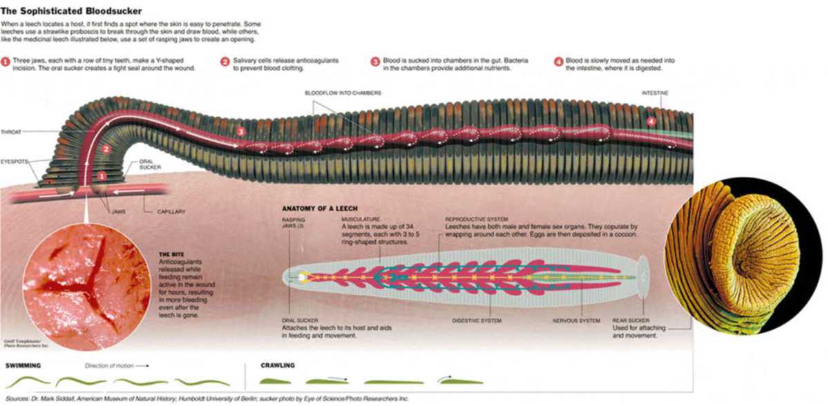
Leech has two suckers, each is located in its body end. In the anterior sucker, there are a mouth and jaws which are used to slice the skin and suck the blood of the victim mammal. The posterior sucker has no jaw and is used to attach the leech body to the skin surface of the victim and help movement during the leech is sucking the blood of its victim. In the anterior sucker, there is a mouth within which there are three jaws. The sides of the jaws form a 120° angle making a shape like a Mercedes-Benz logo. Each blade-shaped jaw contains 60-100 small teeth alongside its edge7. The penetration of these jaws into the victim’s skin leaves a star-shaped mark on the skin Fig. 1.
Mechanism of blood-sucking and leech saliva infusion: The way a leech in nature gets its victim and sucks blood from its victim is well described9. In their habitat, hungry leeches will sensitively be reactive to any water surface movement. These animals will immediately approach the source of movement to seek the victim. It is suspected that the sweat of the victim animal attracts leech attention to approach the prospective victim animal. This was proven by the finding that solution containing NaCl and arginine was able to stimulate leeches to find, attach themselves to the skin of and suck blood from the victim animal. Both chemical compounds are the components of the skin secretion of a mammal animal10.
 |
| Fig. 1: | Leech body anatomy Source: http://www.nytimes.com/imagepages/2006/02/06/science/20060207_LEECH_GRAPHIC.html |
| Table 1: | Leech taxonomy |
| Class | Hirudinea |
| Order | Euhirudinea |
| Suborder | Hirudiniformes |
| Family | Hirudinidae |
| Species | Hirudo medicinalis |
| Hirudo verbana | |
| Hirudinaria manillensis |
After it attaches its anterior sucker to the skin of its victim, the leech immediately starts moving its teeth in a sawing movement to slice the skin of the victim animal. Simultaneously, pharyngeal muscles perform a peristaltic movement so that the blood from the tore blood vessel is sucked and transported into the leech crop9. As this pumping movement of pharyngeal muscles is occurring, saliva glands located along the body segment 3-9 produce saliva. This saliva is transported through interdental pores in leech jaws in Fig. 2a and b to wound location on the skin tissue where the saliva then penetrates the blood vessel of the victim animal7.
Figure 3 shows how leeches saliva is transported to the blood vessel of the victim animal through interdental pores. Some of the various chemical compounds contained in leech saliva are absorbed by the blood vessel surface of the wounded victim animal or patient and some are mixed with blood oozing out from the wounded area11. First, hirudin inhibits blood coagulation in the wounded area. Simultaneously, calin binds with and deactivates the von Willebrand factor so that the wound will remain open for up to about 12 hrs. Von Willebrand factor is a glycoprotein compound contained in blood plasma that plays a role in mediating platelet attachment to subendothelial connective tissue and binds with blood coagulation factor VIII12 in Fig. 3 left. Then, hyaluronidase, another active compound, plays its role as a spreading factor in Fig. 4 right. This enzyme facilitates the cleansing of mucopolysaccharides within an intercellular cavity to make the tissue more permeable to any fluid and open the way for other chemical substances to enter the cells. Hyaluronidase facilitates the penetration and diffusion of other active compounds contained in saliva9,13. Further, anti-carboxypeptidase-A inhibiting compound and histamine like substance dilate capillaries around the wound to increase blood flow to the wound area. Finally, anti-inflammation substances including Eglin and bdellin accelerate wound healing.
It may take about 30 min for a leech to suck the blood of its victim animal. The amount of blood sucked can be about 10 mL or 10 times of leech's initial body weight. Blood entering the leech intestine is thickened by the leech by secreting blood plasma out through transcutaneous pores. This process is observed as a sweating process shown by a leech during a blood-sucking period. This process results in thickened blood and concentrated albumin and red blood cells. Satiated leeches may survive without feeding for up to 1 year14.
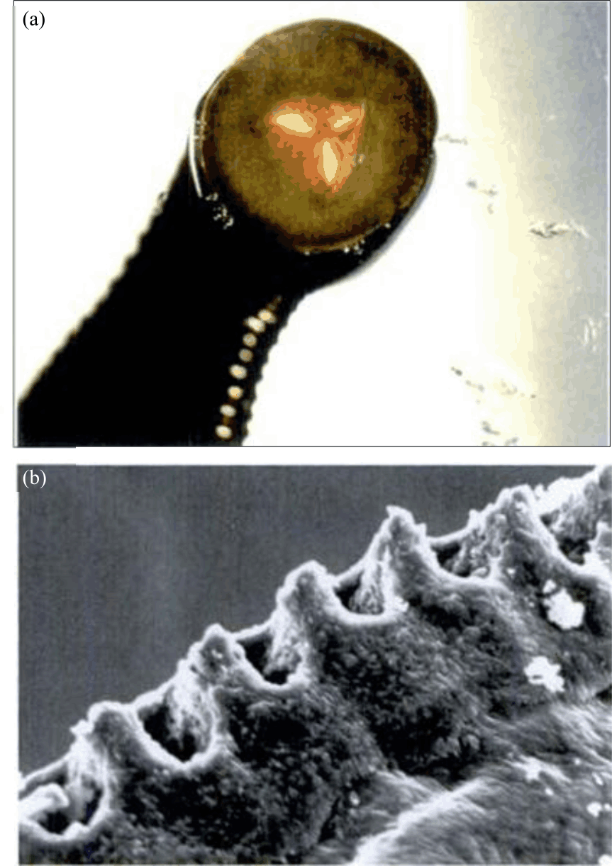 |
| Fig. 2(a-b): | Anterior sucker and (a) Pharyngeal muscle and (b) Interdental pores Source: Roth7 |
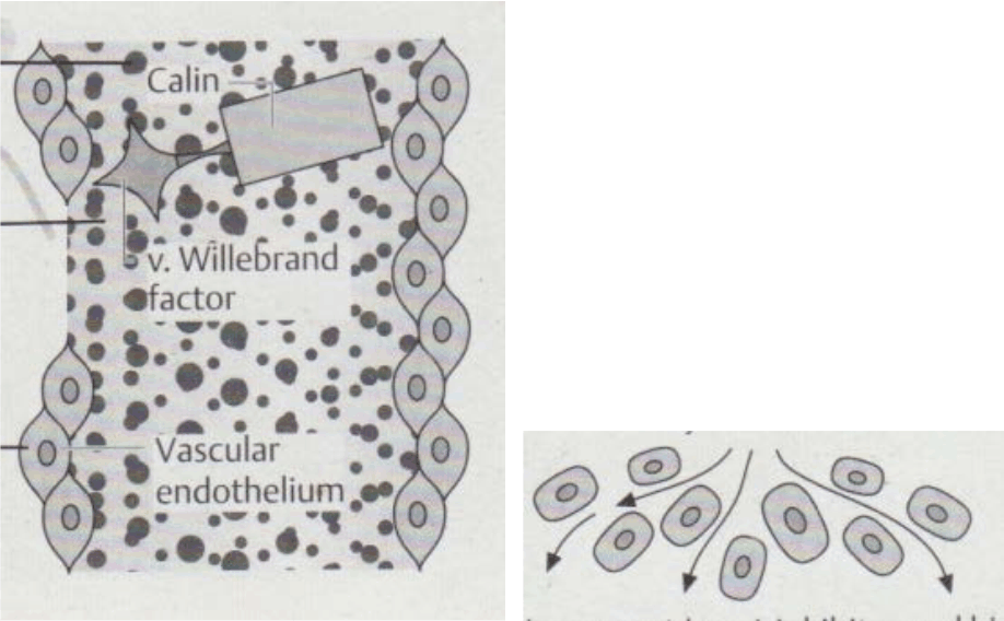 |
| Fig. 3: | Mechanism of leech saliva active compounds entering victim animal’s body |
Eating pattern: Every living creature has its habitat and eating pattern. Leeches in nature are found in muddy eutrophic ponds with littoral vegetations and warm temperatures where frogs and lizards commonly breed2. In their very first days, early hatched young leeches feed on protein fluid found in their cocoons. Once they hatch from their cocoons, young leeches feed on benthos and planktons7. Later, these young leeches feed mainly on the body fluid of amphibians whose skin is so soft that it can be penetrated by the teeth of young leeches9. As the body size of leeches grows, their feed changes into mammalian blood. In addition to sucking the blood of mammals including cattle, horses, deer and humans, mature leeches also feed on the blood of fish, water birds and amphibians2.
It takes 14 months with 5-6 times of blood-sucking for a leech of H. manillensis to reach its sexual maturity. In their juvenile phase, leeches will have 3 times of bloodsucking. In their 4th blood-sucking, leeches will be in their sub mature phase. Leeches will enter their sexually mature phase after they have their 5th blood sucking and their life cycle is completed15.
Biochemical activity of leech saliva
Chemical components in leech saliva: When a leech bites the skin of its victim animal, this annelid secretes a complex mixture of various biologically and pharmacologically active compounds into the bite wound. These various active compounds bring health benefits to humans having leech therapy. In fact, from the perspective of leech interests, these compounds are produced by leech to facilitate its activities of seeking for and obtaining food by sucking the blood of victim animals9. For instance, to make a victim animal not realize the leech attack when the leech starts sucking blood, the leech produces compounds with analgesic, anaesthetic and anti-inflammatory properties. To ensure that the blood of the victim animal does not coagulate and flows smoothly to the leech intestine, leech saliva contains anticoagulants such as hirudin. Then, to ensure that the blood flowing through the leech’s intestines carries no microorganisms, leech saliva contains compounds having antimicrobial properties. Other compounds contained in leech saliva also bring benefits to leech.
The saliva of H. medicinalis commonly used in health therapy is known to contain various kinds of proteins and peptides. Table 2 shows some leech saliva components successfully identified and known to give an effect on the body of victim animals or humans are listed. Calin is one of the compounds contained in leech saliva. This compound is a protein with a molecular mass of 65 kD. Calin is an inhibitor of platelet aggregation and attachment to collagen tissues by binding the collagen and von Willebrand factor in a very quick time (1-10 min). This work of calin makes the bite wounds stay open for about 12 hrs. This prolonged bleeding is useful for cleaning the wound to avoid sepsis which may be dangerous for the victim animal or patient12.
| Table 2: | Components of leech saliva known to give effects on the body of victim animal or patient |
| Substance | Effects on the body of victim animal or patient |
| Hirudin | Avoid blood coagulation by binding with thrombin |
| Calin (saratin) | Avoid blood coagulation by inhibiting von Willebrand-collagen binding |
| Avoid platelet aggregation mediated by collagen | |
| Destabilase | Involve in monomerizing activity |
| Dissolve fibrin | |
| Have thrombolytic effects | |
| Hirustasin | Inhibit kallikrein, trypsin, chymotrypsin and neutrophilic cathepsin G |
| Bdellins | Have anti-inflammatory property |
| Inhibit trypsin, plasmin and acrosin | |
| Hyaluronidase | Increase interstitial viscosity |
| Have antibiotic property | |
| Eglin’s | Have anti-inflammatory property |
| Inhibit activities of α-chymotrypsin, chymase, subtilisin, elastase and cathepsin G | |
| Histamine-like substances | Vasodilator, increase blood flow in the biting site |
| Acetylcholine | Vasodilator |
| Anaesthetic substances | Have anaesthetic property |
| Source: Gross and Roth12 | |
Destabilase is an enzyme that has a glycosidase8 and can perform monomerization and dissolve fibrin12. This enzyme is also able to specifically break the isopeptide bond at fibrin monomer molecules16. Hyaluronidase is an enzyme that works on the degradation of hyaluronan, a multifunctional polysaccharide with high molecular mass found in the extracellular matrix of connective tissues. Hyaluronan is involved in various biological processes including wound healing which makes hyaluronan existence increase. Degradation of hyaluronan by hyaluronidase increases the permeability of connective tissue and lowers the viscosity of body fluid17. About the process of leech saliva and its components entering the body of a victim animal or patient, hyaluronidase plays a role as a spreading factor that opens the interstitial so that active substances in leech saliva can penetrate the tissue deeper. Other active substances including bdellin and Eglin work as anti-inflammation. Bdellins inhibit trypsin, plasmin and acrosin. Eglin is an inhibitor of α-chymotrypsin, trypsin, chymase and subtilisin activities12.
The lipid content of leech saliva has also been isolated. An analysis of the lipid content of H. medicinalis saliva was performed and about 3 mg of lipids were found in every 100 mL of leech saliva. It was also found that free fatty acids (67%) and phosphatidic acid (3%) were the main components of lipid in leech saliva18. In another study, it was revealed that 20% of total leech saliva weight was lipid. Lipid contained in leech saliva was mostly steroids including steroid hormones such as cortisol, progesterone, testosterone, estradiol and dehydroepiandrosterone. In addition, histamine and serotonin were also identified. These lipid components were suspected to be strong phospholipase and lipase used by leech to avoid wound covering and healing when the leech is biting its victim animal or patient19.
Hirudin and anticoagulant activity: Blood coagulation is a mechanism of blood balance maintenance (hemostasis) in the body. When there is a wound, hemostasis is disrupted and to return hemostasis, processes including blood vessel contraction, platelet clotting, blood coagulation and fibrinolysis immediately occur20. The coagulation phase in a hemostasis system is aimed at forming fibrin fibres to bind with and stabilize weak platelets21. Imbalances in these processes may result in the inability of blood to coagulate which in turn may lead to excessive bleeding or unexpected blood coagulation which may cause death and various cardiovascular diseases20.
Anticoagulant activity of leech saliva is related to hirudin which is the most known component and often used to name all active substances contained in leech saliva. Hirudin is only one among the other active substances. The name hirudin was first given by Jakobj in 1903-4. Fritz Markwardt from Germany was the first who successfully isolated hirudin from H. medicinalis saliva in 195512.
Hirudin is a polypeptide containing 62-66 amino acid residues with a molecule weight of 7000-7500 Da. This polypeptide has three disulfide bridges namely Cys6’-Cys14’, Cys16’-Cys28’ and Cys22’-Cys39’. These amino acid sequences are rich in acid residues as represented by the fact that 6 out of the last 13 amino acids were acidic. Besides, Tyr63’ is a sulphated amino acid that increases the number of amino acids with negative charge22,23. The sequences of amino acids of hirudin are depicted in Fig. 4.
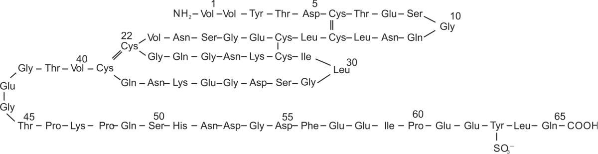 |
| Fig. 4: | Sequence of hirudin amino acid components Source: https://pubmed.ncbi.nlm.nih.gov/2294999/ |
 |
| Fig. 5: | Blood coagulation inhibition by hirudin Source: Gross and Roth12 |
In an anticoagulant activity, hirudin avoids blood coagulation by selectively binding with thrombin in the blood of the victim animal. Based on their anticoagulating mechanism, anticoagulant compounds produced by blood-fed animals can be classified into thrombin inhibitors, factor Xa inhibitors, tenase complex extrinsic inhibitors and tenase complex intrinsic inhibitor20. Hirudin is an anticoagulant compound with a direct target on thrombin at a very low concentration (picomolar)16,24.
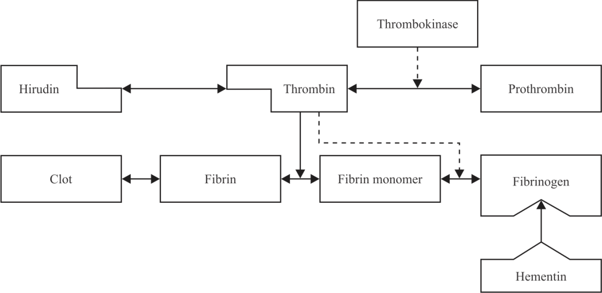
Figure 5 shows how hirudin works on avoiding blood coagulation. With the help of the thrombokinase enzyme, prothrombin changes into thrombin. The formed thrombin then changes fibrin monomers into fibrin polymers which later form new peptide bonds. This peptide bond formation leads to the occurrence of blood coagulation. When there is hirudin, thrombin will bind with hirudin to form inactive complex compounds. As a result, the fibrin polymer formation process does not occur and blood coagulation can be avoided12.
In an anticoagulation mechanism, hirudin is similar to heparin. Before heparin was invented, hirudin was the only compound used as an anticoagulant. Later, heparin became the most used anticoagulant compound as hirudin was more difficult to obtain. Hirudin has some superiorities over heparin. First, hirudin is an anticoagulant with a higher level of stability. Hirudin is not inactivated by platelet or other proteins which neutralize heparin25. Second, hirudin directly inhibits thrombin which does not bind with protein plasma or endogenous cofactor24. This results in responses that are easier to predict and more effective. In contrast, heparin requires a plasma cofactor in the form of antithrombin (AT) to express its anticoagulant activity. Heparin-AT complex compound is very big making its anticoagulant activity less effective26-28. Another superiority of hirudin is that this anticoagulant is not susceptible to peptides and enzymes in blood and hence, it can be released from the body through urine in an intact form without being metabolized in the liver12.
Behind its superiorities, there are some drawbacks of hirudin. This anticoagulant does not have any antagonist making its excessive use not easy to overcome. However, excessive use of hirudin can be avoided as long as it is used in a recommended amount12. It was revealed in a study that the administration of hirudin in patients with kidney failure should be accompanied by strict rate determination and monitoring. Bleeding and symptoms of the formation of IgG, IgM and IgA, but not IgE antibodies were also abserved29.
Antioxidative activity: In a normal physiological condition, there is a homeostasis balance between radical oxygen formation and its removal by endogenous antioxidant scavengers. The oxidant is a normal product of aerobic metabolism occurring in the body of living creatures. However, in a pathological condition, oxidant production increases destructs homeostasis balance and triggers the occurrence of oxidative stress. Oxidative stress is a condition when the number of oxidants is higher than that of antioxidants. It is known that some diseases including hyperglycemia, hypertension and dyslipidemia may trigger oxidative stress30.
Effects of antioxidant consumption on the reduction of coronary heart disease risk have been extensively studied31. However, although leech therapy has been long done on patients with cardiovascular diseases13, information about the antioxidant content and antioxidative activity of leech saliva is still very limited. This notion has made studies on antioxidative activity more interesting to do especially after an increased level of glutathione peroxidase and superoxide dismutase (SOD) in patients with chronic sialadenitis and sialadenosis (salivary gland diseases) having a leech therapy was observed32 and antioxidative effect of leech therapy was found to reduce testicular damage in rats suffering from testicular ischemia33. Besides, in another study, leech saliva was found as a promising antioxidant as it had free radical scavenging activity with a higher IC10 levels (7.282 μg mL–1) than vitamin C (5.803 μg mL–1)4. The antioxidative activity was also shown in the saliva of Nigerian34 leeches.
Antimicrobial activity: Multicellular organisms keep surviving themselves from parasite attacks endangering their lives. Epithelial tissues (skin, eyes, nose, etc.) are points where contact with microbes commonly occurs35. It is suspected that antimicrobial activity is owned by annelids in the form of various kinds of antimicrobial peptides found in the body fluid of annelids. Peptide B are antimicrobial peptide found in the body of H. medicinalis and Theromyzon tessulatum36,37. These antimicrobial peptides were found to have antibacterial activity on gram-positive bacteria36.
An interesting finding in leech is that the number of bacterial colonies in the leech digestive tract is very limited. This is different from the condition of humans or other animals. About 300 species of microflora are found in human feces38 and microfloral colonies in the mosquito, another blood-fed animal, vary widely39. In contrast, in the digestive tract of H. medicinalis only a pure colony of Aeromonas spp., was found40. Bacteria isolated from the leech digestive tract were found to be positive for beta-hemolysis and able to produce extracellular protease and lipase. As these enzymes play an important role in blood digestion, these bacteria were categorized as symbions41.
The exact role of leech digestive tract bacteria in symbiosis is not fully revealed. Yet, three possibilities, namely that bacteria in the leech digestive tract (1) Help digest blood consumed by a leech, (2) Produce nutrients needed by a leech and (3) Avoid the growth of other types of bacteria, were proposed41. Concerning the third possibility, in a study, leeches were fed with blood containing Escherichia coli, Pseudomonas aeruginosa and Staphylococcus aureus isolates40. It was found that over time the concentration of E. coli in the leech intestine decreased. Part of the blood sample containing E. coli was not given to the leeches, but it was incubated in vitro as a control. A decreased E. coli concentration was also found in this control blood sample. This indicated that the antimicrobial substance in the blood was still active. Meanwhile, the other two bacteria, P. aeruginosa and S. aureus in the leech intestine still survived although with an extremely inhibited growth rate. In a review on this subject, it was concluded that there were certain substances in the leech digestive tract which inhibited or even killed bacteria. These substances might be produced by the leech itself, symbionts, or already available in blood consumed by the leech41.
Antimicrobial property is also found in leech saliva and it is attributed to the hyaluronidase enzyme. This enzyme functions as a ‘spreading factor’, meaning that by degrading hyaluronic acid, this enzyme opens a way for other compounds in leech saliva to reach deeper tissues12. In addition, in a trial with mice, hyaluronidase, by attacking streptococci mucous capsules, was also suspected to have some antibiotic property12. Hyaluronidase was also found in insects, arthropods, mammals and bacteria42. The saliva of local Malaysian leeches (Hirudinaria manillensis) was also found to have antimicrobial property. The saliva of these leeches which were fasted for 14 weeks had wide spectrum of antibacterial activities on gram-positive (S. aureus) and gram-negative (Salmonella typhi and E. coli ) bacteria5.
Mastitis and its potential control by leech saliva
Mastitis in animal production: Mastitis is an inflammation of the milk gland parenchymal caused by bacteria and their toxins43,44. This disease is characterized by physical, chemical and bacteriological changes in milk and pathological changes in udder gland tissues45. The most frequently found bacteria causing mastitis in dairy cows to include Staphylococcus aureus and Escherichia coli 46.
| Table 3: | Relation between LS, SCC and milk production loss | ||
| SCC (1000 cells mL–1) | |||
| LS | Mean value | Range | Milk production loss (kg/305 days) |
| 0 | 12.5 | 0-17 | 0 |
| 1 | 25 | 18-34 | 0 |
| 2 | 50 | 35-70 | 0 |
| 3* | 100 | 71-140 | 882 |
| 4 | 200 | 141-282 | 1,764 |
| 5 | 400 | 283-565 | 2,646 |
| 6 | 800 | 566-1,130 | 3,528 |
| 7 | 1,600 | 1,131-2,263 | 4,410 |
| 8 | 3,200 | 2,263-4,525 | 5,292 |
| *Production loss is assumed to start at SCC 100,000 cells/ml, LS: linear score, SCC: somatic cell count, Source: Sharma et al.44 | |||
In dairy farms, mastitis is an important disease to be controlled. The prevalence of this disease is 29-78.5%44 while in Indonesia it is 46-68%, mostly in the form of subclinical mastitis46,47. Economic loss caused by this disease is highly significant and it comes from reduced milk production (up to 70%), removed milk after treatment (9%), medicine cost (7%) and premature animal culling (14%)48,49.
Mastitis is also a serious problem in small ruminant animals including dairy goats and sheep. Results of a study in Croatia showed that 211 of 1060 (20%) French alpine were indicated to suffer from mastitis with a prevalence rate of 6-47%50. A relatively similar intramammary infection rate (22.7%) was found in the dairy goat population in Sardinia, Italy51. Furthermore, results of a mastitis test on 38 Saanen and Etawah goat breeds in Bogor, Indonesia showed that 71% of this goat population suffered from mastitis52.
Based on its clinical symptoms, there are two types of mastitis, namely clinical and subclinical mastitis. Clinical mastitis is characterized by swollen and reddish udders. The infected animals were also shown to experience pain, fever, anorexia and depression. Milk is clotted, broken and watery and its secretion is disturbed and reduced53,54.
Meanwhile, in subclinical mastitis, no physical changes in udder and milk are seen. Inflammation cannot be detected by a clinical examination method including palpation and organoleptic evaluation. Yet, milk production decreases and somatic cell counts (SCC) in milk increase. Therefore, diagnosis of subclinical mastitis is based more on SCC, mastitis test and bacteriological test on milk50,54,55.
Even though subclinical mastitis infected animals do not show clear clinical symptoms, this disease should not be underestimated for some reasons. First, the subclinical mastitis prevalence rate is 15-40 times higher than that of clinical mastitis. Second, subclinical mastitis often develops into clinical mastitis. Also, subclinical mastitis is more difficult to detect and may last longer. In addition, subclinical mastitis gives negative effects on milk production and quality. Animals suffering from this type of mastitis may transfer the disease to other animals in the herd53.
The SCC is a way to predict the subclinical mastitis infection rate on a dairy farm. Milk somatic cells are epithelial cells (25%) shedding from milk glands and white blood cells (leukocyte) (75%) which include macrophages, lymphocytes, neutrophils and erythrocytes. SCC increase as milk gland inflammation occurs as a result of neutrophil flow to the milk gland to fight the infection43,44. Therefore, SCC can be used to indicate any infection in the milk gland.
Most economic loss caused by mastitis occurs as a result of reduced milk production. Consequently, it will be useful to use SCC for estimating the level of economic loss resulting from reduced milk production. Researchers have been successful in determining a linear relation between milk production loss and the SCC logarithm. This relation is called a linear score (LS)44 as listed in Table 3.
Besides, SCC can also be used as a standard of consumable milk for humans. Many countries have set SCC limits for quality milk. The USA set a very high standard, SCC 750,000 cells mL–1 for cow milk of A quality. Canada adopted SCC 500,000 cells mL–1 and most countries in Europe, Australia and New Zealand used SCC44 400,000 cells mL–1.
In dairy goats, standard SCC values for cow milk are inappropriate as normal goat milk tends to have higher SCC than normal cow milk does. This is caused by the finding that milk secretion in goats is apocrine while that in cows is merocrine. As a result, the concentration of cytoplasmic particles in goat milk (150,000 cells mL–1) is relatively higher than that in cow and ewe milk (15,000 cells mL–1). These cytoplasmic particles have a size which is similar to that of somatic cells. In animals free from intramammary infection, SCC in goat milk is 270,000-2,000,000 cells mL–1 and in cow and ewe milk is 10,000-200,000 cells mL–1. Therefore, the USA sets a higher legal limit of SCC in goat milk (1,000,000 cells mL–1) than in cow milk (750,000 cells mL–1)56. Meanwhile, an SCC of over 1,500,000 cells mL–1 is used as an indicator that mastitis infection starts occurring in dairy goats53. In the Bogor area of Indonesia, it was shown that 17 of 38 (45%) goats of Ettawa Breed which were clean from mastitis infection produced milk with SCC value over52 1,000,000 cells mL–1.
Control of mastitis through nutrition: Nutrition factor is closely related to the susceptibility of dairy animals to mastitis infection. Poor nutritional status gives a negative effect on animal immunity status which in turn makes the animal more susceptible to diseases. In terms of mastitis, animals having poor body immune are easier to be infected by mastitis-causing bacteria. Therefore, nutrition can complement good management practices to control mastitis57.
Studies have been conducted and their results showed the importance of vitamins and minerals to maintain immunity and the health of milk glands in dairy animals. These vitamins and minerals include vitamin E, selenium (Se), copper (Cu) and zinc (Zn)58. In another study vitamin E (1.06 mg/hr/day) and Se (0.1 mg kg–1 b.wt.,) supplementations were given through injection to cows 21 days before parturition. Results of this study showed that compared to that in the control group, a lowered occurrence of mastitis infection was found in cows supplemented with vitamin E (37%) and Se (12%). Besides, the infection period was shortened by 44% (from 29 to 16 days) in cows supplemented with vitamin E and 46% (from 29 to 15 days) in those given Se supplementation. Vitamin E and Se supplementation combination gave the best results as it lowered infection occurrence by 37% and shortened infection period by 62% (from 29 to 11 days)59. Positive results in the form of lowered SCC and less occurrence of udder infection were also obtained in dairy goats given barium selenate (Se) supplementation before mating in regions deficient in Se60.
The Cu supplementation (20 ppm) was given in the last 60 days before parturition to pregnant Holstein heifers fed basal diets containing Cu 6.5 ppm. After parturition, the animals were challenged with Escherichia coli. It was found that Cu supplementation lowered SCC and udder infection scores although the infection period did not change61. The effect of Cu was suspected to be related to its ability to increase phagocyte function which in turn affected the immune system in cows58.
Today dairy animals, cows and goats, are obtained through genetic selection and they are of high productivity. These animals are demanded to perform a highly intensive cell metabolism process to produce milk in extremely high quantities. One of the end products of this very active cell metabolism is free radicals. These free radicals can interfere with homeostasis balance in the body which may lead to oxidative stress conditions. Oxidative stress in animals is an important factor affecting the body's immune system. In dairy animals, this condition is usually manifested in the form of udder inflammation62.
Mastitis is a very complex disease that is not possible to control simply. The use of antibiotics to reduce the loss caused by this disease has been long practised. Nevertheless, excessive use of antibiotics may cause resistance and residue problems in milk. Hence, alternative approaches to improving the body immune system become promising choices. In this context, the use of antioxidants in mastitis control is important45.
A proof of close relation between excess oxidation and udder infection in dairy cows was pointed out63. Some blood antioxidative profiles were for to change an amount in dairy cows with mastitis. Superoxide dismutase (SOD) concentration dropped from 0.58 μmol mg–1 in healthy cows to 0.45 μmol mg–1 in infected cows. Glutathione (GSH) also dropped from 0.109 to only 0.069 μmol mg–1. Similar results that GSH, SOD and catalase concentration in dairy goats suffering from gangrenous mastitis were lowered than that in healthy goats were also noted64.
Positive effects of vitamin E, Se and Cu supplementations on animals with mastitis discussed above are related to the role of this vitamin and minerals as antioxidants which improve the body's immune system. Vitamin E and Se are basic components of antioxidant defence in body tissues and cells. Vitamin E takes a role in protecting the lipid membrane from attack from reactive oxygen species65. Selenium is an important component of glutathione peroxidase and thioredoxin reductase enzymes which are found in the cytosol and plays a role in avoiding the occurrence of oxidative stress. Selenium was also found to increase the amount of neutrophil which fights bacteria causing udder inflammation57,58.
The prevalence of mastitis in dairy animals can also be controlled by dietary plant secondary compounds, particularly tannin66. Tannin is a natural polyphenolic compound and has a wide spectrum of antimicrobial activity67-69. Apart from its potential use to control mastitis, tannin possesses other beneficial effects for ruminant animals, including meat producing- and milk producing-animals. The ability of tannin to interact with dietary protein makes the nutrient by-pass from microbial degradation in the rumen and supplies amino acids in the small intestine for further absorption and utilization by the ruminants, such mechanisms may lead to an enhanced animal productivity70,71. Tannin is also beneficial in terms of mitigating environmental pollution since it can decrease enteric methane emission72,73, a second major greenhouse gas in the atmosphere after carbon dioxide. Furthermore, tannin partially inhibits the biohydrogenation process of polyunsaturated fatty acids in the rumen74-76 and therefore elevates their deposition in animal products76, creating healthier meat and milk products for human consumption.
Disseminated intravascular coagulation: Disseminated intravascular coagulation (DIC) is a condition of coagulation abnormality accompanying a clinical and subclinical disease. DIC is characterized by disseminated coagulation causing the formation of fibrin in big and small blood vessels. This condition may block the blood supply to various body organs which may further lead to the failure of many body organs. Simultaneously, excessive bleeding may occur as a result of decreased platelet and coagulation protein (protein C) levels77,78.
The DIC can be caused by various types of viruses, gram-positive and gram-negative bacteria, protozoa, helminths, neoplasia and toxins. In animals, DIC has been found in chickens infected by Erysipelothrix rhusiopathiae 79, dogs with tumor80, cattle with abomasum displacement81, pigs with acute lung wounds and other animals. In dairy cows, DIC was found in cows with mastitis caused by Staphylococcus aureus and Escherichia coli 82.
Hirudin and recombinant hirudin are known as selective blood coagulation inhibitors that directly bind with thrombin. Hirudin binds with thrombin to form inactive complex compounds so that the formation of polymer fibrin does not occur and blood coagulation can be avoided12. This phenomenon makes the use of crude leech saliva extract (CLSE) to overcome DIC which may occur in animals with mastitis an interesting subject of study. Besides, its antioxidant property4 make CLSE a potential substance worth studying concerning the ability of antioxidants to repair secretory cell damages which hamper milk production in mastitis infected dairy cows. This notion is supported by the findings that in the blood of mastitis infected dairy cows, superoxide dismutase (SOD) and glutathione (GSH) enzymes were lowered while peroxidase content of erythrocyte fat increased63.
CONCLUSION
Leech is a biological diversity whose potential has not yet been fully explored in animal health and production. Leech and active compounds contained in it have been studied and utilized in health therapy for humans. The saliva of leech has antimicrobial and antioxidant activities has made the utilization of active compounds contained in leech saliva as a control for subclinical mastitis and other animal diseases worth an implementation.
SIGNIFICANCE STATEMENT
This study discovered the potential use of leech saliva that can be beneficial for animal health and production, particularly dairy animals. This study will help researchers to uncover the critical areas of mastitis control that many researchers were not able to explore. Thus a new theory on the complexity of mastitis control may be arrived at.
REFERENCES
- Wittke-Michalsen, E., 2007. The History of Leech Therapy. In: Medicinal Leech Therapy, Michaelsen, A., M. Roth and G. Dobos (Eds.), Georg Thieme Verlag, Stuttgart, Germany, ISBN-13: 9781588905635, pp: 1-3.
CrossRefDirect Link - Elliott, J.M. and U. Kutschera, 2011. Medicinal leeches: Historical use, ecology, genetics and conservation. Freshwater Rev., 4: 21-41.
CrossRefDirect Link - Ghawi, A.M., A.M. Abdulkader, A. Merzouk and M. Alaama, 2012. Season variation and starvation period influence on the antithrombotic acivity of leech saliva extract from the medicinal Malaysian leech, Hirudinaria manillensis. J. Bioequivalence Bioavailability.
CrossRefDirect Link - Ghawi, A.M. and A.M. Abdualkader, 2012. Free radical scavenging activity of the medicinal Malaysian leech saliva extract, Hirudinaria manillensis. J. Bioequivalence Bioavailability.
CrossRefDirect Link - Abdualkader, A.M., A. Merzouk, A.M. Ghawi and M. Alaama, 2011. Some biological activities of Malaysian leech saliva extract. IIUM Eng. J., Vol. 12.
CrossRefDirect Link - Merzouk, A., A.M. Ghawi, A.M. Abdualkader, A.D. Abdullahi and M. Alaama, 2012. Anticancer effects of medical Malaysian leech saliva extract (LSE). Pharma. Anal. Acta.
CrossRefDirect Link - Roth, M., 2007. The Biology of Leeches. In: Medicinal Leech Therapy, Michaelsen, A., M. Roth and G. Dobos (Eds.), Georg Thieme Verlag, Stuttgart, Germany, ISBN: 9781588905635, pp: 13-15.
CrossRefDirect Link - Zaidi, S.M., S.S. Jameel, F. Zaman, S. Jilani, A. Sultana and S.A. Khan, 2011. A systematic overview of the medicinal importance of sanguivorous leeches. Altern. Med. Rev., 16: 59-65.
PubMedDirect Link - Hildebrandt, J.P. and S. Lemke, 2011. Small bite, large impact-saliva and salivary molecules in the medicinal leech, Hirudo medicinalis. Naturwissenschaften, 98: 995-1008.
CrossRefDirect Link - Amani, L., F. Fadaei, M.S. Ardakani, M.M. Ardakani, S.N.S. Lamardi and L. Shirbeigi, 2020. Leech therapy in skin conditions from the viewpoints of Avicenna and modern medicine: Historical review, current applications, and future recommendations. Iran. J. Dermatol., 23: 168-175.
CrossRefDirect Link - Baskova, I.P. and L.L. Zavalova, 2001. Proteinase inhibitors from the medicinal leech Hirudo medicinalis. Biochem. (Moscow), 66: 703-714.
CrossRefDirect Link - Gross, U. and M. Roth, 2007. The Biochemistry of Leech Saliva. In: Medicinal Leech Therapy. Michaelsen, A., M. Roth and G. Dobos (Eds.), Georg Thieme Verlag, Stuttgart, Germany, ISBN: 9781588905635, pp: 131-133.
CrossRefDirect Link - Michalsen, A., 2007. The Scientific Basis of Leech Therapy. In: Medicinal Leech Therapy, Michalsen, A., M. Roth and G. Dobos (Eds.), Georg Thieme Verlag, Stuttgart, Germany, ISBN: 9781588905635, pp: 115-117.
CrossRefDirect Link - Nowak, G. and K. Schrör, 2007. Hirudin-the long and stony way from an anticoagulant peptide in the saliva of medicinal leech to a recombinant drug and beyond: A historical piece. Thrombosis Haemostasis, 98: 116-119.
CrossRefDirect Link - Enguang, T., 2008. Progress in the study of ecology, zoogeography, group, control repellent and medical usage of Hirudinea in China. Acta Ecol. Sin., 28: 6272-6281.
CrossRefDirect Link - Zavalova, L.L., I.P. Baskova, S.A. Lukyanov, A.V. Sass and E.V. Snezhkov et al., 2000. Destabilase from the medicinal leech is a representative of a novel family of lysozymes. Biochim. Biophys. Acta (BBA) Protein Struct. Mol. Enzymol., 1478: 69-77.
CrossRefDirect Link - Girish, K.S. and K. Kemparaju, 2007. The magic glue hyaluronan its eraser hyaluronidase: A biological overview. Life Sci., 80: 1921-1943.
CrossRefDirect Link - Kabeiseman, E., R. Paulsen and B.D. Burrell, 2020. Characterization of a monoacylglycerol lipase in the medicinal leech, Hirudo verbana. Comp. Biochem. Physiol. Part B: Biochem. Mol. Biol., Vol. 243-244.
CrossRefDirect Link - Baskova, I.P., Z. Ferner, A.S. Balkina, S.A. Kozin, O.V. Kharitonova, L.L. Zavalova and V.G. Zgoda, 2008. Steroids, histamine, and serotonin in the medicinal leech salivary gland secretion. Biochem. (Moscow) Suppl. Ser. B: Biomed. Chem., 2: 215-225.
CrossRefDirect Link - Koh, C.Y. and R.M. Kini, 2008. Anticoagulants from hematophagous animals. Expert Rev. Hematol., 1: 135-139.
CrossRefDirect Link - Walker, C.P.R. and D. Royston, 2002. Thrombin generation and its inhibition: A review of the scientific basis and mechanism of action of anticoagulant therapies. Br. J. Anaesth., 88: 848-863.
CrossRefDirect Link - Zhang, J. and N. Lan, 2018. Hirudin variants production by genetic engineered microbial factory. Biotechnol. Genet. Eng. Rev., 34: 261-280.
CrossRefDirect Link - Montinari, M.R. and S. Minelli, 2022. From ancient leech to direct thrombin inhibitors and beyond: New from old. Biomed. Pharmacother., Vol. 149.
CrossRefDirect Link - Junren, C., X. Xiaofang, Z. Huiqiong, L. Gangmin and Y. Yanpeng et al., 2021. Pharmacological activities and mechanisms of hirudin and its derivatives-A review. Front. Pharmacol., Vol. 12.
CrossRefDirect Link - Wüstenhagen, D.A., P. Lukas, C. Müller, S.A. Aubele, J.P. Hildebrandt and S. Kubick, 2020. Cell-free synthesis of the hirudin variant 1 of the blood-sucking leech Hirudo medicinalis. Sci. Rep., Vol. 10.
CrossRefDirect Link - Hirsh, J., K.A. Bauer, M.B. Donati, M. Gould, M.M. Samama and J.I. Weitz, 2008. Parenteral anticoagulants: American college of chest physicians evidence-based clinical practice guidelines (8th edition). Chest, 133: 141S-159S.
CrossRefDirect Link - Laux, V., E. Perzborn, S. Heitmeier, G. von Degenfeld and E. Dittrich-Wengenroth et al., 2009. Direct inhibitors of coagulation proteins-the end of the heparin and low-molecular-weight heparin era for anticoagulant therapy? Thrombosis Haemostasis, 102: 892-899.
CrossRefDirect Link - Castellone, D.D. and E.M. van Cott, 2010. Laboratory monitoring of new anticoagulants. Am. J. Hematol., 85: 185-187.
CrossRefDirect Link - Greinacher, A. and T.E. Warkentin, 2008. The direct thrombin inhibitor hirudin. Thrombosis Haemostasis, 99: 819-829.
CrossRefDirect Link - Koksal, G.M., 2012. Oxidative stress and its complications in human health. Adv. Biosci. Biotechnol., 3: 1113-1115.
CrossRefDirect Link - Bahoran, T., M.A. Soobrattee, V. Luximon-Ramma and O.I. Aruoma, 2006. Free radicals and antioxidants in cardiovascular health and disease. Internet J. Med. Update, 1: 24-40.
CrossRefDirect Link - Abal'masov, D.V., M.M. Pozharitskaia, L.K. Starosel'tseva and V.V. Afanas'ev, 2004. Study of free-radical processes and antioxidant defense parameters during hirudotherapy of patients with diseases of salivary glands. Stomatologiia, 83: 27-29.
Direct Link - Davoodi, F., S. Taheri, A. Raisi, A. Rajabzadeh, A. Zakian, M.H. Hablolvarid and H. Ahmadvand, 2021. Leech therapy (Hirudo medicinalis) attenuates testicular damages induced by testicular ischemia/reperfusion in an animal model. BMC Vet. Res., Vol. 17.
CrossRefDirect Link - Omalu, I.C.J., E.C. Egwim, C.C. Mgbemena, S.S. Eke, D. Ubanwa, M.B. Busari and P.C. Ossai, 2015. Free radical scavenging activity of the Nigerian Leech (Aliolimnatis michaelseni) saliva extract. J. Pharm. Res. Int., 8: 1-6.
CrossRefDirect Link - Ganz, T., 2003. The role of antimicrobial peptides in innate immunity. Integr. Comp. Biol., 43: 300-304.
CrossRefDirect Link - Tasiemski, A., F. Vandenbulcke, G. Mitta, J. Lemoine, C. Lefebvre, P.E. Sautière and M. Salzet, 2004. Molecular characterization of two novel antibacterial peptides inducible upon bacterial challenge in an annelid, the leech Theromyzon tessulatum. J. Bio. Chem., 279: 30973-30982.
CrossRefDirect Link - Tasiemski, A., 2008. Antimicrobial peptides in annelids. Invertebr. Survival J., 5: 75-82.
Direct Link - Bae, C.H., M.S. Park, G.E. Ji and H.D. Park, 2013. Effects of phosphorylated cross-linked resistant corn starch on the intestinal microflora and short chain fatty acid formation during in vitro human fecal batch culture. Food Sci. Biotechnol., 22: 1649-1654.
CrossRefDirect Link - Osei-Poku, J., C.M. Mbogo, W.J. Palmer and F.M. Jiggins, 2012. Deep sequencing reveals extensive variation in the gut microbiota of wild mosquitoes from Kenya. Mol. Ecol., 21: 5138-5150.
CrossRefDirect Link - Indergand, S. and J. Graf, 2000. Ingested blood contributes to the specificity of the symbiosis of Aeromonas veronii biovar sobria and Hirudo medicinalis, the medicinal leech. Appl. Environ. Microbiol., 66: 4735-4741.
CrossRefDirect Link - Grof, J., 2007. Bacterial Flora of the Medicinal Leech (Hirudo medicinalis). In: Medicinal Leech Therapy, Michalsen, A., M. Roth and G. Dobos (Eds.), Georg Thieme Verlag, Stuttgart, Germany, ISBN: 9781588905635, pp: 141-143.
CrossRefDirect Link - Černá, P., L. Mikeš and P. Volf, 2002. Salivary gland hyaluronidase in various species of phlebotomine sand flies (Diptera: Psychodidae). Insect Biochem. Mol. Biol., 32: 1691-1697.
CrossRefDirect Link - Zavalova, L.L., A.V. Basanova and I.P. Baskova, 2002. Fibrinogen-fibrin system regulators from bloodsuckers. Biochem. (Moscow), 67: 135-142.
CrossRefDirect Link - Sharma, N., N.K. Singh and M.S. Bhadwal, 2011. Relationship of somatic cell count and mastitis: An overview. Asian-Aust. J. Anim. Sci., 24: 429-438.
CrossRefDirect Link - Sharma, N., 2007. Alternative approach to control intramammary infection in dairy cows: A review. Asian J. Anim. Vet. Adv., 2: 50-62.
CrossRefDirect Link - Jensen, K., J. Günther, R. Talbot, W. Petzl and H. Zerbe et al., 2013. Escherichia coli-and Staphylococcus aureus-induced mastitis differentially modulate transcriptional responses in neighbouring uninfected bovine mammary gland quarters. BMC Genomics, Vol. 14.
CrossRefDirect Link - Khasanah, H., H.B. Setyawan, R. Yulianto and D.C. Widianingrum, 2021. Subclinical mastitis: Prevalence and risk factors in dairy cows in East Java, Indonesia. Vet. World, 14: 2102-2108.
CrossRefDirect Link - Sharma, N., G.J. Rho, Y.H. Hong, T.Y. Kang, H.K. Lee, T.Y. Hur and D.K. Jeong, 2012. Bovine mastitis: An asian perspective. Asian J. Anim. Vet. Adv., 7: 454-476.
CrossRefDirect Link - Zhao, X. and P. Lacasse, 2008. Mammary tissue damage during bovine mastitis: Causes and control. J. Anim. Sci., 86: 57-65.
CrossRefPubMedDirect Link - Kostelič, A., M. Cergolj, B. Tariba, V. Rupič, M. Benič, V. Gantner and I. Štokovič, 2009. Prevalence and aetiology of subclinical mastitis in goats. Ital. J. Anim. Sci., 8: 134-136.
CrossRefDirect Link - Marogna, G., C. Pilo, A. Vidili, S. Tola, G. Schianchi and S.G. Leori, 2012. Comparison of clinical findings, microbiological results, and farming parameters in goat herds affected by recurrent infectious mastitis. Small Ruminant Res., 102: 74-83.
CrossRefDirect Link - Petlane, M., R.R. Noor and R.R.A. Maheswari, 2013. Relationship between somatic cell counts, mastitis, and milk quality in Ettawah grade and PESA goats. Walailak J. Sci. Technol., 10: 607-613.
Direct Link - Shearer, J.K. and B. Harris, Jr., 2003. Mastitis in Dairy Goats. University of Florida Cooperative Extension Service, Institute of Food and Agriculture Sciences, Gainesville, Florida, Pages: 7.
Direct Link - Khan, M.Z. and A. Khan, 2006. Basic facts of mastitis in dairy animals: A review. Pak. Vet. J., 26: 204-208.
Direct Link - Halasa, T. and C. Kirkeby, 2020. Differential somatic cell count: Value for udder health management. Front. Vet. Sci., Vol. 7.
CrossRefDirect Link - Paape, M.J., B. Poutrel, A. Contreras, J.C. Marco and A.V. Capuco, 2001. Milk somatic cells and lactation in small ruminants. J. Dairy Sci., 84: E237-E244.
CrossRefDirect Link - Dey, D., B. Sharma and S. Mondal, 2019. Review: Nutritional approach to prevent mastitis of dairy cattle. Environ. Ecol., 37: 344-348.
Direct Link - Heinrichs, A.J., S.S. Costello and C.M. Jones, 2009. Control of heifer mastitis by nutrition. Vet. Microbiol., 134: 172-176.
CrossRefPubMedDirect Link - Hoque, M.N., Z.C. Das, A.N.M.A. Rahman and M.M. Hoque, 2016. Effect of administration of vitamin E, selenium and antimicrobial therapy on incidence of mastitis, productive and reproductive performances in dairy cows. Int. J. Vet. Sci. Med., 4: 63-70.
CrossRefDirect Link - Sanchez, J., P. Montes, A. Jimenez and S. Andres, 2007. Prevention of clinical mastitis with barium selenate in dairy goats from a selenium-deficient area. J. Dairy Sci., 90: 2350-2354.
CrossRefDirect Link - Scaletti, R.W., D.S. Trammell, B.A. Smith and R.J. Harmon, 2003. Role of dietary copper in enhancing resistance to Escherichia coli mastitis. J. Dairy Sci., 86: 1240-1249.
CrossRefPubMedDirect Link - Turk, R., M. Koledić, N. Maćešić, M. Benić and V. Dobranić et al., 2017. The role of oxidative stress and inflammatory response in the pathogenesis of mastitis in dairy cows. Mljekarstvo: J. Dairy Prod. Process. Improv., 67: 91-101.
CrossRefDirect Link - Jhambh, R., U. Dimri, V.K. Gupta and R. Rathore, 2013. Blood antioxidant profile and lipid peroxides in dairy cows with clinical mastitis. Vet. World, 6: 271-273.
CrossRefDirect Link - El-Deeb, W.M., 2013. Clinicobiochemical investigations of gangrenous mastitis in does: Immunological responses and oxidative stress biomarkers. J. Zhejiang Univ. Sci. B, 14: 33-39.
CrossRefDirect Link - Abd Ellah, M.R., 2013. Role of free radicals and antioxidants in mastitis. J. Adv. Vet. Res., 3: 1-7.
Direct Link - Prapaiwong, T., W. Srakaew, C. Wachirapakorn and C. Jarassaeng, 2021. Effects of hydrolyzable tannin extract obtained from sweet chestnut wood (Castanea sativa Mill.) against bacteria causing subclinical mastitis in Thai Friesian dairy cows. Vet. World, 14: 2427-2433.
CrossRefDirect Link - Jayanegara, A., F. Leiber and M. Kreuzer, 2012. Meta-analysis of the relationship between dietary tannin level and methane formation in ruminants from in vivo and in vitro experiments. J. Anim. Physiol. Anim. Nutr., 96: 365-375.
CrossRefPubMedDirect Link - Kaczmarek, B., 2020. Tannic acid with antiviral and antibacterial activity as a promising component of biomaterials-A Minireview. Materials, Vol. 13.
CrossRefDirect Link - Firmansyah, M.A., Erfiani, A. Jayanegara, A. Achmad and N. Wijayanto, 2020. In vitro biological control of Ceratobasidium ramicola by using tannin extracts from Acacia villosa, Myristica fragrans, Acacia mangium, and Calliandra calothyrsus leaves. Braz. J. Biol., 80: 235-239.
CrossRefDirect Link - Yanza, Y.R., A. Fitri, B. Suwignyo, Elfahmi and N. Hidayatik et al., 2021. The utilisation of tannin extract as a dietary additive in ruminant nutrition: A meta-analysis. Animals, Vol. 11.
CrossRefDirect Link - Kondo, M., M. Hidaka, Y. Hirano, K. Kita, A. Jayanegara and H.O. Yokota, 2022. Nutrient digestibility, fecal output of fractionated proteins, and ruminal ermentation parameters of goats fed a diet supplemented with spent green tea and black tea leaf silage. Anim. Sci. J., Vol. 93.
CrossRefDirect Link - Jayanegara, A., H.P.S. Makkar and K. Becker, 2015. Addition of purified tannin sources and polyethylene glycol treatment on methane emission and rumen fermentation in vitro. Media Peternakan, 38: 57-63.
Direct Link - Jayanegara, A., G. Goel, H.P.S. Makkar and K. Becker, 2015. Divergence between purified hydrolysable and condensed tannin effects on methane emission, rumen fermentation and microbial population in vitro. Anim. Feed Sci. Technol., 209: 60-68.
CrossRefDirect Link - Jayanegara, A., M. Kreuzer, E. Wina and F. Leiber, 2011. Significance of phenolic compounds in tropical forages for the ruminal bypass of polyunsaturated fatty acids and the appearance of biohydrogenation intermediates as examined in vitro. Anim. Prod. Sci., 51: 1127-1136.
CrossRefDirect Link - Jayanegara, A., M. Kreuzer and F. Leiber, 2012. Ruminal disappearance of polyunsaturated fatty acids and appearance of biohydrogenation products when incubating linseed oil with alpine forage plant species in vitro. Livest. Sci., 147: 104-112.
CrossRefDirect Link - Niderkorn, V. and A. Jayanegara, 2021. Opportunities offered by plant bioactive compounds to improve silage quality, animal health and product quality for sustainable ruminant production: A review. Agronomy, Vol. 11.
CrossRefDirect Link - Levi, M., 2018. Pathogenesis and diagnosis of disseminated intravascular coagulation. Int. J. Lab. Hem., 40: 15-20.
CrossRefDirect Link - Franchini, M., G. Lippi and F. Manzato, 2006. Recent acquisitions in the pathophysiology, diagnosis and treatment of disseminated intravascular coagulation. Thrombosis J., Vol. 4.
CrossRefDirect Link - Wattrang, E., H. Eriksson, T. Jinnerot, M. Persson and E. Bagge et al., 2020. Immune responses upon experimental Erysipelothrix rhusiopathiae infection of naïve and vaccinated chickens. Vet. Res., Vol. 51.
CrossRefDirect Link - Maruyama, H., T. Miura, M. Sakai, H. Koie and Y. Yamaya et al., 2004. The incidence of disseminated intravascular coagulation in dogs with malignant tumor. J. Vet. Med. Sci., 66: 573-575.
CrossRefDirect Link - Irmak, K. and K. Turgut, 2005. Disseminated intravascular coagulation in cattle with abomasal displacement. Vet. Res. Commun., 29: 61-68.
CrossRefDirect Link - Ismail, Z.A.B. and C. Dickinson, 2010. Alterations in coagulation parameters in dairy cows affected with acute mastitis caused by E. coli and S. aureus pathogens. Vet. Res. Commun., 34: 533-539.
CrossRefDirect Link