
Lamiaa Ibrahim Ahmed
Department of Food Hygiene and Control, Faculty of Veterinary Medicine, Cairo University, Giza, Egypt
LiveDNA: 20.25297
Karima Mogahed Fahim
Department of Food Hygiene and Control, Faculty of Veterinary Medicine, Cairo University, Giza, Egypt
International Journal of Dairy Science
Year: 2018 | Volume: 13 | Issue: 1 | Page No.: 30-39


ABSTRACT
Background and Objective: Subclinical mastitis is considered as one of the most costly diseases in dairy production throughout the world and the second problem in dairy Egyptian farms after laminitis, therefore this study focused on determination of the prevalence of subclinical mastitis (SCM) in two dairy herds located in Ismailia and El-Sharkia governorate, Egypt with special reference to Lactate Dehydrogenase (LDH) enzyme as a biomarker with isolation of some of the etiological agents. Materials and Methods: About 230 quarter milk samples (QMS) were collected from apparently healthy 60 cows and subjected to California Mastitis Test (CMT). The positive CMT samples were subjected to some of the screening tests (Milk Somatic Cell Count [MSCC], Koestler value and Lactate Dehydrogenase (LDH) enzyme level in milk and blood serum samples and isolation and identification of some of the etiological agents. Result: The CMT revealed that 43.5% of the examined samples had subclinical mastitis, the mean values of Milk Somatic Cell Count [MSCC], Koestler value and Lactate Dehydrogenase (LDH) enzyme level in milk and blood serum samples were 1.1×10⁶±61554.12 cells mL–1, 4.62±0.08, 621.15±55.45 and 633.17±30.20 U L–1, respectively. Bacteriological examination showed the isolation of Streptococci and Coliforms from 88 and 100% of the examined samples, respectively, Polymerase Chain Reaction (PCR) revealed the isolation of Sreptococcus agalactiae and E. coli strains with percentages of 21.74 and 1.36%, respectively. Conclusion: The poor management and udder health practices and inadequate screening tests applying by most farmers in the study area resulted in high prevalence rate of SCM with isolation of pathogenic E. coli.
PDF Abstract XML References Citation
Received: November 04, 2018;
Accepted: November 30, 2018;
Published: January 05, 2019
Copyright: © 2018. This is an open access article distributed under the terms of the creative commons attribution License, which permits unrestricted use, distribution and reproduction in any medium, provided the original author and source are credited.
How to cite this article
Lamiaa Ibrahim Ahmed and Karima Mogahed Fahim, 2018. Incidence of Subclinical Mastitis with Special Reference to
Lactate Dehydrogenase (LDH) Enzyme as a Biomarker. International Journal of Dairy Science, 13: 30-39.
DOI: 10.3923/ijds.2018.30.39
URL: https://scialert.net/abstract/?doi=ijds.2018.30.39
DOI: 10.3923/ijds.2018.30.39
URL: https://scialert.net/abstract/?doi=ijds.2018.30.39
INTRODUCTION
Mastitis is the most common inflammatory diseases of mammary gland in ruminants, which leads to physical, chemical and microbiological changes in milk of dairy animals1. The disease is usually classified into two forms, clinical in which the disease is diagnosed clinically and subclinical which is mainly diagnosed via assessment of somatic cell count and bacterial examination2.
Over 100 different micro-organisms have been recorded as a cause of intra mammary infection in dairy cow, the major causative micro-organisms were classified into contagious pathogens including (Staphylococcus aureus and Streptococcus agalactiae) and environmental pathogens most frequently encountered are species of streptococci other than Streptococcus agalactiae and Gram-negative bacteria such as E. coli and Klebsiella3,4.
Subclinical mastitis has been considered as a major problem for the dairy industry, the infected quarters look apparently normal and could act as an invisible source of infection among the dairy cattle5. This disease inflicts heavy economic losses on account of reduced milk production, treatment costs, increased labor, milk with held for human consumption following treatment and premature culling, therefore early detection of mastitis at the subclinical stage is important for most dairy farmers to take appropriate measures toward treatment and prevention of transmission of infection to other healthy one in the herd and hence reduce the production losses.
Diagnosis of mastitis according to the IDF recommendations is based on the SCC and microbiological status of the quarter. Bacteriological culture of milk samples is the standard method for identifying mastitis6 but it consumes a long time. Subclinical mastitis milk appears normal but usually has an elevated somatic cell count7, animals are considered to have a subclinical mastitis if milk contains a threshold more than8,9 2×10⁵ cells mL–1. The use of advanced tests like real time PCR and acute phase protein estimation as routine tests are costly. Therefore, other inflammatory markers with high indicative value and faster times are necessary for early diagnosis of SCM, from these markers lactate dehydrogenase (LDH)) enzyme which increases during inflammation of mammary glands so it has the potential to be used as a screening test and biomarkers for detection of subclinical mastitis10.
Due to the undesirable effect of subclinical mastitis economically and on the public health, this study was planned to determine the prevalence of subclinical mastitis (SCM) in two dairy herds located in Ismailia and El-Sharakia governorates, Egypt with special reference to Lactate Dehydrogenase (LDH) enzyme as a biomarker and isolation and identification of some of the etiological agents for control these big problem.
MATERIALS AND METHODS
Collection of samples11: This study was carried out on two dairy herds located in Ismailia and El-Sharakia Governorates during the period from October, 2016 to August, 2017. About 230 quarters milk samples (QMS) were collected from 60 apparently healthy (Holstein-Friesian) cows at all stages of lactation. All cows were milked twice daily mechanically. Teats were washed, dried and sterilized with cotton soaked in 70% ethyl alcohol, the first 3-4 streams of milk were discarded and milk samples were collected from each quarter into sterile vials. Collected samples were transferred to the laboratory in an insulating ice box with a minimum of delay to be immediately examined.
Screening tests:
| • | California Mastitis Test (CMT) was adopted according to APHA11 |
All samples which showed positive CMT were subjected to the following tests:
| • | Milk Somatic Cell Count (MSCC) was determined according to Zecconi et al.12 |
| • | Koestler value was cited after Kifaro et al.13 |
| • | Evaluation of enzymatic activity (lactate dehydrogenase, LDH) according to Bergmeyer14 |
Bacteriological examination:
| • | Isolation of Streptococci was adopted according to APHA11 |
| • | Biochemical identification of the isolated organisms was determined according to Quinn et al. 15 and BAM 16 |
| • | Molecular identification of Streptococcus agalactiae by polymerase chain reaction (PCR) was adopted according to Ke et al.17 |
| • | S. agalactiaec fb TTTCACCAGCTGTATTAGAAGTA 53 bp |
| • | GTTCCCTGAACATTATCTTTGAT |
| • | Isolation of coliform organisms was carried out according to BAM16 |
| • | Biochemical identification of the isolated organisms was adopted according to Quinn et al. 15 and BAM16 |
| • | Serological identification of E.coli was adopted according to Kok et al.18 |
| • | Molecular identification of E. coli by polymerase chain reaction (PCR) was adopted according to Louie et al.19 |
| • | Strain O157....... rfb Gene.....CGGACATCCATGTGATATGG (259 Bb) |
| • | TTGCCTATGTACAGCTAATCC |
| • | Strain O25.... wzy Gene... AGAGATCCGTCTTTATTTCTTCGC (230 Bb) |
| • | GTTCTCGATACCTAACGCAATACCC |
Statistical analysis: Results were expressed as Mean±Standard Deviation with correlation matrix between the chemical parameters were investigated by ANOVA. Statistical analysis were performed using the program Statistical Package for Social Science (SPSS), version 17.
RESULTS
California mastitis test (CMT): Data illustrated in Fig. 1 Showed that 230 quarter milk samples were examined by CMT and found that 43.5% of the examined samples reacted positively to CMT.
Milk somatic cell count (MSCC): Study showed that Milk Somatic Cell Count ranged from 263×103-263×103 cells mL–1 with a mean count of 1.1×10⁶±61554.12 cells mL–1 in the positive CMT quarter milk samples (Table 1). Egyptian standards considered udder is affected with subclinical mastitis when MSCC exceed 500.000 cells mL–1 (Egyptian Standards for raw milk)20 according to these Standards, the acceptable samples represent 9.00% of the examined samples, while the unacceptable samples that had subclinical mastitis were 91.00% (Fig. 2).
Koestler value: Data illustrated in Table 1 revealed that the minimum Koestler value was 3.12, the maximum was 7.39 with a mean value of 4.62±0.08.
Lactate dehydrogenase enzyme (LDH): Results represented in Table 1 revealed that the LDH level in the examined milk samples ranged from 236.00-1033.00 (U L–1) with mean value of 621.15±55.45 (U L–1), while its minimum, maximum and average value in blood serum samples were 496.00, 867.00 and 633.17±30.20 U L–1, respectively.
Correlation matrix between the examined chemical parameters: Data of the correlation matrix of the examined parameters showed that Koestler’s value and LDH activity were positively correlated with SCC.
 | |
| Fig. 1: | Incidence of subclinical mastitis in the examined Quarter Milk Samples (QMS) using California Mastitis Test (CMT) |
 | |
| Fig. 2: | Acceptability of the examined quarter milk samples in relation to SCC |
| Source: Egyptian standards of Raw milk20 | |
The LDH of blood serum had a strong significant positive correlation with SCC (p<0.01).
Incidence of the isolated micro-organisms from the examined samples: This study conducted a bacteriological examination to 100 quarter milk samples collected from sub clinically mastitis cows using CMT. The obtained results showed that Streptococci and Coliforms were present in 88 and 100% of the examined positive CMT quartet milk samples, respectively (Table 2).
Prevalence of Streptococcus agalactiae in the examined QMS: The results obtained in Table 3 showed that the incidence of contamination of the examined positive CMT quarter milk samples with S. agalactiae biochemically was 17.7% (23 isolates) among 130 isolated strains of streptococci, 21.74% of the isolated S. agalactiae identified biochemically were examined by PCR and the obtained results confirmed positive amplification of 153 bp in all of the examined isolates (21.74%) (Fig. 3).
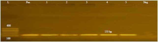 | |
| Fig. 3: | PCR results for S. agalactiaec fb gene showing positive amplification of 153 bp in the 5 tested samples |
| Lanes 1: 5 represent 5 tested samples, Pos: Positive control, Neg: Positive control, L [Gelpilot100 bp ladder (Qiagen, 100-600 bp)] | |
 | |
| Fig. 4: | PCR results for E. coli genes (rfb Gene and wzy Gene) |
| Lane 1: 1 kb DNA marker Fermentas Lane 4: Sample 2 (E. coli O25), Lane 2: Sample 1 (E. coli O157) Lane 5: Control negative, Lane 3: Control negative | |
| Table 1: | Statistical analytical results of screening tests in the examined positive CMT quarter milk samples |
 | |
| Table 2: | Prevalence of the isolated micro-organisms in the examined positive CMT quarter milk samples (QMS) |
 | |
Coliforms: Regarding the results represented in Table 4 Coliforms could be detected in all of the examined positive CMT quarter milk samples with isolation of 147 strains, the biochemical identification of coliform organisms revealed that Citrobacter diversus 82 (55.78%) was the most common one followed by Serratia fonticola 18 (12.25%) and Citrobacter freundii 13(8.84%) then Klebsiella oxytoca 9 (6.13%) and Enterobacteraerogenes 8 (5.44%), E. coli 7 (4.76%) and Enterobacter intermedius 7 (4.76%), finally Klebsiella Pneumoniae subsp. Azoenae 3 (2.04%).
| Table 3: | Incidence of isolated Streptococcus agalactiae in the positive CMT quarter milk samples (total streptococcus isolates = 130) |
 | |
Incidence of Escherichia coli in the examined QMS: Results illustrated the serological identification of the suspected isolates of E. coli by slide agglutination test. The results showed that two (1.36%) isolates out of 7 (4.76%) were positive and they were E. coli O25 and E. coli O157, when these isolates 2 (1.36%) were investigated by PCR, the results confirmed the obtained results serologically (Fig. 4, 5).
 | |
| Fig. 5: | Prevalence of E. coli using different methods of identification |
| Table 4: | Incidence of coliform isolates obtained from the positive CMT quarter milk samples |
 | |
DISCUSSION
Subclinical mastitis milk appears grossly normal and there are no visible signs of inflammation in the udder. However there are many changes encountered in milk from udder with subclinical infection, cellular, biochemical and enzymatic changes are present in such milk. These changes are detected in most of screening and routine tests.
The CMT is the most common test used for diagnosis of subclinical cases depending on detection of increased leukocyte count in milk. Data illustrated in Fig. 1 showed that 43.5% of the examined samples had subclinical mastitis and these results were in accordance with that reported by Ayano et al.21, who evaluated 546 milking cows by CMT and found that 41.02% of the examined samples were positive for subclinical mastitis, while lower than that obtained by Mureithi and Njuguna22, who found that 64% of samples had SCM, where as higher than that recorded by Sanotharan et al.23, who illustrated that 19.1% of the examined quarters milk samples were positive to CMT. This difference in results may be attributed to kind of milking (manual, automatic), numbers of milking time, farm hygiene, milking cows like (age, climate, state of udder and numbers of lactation) and labor hygiene.
Somatic Cell Count (SCC) has been recognized as an available tool for identifying cows with intra mammary infection (IMI) with major pathogens or for monitoring the udder health at the herd level24, also there are some other factors have a direct and dramatic effect on SCC as: Stresses, seasonal variations, age of cow, stage of lactation and time of milking25. In healthy udder, Milk Somatic Cell Count (MSCC) usually ranged between 50,000 and 100,000 cells mL–1 and in some cases up to 200,000 cells mL–1. If the SCC exceeded 200,000 cells mL–1, animal is considered affected with subclinical mastitis26, while Egyptian standards considered udder is affected with subclinical mastitis when MSCC exceed20 500.000 cells mL–1.
Results of MSCC that recorded in Table 1 were nearly similar to that mentioned by Mohamed et al.27, who found that the average count of somatic cell were 9.24±2.0 (×105 cells mL–1), while higher than that reported by Andrea et al.28, who showed that the average SCC count was 721,000 cells mL–1 in the examined milk samples indicating presence of subclinical mastitis. The obtained results proved the presence of good correlation between CMT and SCC as a tool for monitoring subclinical mastitis in dairy farms and these results were agreed with that reported by Abd El-Fatah29 and Khalil30.
Subclinical Mastitis declined milk lactose in the affected quarters due to the damage in the alveolar epithelial cells, also had markedly changed the ionic environment, sodium and chloride are increased, while potassium levels was decreased, by necessity to maintain udder osmotic equilibrium31,32 and this resulting in higher Koestler values which computed by using percentages of chloride and lactose, showing an increase in values with severity of mastitis13 and this was in accordance with that reported by Morsi et al.33, who proved that the decrease in milk lactose content and/or the increase in milk chloride content can be used as an index for suspecting the presence of sub-clinical mastitis.
Results illustrated in Table 1 which revealed that the mean Koestler value were 4.62±0.08 were lower than that mentioned by Kifaro et al.13, who stated that the mean Koestler value in its study was very high (6.4).
Although CMT and SCC are field, rapid and cheap tools helping as a screening tests for detection of subclinical mastitis34, CMT is unsuitable in early lactation or in dry period and SCC lacks the needed specificity because its affected by many other factors such as the number of lactations, stage of lactation, level of milk production, season, age and breed of cattle35,36.
The detection of milk enzymatic activities of lactate dehydrogenase enzyme (LDH) might represent a reliable diagnostic method for identifying subclinical mastitis in early lactation and dry period37.
Some enzyme levels increase in milk during intra mammary infections38, 39, one of these enzymes is LDH which is a cytoplasmic enzyme that has been proposed as a biomarker for udder health check, it gets released into milk from ruptured mammary epithelial cells, phagocytes and from serum, the activity of this enzyme significantly increase in milk obtained from quarters with subclinical mastitis and its activity has a high and positive correlation with SCC especially in infected quarters40-42.
Results obtained in Table 1 were in accordance with Batavani et al.43, who compared the level of LDH in milk samples from healthy quarters and milk from quarters with subclinical mastitis and found that the activity of LDH elevated in quarters affected with subclinical mastitis (1524.04 vs. 485.94 U L–1) and Mohammadian40, Kalantari et al.42 and Sorensen et al.44, who reported marked elevation in LDH activity in milk serum from sub clinically infected bovine mammary gland but Babaei et al.37 reported that LDH was not a sensitive marker and the blood LDH may be transferred to milk due to damage of blood-milk barrier.
Measuring enzymatic activities in milk which is both easy and of low cost compared to other methods could be used as a diagnostic test with acceptable sensitivity and specificity for detecting quarters with subclinical mastitis and these was assured by Kalantari et al.42, who reported that Milk LDH had the most clinical accuracy with 94.8 sensitivity and 94.1% specificity.
The positive correlation between SCC and Koestler value was agreed with that cited by Kifaro et al.13, who reported that lactose content tended to decrease with a tendency for chloride content to increase with increase in CMT score. The strong positive correlation of blood serum LDH with SCC (p< 0.01) was approved by Chagunda et al.45, Hiss et al.46 and Guha et al.47, who reported that LDH is positively correlated with SCC. Kalantari et al.42 showed that there is a significant positive correlation between LDH in milk and somatic cells and oppositely no significant increase was seen in the activity of these enzymes in the blood serum of dairy cows with subclinical mastitis compared to the healthy cows.
From the results, study showed a high prevalence of SCM which could be explained by the poor management and udder health practices, inadequate milking procedures, non-use of teat dips and other mastitis control techniques and inadequate screening tests for sub clinical mastitis detection at earlier stages.
In order to establish a specific and efficient management of dairy herd and to avoid the development of clinical mastitis, identification of micro-organisms responsible for subclinical mastitis is significant48.
Bacteria that commonly cause mastitis are generally classified into contagious and environmental pathogens depending on the source of the pathogen and mode of transmission. Streptococcus agalactiae and Staphylococcus aureus are considered as typical contagious pathogens, these pathogens have adapted to survive within the mammary gland and are spread from cow to cow at or around the time of milking. Typical environmental organisms are called environmental streptococci (Streptococci other than Streptococcus agalactiae) and the enterobacteriaceae49.
The obtained results of Streptococci showed in Table 2 were nearly similar to that obtained by Merl et al.50, while lower results were determined by Hanan et al.51 and Mpatswenumugabo et al.52, where as lower findings of Coliforms were reported by Ferguson et al.53 and Mbuk et al.54.
El-Attar et al.55 considered these micro-organisms as major causative agents of sub clinical mastitis worldwide due to teat-to-teat and cow-to-cow spread, feasibly via milking machines and the milker’s hands under the lack of hygiene and this opinion was proven by Radostits et al.56, who cited that Streptococcus spp. was the most prevalent bacteria along with Staphylococcus spp.
It is important to identify the presence of S. agalactiae in a herd with the appearance of the first infected animal as this pathogen is highly infectious bovine mastitis pathogen that can rapidly spread throughout a herd from a single infected animal57. Moreover mastitis caused by S. agalactiae is extremely costly, once these bacteria appear usually infect many quarters of the mammary gland causing extensive damage to the alveolar cells and resulting in decrease of milk production58,59.
The results obtained in Table 3 showed the incidence of contamination of the examined positive CMT quarter milk samples with S. agalactiae biochemically were in accordance with that obtained by Bhagat et al.60, who found that 22% of the isolated strains were Streptococcus agalactiae, whereas Lower results were determined by Mpatswenumugabo et al.52, who found that Streptococcus agalactiae were isolated from 5.8% of the examined isolates. The isolated S. agalactiae were confirmed by PCR as an accurate and rapid molecular technique in comparison to conventional biochemical test61,62.
The primary source of environmental pathogens is the bedding used to house cattle but contaminated teat dips, intramammary infusions, water used for udder preparation before milking, water ponds or mud holes, skin lesions, teat trauma and flies have all been incriminated as sources of infection63,64. Coliforms are able to produce subclinical infections persist for longer periods of time65.
Recently, all species and all serotypes of Klebsiella, Citrobacter, Enterobacter and Serratia are listed by the United States Public Health Service, Department of Health and Human Services bioterrorism list of dangerous biological agents that have the potential to pose a severe threat to public health and safety to animal health, plant health or to animal and plant products66.
The E. coli is considered an environmental pathogen and one of the most important causes of bovine mastitis, which is mostly observed in the early lactation period and in high-producing cows with low somatic cell counts67 and one of the major sources of economic loss in the dairy industry due to reduced milk production, treatment costs, discarded milk and occasional fatal disease68.
Results presented in Fig. 5 illustrated the serological identification of the suspected isolates of E. coli by slide agglutination test and confirmed by PCR were nearly similar to that reported by Vasquez-Garcia et al.28, who isolated Escherichia coli with the percentage of 4.5% from subclinical mastitis milk, higher obtaining were recorded by El-Bagory and Zayda69, who cited that E. coli could be isolated from cows suffered from subclinical mastitis with the percentages of 15 and 2% and Singh et al.70, who stated that out of 24 isolates, 4 isolates (16.66%) proved to be E. coli, while lower results were reported by Mpatswenumugabo et al.52 who could isolate Escherichia coli with the percentage of 1.5%.
Milk collected for human consumption can become contaminated with E.coli directly via animal faeces and infected udder or indirectly via contaminated farm and dairy parlor environments, equipment and workers. Moststrains of E. coli are harmless commensals, some area bletocause human gastro in test inaldisease with mild to severe symptoms that may progress to long-term squealorfatal out comes in high-risk individuals. Also E. coli was found to be responsible for cases of cystitis, pyelitis, pyelonephritis as well as appendicitis and peritonitis71,72.
Finally subclinically infected udder quarters can develop clinical mastitis with high rate of infection73, cows with subclinical mastitis maintain a reservoir of infection within the dairy herd and increase the potential exposure of uninfected cows to contagious pathogens74. Milk and dairy products made from these sub clinical mastitis milk may cause food borne diseases75,76, so early detection of this disease at the sub clinical stage in very important for control and prevention.
CONCLUSION
To reduce the prevalence of SCM which is a hidden problem facing dairy herds proper sanitation and hygiene during milking procedures and management practices of dairy cows, application of screening tests to detect SCM at earlier stages, High correlation between LDH and SCC in the present study advised that LDH activity which is both easy and cost effective with high sensitivity and specificity can substitute SCC for detection of SCM, establishment of correct in vitro anti biogram that were the main prerequisites for implementation of effective treatment of SCM with timely treatment might prevent the occurrence of clinical mastitis and transmission to herd mates with improvement in milk production and heat treatment of raw milk before human consumption.
SIGNIFICANCE STATEMENT
The high incidence of Subclinical Mastitis which is the major hidden problem in the dairy farms can be reduced by many ways, the most important one is the early detection of SCM by using more recent screening methods as the determination of the enzymatic activity of LDH (Lactate Dehydrogenase enzyme) as well as the isolation of the etiological agents of SCM using culture methods.
ACKNOWLEDGMENT
First and before all thanks god the most graceful and the most merciful. I would like to express my gratitude to Professors Doctor Ragaa Shehata Hafez and Professors Doctor Sabry Darwish Morgan, the professors of Milk Hygiene and Control, Department of Food Hygiene and Control, Faculty of Veterinary Medicine, Cairo University, Egypt for their valuable ideal guidance and constructive criticism.
REFERENCES
- Eshratkhah, B., R. Beheshti and J. Shayegh, 2012. Variation of some minerals values in subclinical mastitic milk of buffaloduring different ages and lactation stages. Global Vet., 8: 333-337.
Direct Link - Fox, L.K. and J.M. Gay, 1993. Contagious mastitis. Vet. Clin. N. Am.: Food Anim. Pract., 9: 475-487.
CrossRefDirect Link - Smith, T.H., L.K. Fox and J.R. Middleton, 1998. Outbreak of mastitis caused by one strain of Staphylococcus aureus in a closed dairy herd. J. Am. Vet. Med. Assoc., 212: 553-556.
Direct Link - Guha, A., S. Gera and A. Sharma, 2010. Assessment of chemical and electrolyte profile as an indicator of subclinical mastitis in riverine buffalo (Bubalus bubalis). Haryana Vet., 49: 19-21.
Direct Link - Forsback, L., H. Lindmark-Mansson, A. Andren and K. Svennersten-Sjaunja, 2010. Evaluation of quality changes in udder quarter milk from cows with low-to-moderate somatic cell counts. Animal, 4: 617-626.
CrossRefDirect Link - Idriss, S.E.A.B., V. Tancin, V. Foltys, K. Kirchnerova, D. Tancinova and M. Vrskova, 2013. Relationship between mastitis causative pathogens and somatic cell counts in dairy cows. Potravinarstvo Slovak J. Food Sci., 7: 207-212.
Direct Link - Yang, M., J. Shi, J. Tian, J. Tao and M. Chai et al., 2017. Exogenous melatonin reduces somatic cell count of milk in Holstein cows. Scient. Rep., Vol. 7.
CrossRefDirect Link - Zecconi, A., L. Cesaris, E. Liandris, V. Dapra and R. Piccinini, 2006. Role of several Staphylococcus aureus virulence factors on the inflammatory response in bovine mammary gland. Microb. Pathog., 40: 177-183.
CrossRefPubMedDirect Link - Kifaro, G.C., N.G. Moshi and U.M. Minga, 2009. Effect of sub-clinical mastitis on milk yield and composition of dairy goats in Tanzania. Afr. J. Food Agric. Nutr. Dev., 9: 622-634.
CrossRefDirect Link - Ke, D., C. Menard, F.J. Picard, M. Boissinot, M. Ouellette, P.H. Roy and M.G. Bergeron, 2000. Development of conventional and real-time PCR assays for the rapid detection of group B streptococci. Clin. Chem., 46: 324-331.
Direct Link - Louie, M., S. Read, A.E. Simor, J. Holland and L. Louie et al., 1998. Application of multiplex PCR for detection of non-O157 verocytotoxin-producing Escherichia coli in bloody stools: Identification of serogroups O26 and O111. J. Clin. Microbiol., 36: 3375-3377.
Direct Link - Ayano, A.A., F. Hiriko, A.M. Simyalew and A. Yohannes, 2013. Prevalence of subclinical mastitis in lactating cows in selected commercial dairy farms of Holeta district. J. Vet. Med. Anim. Health, 5: 67-72.
Direct Link - Mureithi, D.K. and M.N. Njuguna, 2016. Prevalence of subclinical mastitis and associated risk factors in dairy farms in urban and peri-urban areas of Thika Sub County, Kenya. Development, Vol. 28.
Direct Link - Sanotharan, N., M. Pagthinathan and M.S.M. Nafees, 2016. Prevalence of bovine subclinical mastitis and its association with bacteria and risk factors in milking cows of Batticaloa district in Sri Lanka. Int. J. Scient. Res. Innov. Technol., 3: 137-150.
Direct Link - Coulon, J.B., P. Pradel, T. Cochard and B. Poutrel, 1998. Effect of extreme walking conditions for dairy cows on milk yield, chemical composition and somatic cell count1. J. Dairy Sci., 81: 994-1003.
CrossRefDirect Link - Skrzypek, R., J. Wojtowski and R.D. Fahr, 2004. Factors affecting somatic cell count in cow bulk tank milk- A case study from Poland. J. Vet. Med. A, 51: 127-131.
CrossRefDirect Link - Mohamed, A.A., A.K. Wahba and R.A. Faisal, 2013. Some bacteriological and biochemical studies on subclinical mastitis in dairy cows. NY Sci., 6: 71-79.
Direct Link - Vasquez-Garcia, A., T.D.S. Silva, S.R.D. Almeida-Queiroz, S.H. Godoy, A.M. Fernandes, R.L. Sousa and R. Franzolin, 2017. Species identification and antimicrobial susceptibility profile of bacteria causing subclinical mastitis in buffalo. Pesquisa Vet. Brasil., 374: 447-452.
CrossRefDirect Link - Bruckmaier, R.C., JB. Ontsouka and J.W. Blum, 2004. Fractionized milk composition in dairy cows with subclinical mastitis. Vet. Med. Czeh., 8: 283-290.
Direct Link - Norberg, E., H. Hogeveen, I.R. Korsgaard, N.C. Friggens, K.H.M.N. Sloth and P. Lovendahl, 2004. Electrical conductivity of milk: Ability to predict mastitis status. J. Dairy Sci., 87: 1099-1107.
CrossRefDirect Link - Morsi, N.M., Y. Saleh, H. El-Gazzar and A. Hanafi, 2000. Effect of mastitis on milk fat content. Pak. J. Biol. Sci., 3: 196-200.
CrossRefDirect Link - Abdel-Rady, A. and M. Sayed, 2009. Epidemiological studies on subclinical mastitis in dairy cows in Assiut Governorate. Vet. World, 2: 373-380.
Direct Link - Pyorala, S., 2003. Indicators of inflammation in the diagnosis of mastitis. Vet. Res., 34: 565-578.
Direct Link - Babaei, H., L. Mansouri-Najand, M.M. Molaei, A. Kheradmand and M. Sharifan, 2007. Assessment of lactate dehydrogenase, alkaline phosphatase and aspartate aminotransferase activities in cow's milk as an indicator of subclinical mastitis. Vet. Res. Commun., 31: 419-425.
CrossRefDirect Link - Zhao, X. and P. Lacasse, 2008. Mammary tissue damage during bovine mastitis: Causes and control. J. Anim. Sci., 86: 57-65.
CrossRefPubMedDirect Link - Andrei, S., S. Matei, N. Fit, C. Cernea, S. Ciupe, S. Bogdan and I.S. Groza, 2011. Glutathione peroxidase activity and its relationship with somatic cell count, number of colony forming units and protein content in subclinical mastitis cows milk. Romanian Biotechnol. Lett., 16: 6209-6217.
Direct Link - Larsen, T. and K. Aulrich, 2012. Optimizing the fluorometric β-glucuronidase assay in ruminant milk for a more precise determination of mastitis. J. Dairy Res., 79: 7-15.
CrossRefDirect Link - Kalantari, A., S. Safi and F.A. Rahimi, 2013. Determination of the diagnostic value of milk lactate dehydrogenase and alkaline phosphatase in bovine subclinical mastitis. J. Vet. Clin. Res., 3: 235-247.
Direct Link - Batavani, R.A., S. Asri and H. Naebzadeh, 2007. The effect of subclinical mastitis on milk composition in dairy cows. Iran. J. Vet. Res., 8: 205-211.
CrossRefDirect Link - Sorensen, L.P., R.M. Engberg, P. Lovendahl and T. Larsen, 2015. Effects of Bos taurus autosome 9-located quantitative trait loci haplotypes on enzymatic mastitis indicators of milk from dairy cows experimentally inoculated with Escherichia coli. J. Dairy Sci., 98: 5440-5447.
CrossRefDirect Link - Chagunda, M., T. Larsen, M. Bjerring and K.L. Ingvartsen, 2005. Changes in Lactate Dehydrogenase, N-Acetyl-Beta-D-Glucosaminase and Somatic Cell Count in Relation to Development of Mastitis in Dairy Cows. In: Proceedings of the 4th IDF International Mastitis Conference, Mastitis in Dairy Production, Current Knowledge and Future Solutions, Hogeveen, H. (Ed.)., Wageningen Academic Publishers, Maastricht, The Netherlands.
- Guha, A., S. Gera and A. Sharma, 2012. Evaluation of milk trace elements, lactate dehydrogenase, alkaline phosphatase and aspartate aminotransferase activity of subclinical mastitis as and indicator of subclinical mastitis in riverine buffalo (Bubalus bubalis). Asian-Aust. J. Anim. Sci., 25: 353-360.
CrossRefDirect Link - Cheng, D.R., S.Y. Zhu, Y.H. Yin, W.W. Ding, Z.X. Mu, Z.R. Su and H.C. Sun, 2010. Prevalence of bacterial infection responsible for bovine mastitis. Afr. J. Microbiol. Res., 4: 1110-1116.
Direct Link - Bradley, A.J., 2002. Bovine mastitis: An evolving disease. Vet. J., 164: 116-128.
CrossRefDirect Link - Merl, K., A. Abdulmawjood, C. Lammler and M. Zschock, 2003. Determination of epidemiological relationships of Streptococcus agalactiae isolated from bovine mastitis. FEMS Microbiol. Lett., 226: 87-92.
CrossRefDirect Link - Mpatswenumugabo, J.P., L.C. Bebora, G.C. Gitao, V.A. Mobegi, B. Iraguha, O. Kamana and B. Shumbusho, 2017. Prevalence of subclinical mastitis and distribution of pathogens in dairy farms of Rubavu and Nyabihu districts, Rwanda. J. Vet. Med., Vol. 2017.
CrossRefDirect Link - Ferguson, J.D., G. Azzaro, M. Gambina and G. Licitra, 2007. Prevalence of mastitis pathogens in Ragusa, Sicily, from 2000 to 2006. J. Dairy Sci., 90: 5798-5813.
CrossRef - Mbuk, E.U., J.K.P. Kwaga, J.O.O. Bale, L.A. Boro and J.U. Umoh, 2016. Coliform organisms associated with milk of cows with mastitis and their sensitivity to commonly available antibiotics in Kaduna State, Nigeria. J. Vet. Med. Anim. Health, 8: 228-236.
Direct Link - Keefe, G.P., I.R. Dohoo and E. Spangler, 1997. Herd prevalence and incidence of Streptococcus agalactiae in the dairy industry of Prince Edward Island. J. Dairy Sci., 80: 464-470.
Direct Link - Edmondson, P., 2011. Blitz therapy for the eradication of Streptococcus agalactiae infections in dairy cattle. Practice, 33: 33-37.
CrossRefDirect Link - Keefe, G., 2012. Update on control of Staphylococcus aureus and Streptococcus agalactiae for management of mastitis. Vet. Clin.: Food Anim. Pract., 28: 203-216.
CrossRefDirect Link - Odierno, L., L. Calvinho, P. Traverssa, M. Lasagno, C. Bogni and E. Reinoso, 2006. Conventional identification of Streptococcus uberis isolated from bovine mastitis in Argentinean dairy herds. J. Dairy Sci., 89: 3886-3890.
CrossRefDirect Link - Matofari, J.W., Y. Mario, E.W. Mwatha and P.O. Okemo, 2003. Microorganisms associated with sub-clinical Mastitis in the Kenyan camel (Camelus dromedaries). J. Trop. Microbiol. Biotechnol., 2: 11-16.
Direct Link - Kivaria, F.M., J.P.T.M. Noordhuizen and A.M. Kapaga, 2006. Evaluation of the hygienic quality and associated public health hazards of raw milk marketed by smallholder dairy producers in the Dar es Salaam region, Tanzania. Trop. Anim. Health Prod., 38: 185-194.
CrossRefPubMedDirect Link - Burvenich, C., V. Van Merris, J. Mehrzad, A. Diez-Fraile and L. Duchateau, 2003. Severity of E. coli mastitis is mainly determined by cow factors. Vet. Res., 34: 521-564.
CrossRefPubMedDirect Link - Vangroenweghe, F., L. Duchateau, P. Boutet, P. Lekeux, P. Rainard, M.J. Paape and C. Burvenich, 2005. Effect of carprofen treatment following experimentally induced Escherichia coli mastitis in primiparous cows. J. Dairy Sci., 88: 2361-2376.
CrossRefDirect Link - Singh, N., P. Singh and R.K. Patel, 2016. Isolation and identification of bacterial organisms from mastitic milk. J. Livestock Sci., 7: 46-48.
Direct Link - Fratamico, P.M., C.E. Briggs, D. Needle, C.Y. Chen and C. DebRoy, 2003. Sequence of the Escherichia coli O121 O-antigen gene cluster and detection of enterohemorrhagic E. coli O121 by PCR amplification of the wzx and wzy genes. J. Clin. Microbiol., 41: 3379-3383.
CrossRefDirect Link - Zdunczyk, S., H. Zerbe and M. Hoedemaker, 2003. Importance of estrogens and estrogen-active compounds for udder health in cattle: A review. DTW. Deutsche Tierarztliche Wochenschrift, 110: 461-465.
Direct Link - Hortet, P., H. Seegers, C. Fourichon and F. Beaudeau, 1996. Udder Health Monitoring in Dairy Herds. In: Book of Abstracts of the 47th Annual Meeting of the European Association for Animal Production. Lillehammer, Norway, 25-29 August 1996, Van Arendonk, J.A.M. (Ed.)., Enfield Pub and Distribution Company, UK., pp: 154.
- Twardon, J., B. Sobieszczanska, A. Gonet and M. Blaszkowska, 2005. Epidemiology of Shiga-like Toxin-producing Escherichia Coli strains (STEC). Elect. J. Polish Agric. Uni. Series Vet. Med., Vol. 4.
Direct Link