
A.S.M. Fouzy
Department of Food Toxicology and Contaminants, National Research Center, Cairo, Egypt
M.M. El-Loly
Department of Dairy Science, National Research Center, Cairo, Egypt
Y.A. Ghazi
Department of Animal Reproduction and A.I., National Research Center, Cairo, Egypt
International Journal of Dairy Science
Year: 2008 | Volume: 3 | Issue: 1 | Page No.: 1-10


ABSTRACT
Six mature female Baladi goats were selected from Abou Rawash experimental farm, National Research Center, Egypt, during the period from August 2005 to January 2006. These animals divided to three groups each of them two animals, the first keep as control, the second has orally administered low dose of dioxins (1/13 and 1/27) LD50 and the third has orally administered high dose of dioxins (1/3 and 1/6) LD50. Milk and blood samples were collected after 48 and 96 h from dioxins exposure and after 96 h for protein fractions, to investigate the effect of dioxins on the physical, chemical and bacterial properties of goat`s milk, casein, whey protein fractions and blood picture. After 16 days, two of the goats were slaughtered and samples of the mammary gland were taken for analysis of dioxins residues. The results indicated that the gross chemical composition of goat`s milk with low dioxin doses had higher effect than the high dioxin doses as compared to control samples, especially for fat contents (71 and 65%), respectively. The average percentages of dioxins excretion in milk were 0.0015 and 0.0010 for low and high dioxin doses, while the percentages storage of dioxins in the mammary gland were 0.0011 and 0.0012 for low and high dioxin doses. Goats exposed to dioxins revealed significant decrease in total RBCs count and MCV indicating anemia. Also, the same significant decrease in total WBCs count associated with neutropenia. Goats exposed to dioxins revealed positive result by CMT test after 96 h post-treated. The results of bacteriological examination showed positive result only with the positive milk samples by CMT after 96 h the isolates were Streptococcus dysagalactiae and E. coli which were the most prevalent isolates indicate the presence of environmental mastitis. Generally, the oral ingestion of goats with dioxins resulted in different patterns of milk protein fractions in casein and whey proteins. Several bands were observed for dioxin low doses than dioxin higher doses in most fractions of casein and whey proteins.
PDF Abstract XML References Citation
How to cite this article
A.S.M. Fouzy, M.M. El-Loly and Y.A. Ghazi, 2008. Effect of Dioxins on Properties of Egyptian Goat`s Milk and Blood Profile with Relation to Mastitis. International Journal of Dairy Science, 3: 1-10.
DOI: 10.3923/ijds.2008.1.10
URL: https://scialert.net/abstract/?doi=ijds.2008.1.10
DOI: 10.3923/ijds.2008.1.10
URL: https://scialert.net/abstract/?doi=ijds.2008.1.10
INTRODUCTION
The potential environmental impact and health risk posed by the emission of toxic pollutants, such as Polychlorinated Dibenzo-p-Dioxins (PCDDs), dibenzofurans (PCDFs), Polychlorinated Biphenyls (PCBs) and metals from MSWIs has always been the public`s concern (Cheng et al., 2003).
PCDDs and PCDFs are groups of toxic and persistent polychlorinated chemicals ubiquitous in environment of all industrialized countries, which are toxic and potent carcinogenic compounds formed as contaminants in milk and foods. These components are lipophilic and they bio-accumulate in the food chain and in the human body and finally they represent a potential risk for human health. It has been estimated that more than 90% of non-occupational human exposure to PCDDs and PCDFs come from food chain (Travis and Hattemer-Frey, 1987; Ganesh and Jayaraj, 1995; Liem, 1999).
PCDDs and PCDFs are formed mainly as a result of pyrolytic processes, especially at incomplete combustion of organic materials during industrial and other human activities such as the processing of coal and crude oil, the combustion of natural gas, vehicle traffic, cooking and smoking of tobacco (Zedeck, 1988; Scialli, 2001).
Humans and various animal species exposure to these compounds occurs mainly through food, which causes wide varieties of adverse effects in different body tissue with species specific effect. Due to their physical and chemical properties they migrate through the food chain into lipophilic compartments (Menzie, 1990). Thus they accumulate in lipids at the end of the food chain (Madhavan and Naidu, 1995; Bosset et al., 1998). The knowledge of transfer pathways through the food chain is an important issue in food safety. It is known that dioxins are transferred from feed to milk after oral ingestion by lactating ruminants (Jones et al., 1989).
Mastitis is a disease causing inflammation in the parenchyma of the mammary glands regardless of the cause. It characterized by physical, chemical and usually bacteriological changes in the milk. Goats are much less frequently affected by contagious mastitis than cattle but particular care is needed in the clinical examination of goat`s milk because of its apparent normality when there are severe inflammatory changes in the udder (Radostits et al., 1994).
Several methods have been reported for detecting mastitis, but bacteriological isolation of the causative microorganisms is the most accurate one (Hall and Rycroft, 2007).
The objectives of the current study were to determine the concentrations of transferred dioxins in milk and mammary gland of lactating baladi Egyptian goats and to investigate their effects on the chemical and bacterial properties of milk and its relation to mastitis and their effect on casein and whey protein fractions as well as blood picture.
MATERIALS AND METHODS
Animals and Samples
Six mature female baladi goats were selected from Abou Rawash experimental farm, National Research Center, Egypt, during the period from August 2005 to January 2006, mean body weight: 30 ± 5 kg, age: 3 years ± 6 months. These animals divided to three groups each of them two animals, the first keep as control, the second for low doses and the third for high doses. They stayed one week adaptation period to the open room and fed on commercial concentrate mixture with rice straw and barseem ad libitum. The goats were milked manual. The average milk production during one week before the experiment was about 500 ± 10 mL day-1.
Milk and blood samples were collected after 48 and 96 h from dioxins exposure and after 96 h for protein fractions, to investigate the effect of dioxins on the physical, chemical and bacterial properties of goat`s milk, casein, whey protein fractions and blood picture. Blood samples (7 mL) were immediately centrifuged (3000 rpm/15 min) to obtain plasma. Immediately after complete milking of individual animal, the milk was thoroughly mixed and about 50 mL was taken in a clean stopper sample bottle in an ice box and transferred to the dairy laboratory, National Research Center. The milk samples were analysed for Total Solids (TS) by the drying oven method at 105 °C for 3 h, fat and ash contents as described by AOAC (1990). The titratable acidity, pH value and Total Protein (TP) of milk were determined as given by Ling (1963). Lactose content was colorimetrically determined as described by Barnett and Abd El Tawab (1957). Samples were collected and stored under freezing at 20 °C until analysis of PCDDs and PCDFs residues at the Institute for Hygiene and Food, Federal Research Center for Nutrition and Food Safety-Kiel, Germany.
Hematological Evaluation
Complete blood picture was carried (Jain, 2000).
Dioxin Standard
The stock standard solution contained Pg WHO-TEQ (PCDDs/PCDFs) of 17 congeners Labelled with C13 and 17 native congeners at equal preparation total: 57.7826 Pg WHO. It was obtained from Chemisches und Veterinäruntersuchungsamt Freiburg, Germany-Malisch (Rainer, 2002).
Dioxin Oral Ingestion and Doses Application
At the first day of the experiment after morning milking the oral application of the dioxin (4 and 2 mL of stock standard solution = 57.7826 pg WHO-TEQ μL-1) diluted in 5 mL water was directly injection to the mouth of the animal by a sterile syringe amount of doses the administration was repeated every two days to two of the animals for three times and the two other animals were dosed only one time. The doses were calculated as consider the weight of goats 30 kg and the amount of dioxins 6.9, 3.4, 2.3, 1.1 μg, which represent 0.23, 0.11, 0.077, 0.037 μg/body weight, equal 1/3, 1/6, 1/13, 1/27 of the LD50 for guinea pig (0.06 μg kg-1 body weight).
After 16 days two of the goats were slaughtered and samples of the mammary gland was taken for analysis of PCDDs and PCDFs residues.
Method Used for Determination of Dioxins
Analytical Procedures
Fat is extracted with acetone/petroleum benzene and it is removed by gel permeation chromatography. The following clean up of the extracts is performed with florisil and aluminium oxide. The dioxins are determined by GC/HRMS (Furst et al., 1989).
Gas Chromatography-Mass Spectra (GC/MC)
Finnegan MAT 95/HP Series 5890. Conditions: injector 280 °C; column: DB5 60 m, 0.25 μ film thickness, 0.25 mm ID; temperature programmed: 1 min at 140 °C, in 15 min to 240 °C, in 3.5 min to 300 °C, in 15 min at 300 °C; carrier gas: Helium, 4 mL min-1; SIM: 305.90-471.78; scan time: 0.2 s; SEV: 1.6 kv; ion source pressure: 3x10⁻⁷ pa; system pressure: 1x10⁻¹⁰ pa; transfer line temperature: 280 °C; working resolution between 6000 and 8000 (10% valley).
Validation and Detection Limit
The method was validated by the Fraunhofer Institute für Verfahrenstechnik und Verpackung, Munich Germany. The detection limit for all compounds determined is 0.003 pg g-1 fat.
California Mastitis Test (CMT)
A cell count of greater than 1 millions cells mL-1 can be regarded as positive for mastitis (Schaeren and Maurer, 2006).
Bacteriological Examination of Milk
The milk samples were incubated aerobically at 37 °C for 24 h then centrifuged at 3000 rpm for 20 min. The cream and supernatant fluid were discarded. A loop full from the sediment was streaked on to the surface of blood agar, nutrient agar and MacConkey`s agar. The inoculated plates were incubated at 37 °C and examined for bacteriological growth (Ghazi et al., 2002).
Separation of Milk Protein Fractions by SDS-Polyacrylamide Slab Gel Electrophoresis (SDS-PAGE) and Quantitative Measurement by Scanning
The casein and whey samples were separated from the whole milk and the electrophoresis patterns of these protein fractions were carried out using slab gel electrophoresis technique, where polyacrylamide gel containing 0.1% SDS according to the conventional method of Laemmli (1970). Proteins were denaturated by heating for 5 min in sample buffer containing 1% SDS in a boiling water bath prior to its application to the gel. After electrophoresis, proteins were visualized using coomasie blue 0.1% solution. A photograph of stained gel was scanned with an Image densitometry (Biorad G-70) using Gel Pro Analyzer v.3.0 software. For stained gels, optical transmission was measured at a wavelength of 540 nm. The important parameters in this analysis include molecular weight and the percentage of each separated band.
Statistical Analysis
The obtained data were computed and statistically analysed according to Snedecor and Cochran (1982).
RESULTS AND DISCUSSION
Effect of Dioxins on Gross Chemical Compositions of Goat`s Milk
The average total solids contents of untreated goats milk was 13.11% (Table 1). These results are closely similar to those obtained by El-Alamy et al. (1987) being 13.0-13.8% as well as Kholif and Kholif (2003) being 13.2-13.5%.
Also, the results are higher than those reported by El-Loly (1987), namely, 12.33%, Kholif and Abo El-Nor (1998) being 12.0-12.5%, Kholif (1999) being 11.6-12.2%, Abo El-Nor and Kholif (1999) being 11.9-12.6%, Kholif and El-Shewy (2004) being 12.4-12.6%, Kholif and Abd El-Gawad (2005) being 11.7-11.9% and Kholif et al. (2005) being 11.8-12.8%. The total solids contents were decreased to about of 51 and 47% after oral ingestion with low and high dioxin doses, respectively.
Concerning to the average total protein contents of goat`s milk were 5.32, 3.21 and 3.33% for control, low and high dioxin doses, respectively (Table 1). These results are higher than those reported by El-Alamy et al. (1987), El-Loly (1987), Kholif and Abo El-Nor (1998), Abo El-Nor and Kholif (1999), Kholif (1999) as well as Kholif and Abd El-Gawad (2005). Also, it appears from this table that the average fat contents were 3.95, 1.15 and 1.35% for control, low and high dioxin doses, respectively (Table 1). These results are closely similar to those given by El Zayat et al. (1984), El-Alamy et al. (1987) and El-Loly (1987). While, the data were higher than those obtained by Kholif and Abo El-Nor (1998), Abo El-Nor and Kholif (1999) as well as Kholif (1999).
Regarding to the average lactose contents of goat`s milk were 2.95, 1.35 and 1.76% for control, low and high dioxin doses, respectively. The present data are lower than those obtained by El Zayat et al. (1984), El-Alamy et al. (1987), El-Loly (1987), Kholif and Abo El-Nor (1998), Abo El-Nor and Kholif (1999) and Kholif (1999).
Data concerning the average ash contents of goat`s milk were 0.81, 0.53 and 0.68% for control, low and high dioxin doses, respectively. These results are in according with those reported by El-Alamy et al. (1987), El-Loly (1987), Kholif and Abo El-Nor (1998), Abo El-Nor and Kholif (1999) and Kholif (1999).
| Table 1: | Gross chemical compositions of goat`s milk after 48 and 96 h of oral dioxin doses* |
 | |
| *: Means of three replicates for each sample | |
From the previous results, it is clear that the gross chemical composition of goat`s milk with low dioxin doses had higher effect than the high dioxin doses compared to control samples, especially for fat contents (71 and 65%), respectively. This may be due to their physical and chemical properties of varieties of adverse effects in milk and different body tissues. This finding was similar to those obtained by Jones et al. (1989) and Bosset et al. (1998).
Kholif et al. (1994) reported that the spraying by diazinon to control the external parasites reduced both of total protein and lactose contents in buffaloes and Friesian cows.
Transfer of Dioxins from Food to Milk and Mammary Gland
Data indicated that the average levels of dioxins were 26.10, 29.99 and 32.64 pg WHO-TEQ (PCDDs/PCDFs) for control, low and high dioxin doses (Table 2). The average percentages of dioxins excretion in milk were 0.0015 and 0.0010 for low and high dioxin doses.
Concentrations of PCDD/Fs in the mammary gland were 40.4301 and 85.6664 pg WHO-(PCDD/F)-TEQ/g fat for low and high dioxin doses, respectively (Table 3). While the percentages of dioxins excretion in mammary gland were 0.0011 and 0.0012 for low and high dioxin doses, respectively.
These results do agree with Grova et al. (2002) they reported that a small amount of 2, 3, 7, 8-TCDDs transfers into milk of lactating goats after a single oral ingestion and a large part of 2, 3, 7, 8-TCDDs ingested remained in the organs. Also, Ruoff (1995) who reported about the transfer of polychlorinated dibenzodioxins and furans (PCDDs, PCDFs) into the milk and organs of lactating cows after oral supplementation. The present data were proved by Fouzy and Ruoff (2006) they mentioned the same results in excretion of these components from goats to their milk and mammary gland. On the other hand, these data were proved by Fouzy et al. (2007) they decided that the dioxin residue levels in milk were the same in mammary gland 0.0012% as a ratio between oral dose and residues.
| Table 2: | Transfer PCDDs and PCDFs from goats to milk after 48 and 96 h of oral ddioxin doses |
 | |
| *: Pg WHO-TEQ (PCDDs/PCDFs) of 17 congeners Labeled with 13C and 17 native congeners at equal preparation | |
| Table 3: | Distribution of PCDDs, PCDFs in the mammary gland of Egyptian baladi lactating goats after 16 days of oral dioxin doses |
 | |
| *: Pg WHO-TEQ (PCDDs/PCDFs) of 17 congeners Labeled with 13C and 17 native congeners at equal preparation | |
Effect of Dioxins on Blood Picture
Significant decrease in RBCs counts (96 h) and MCV (48 and 96 h) was noticed post treatment as compared with untreated group. Meanwhile, non significant increase in Mean Corpuscular Hemoglobin (MCH) and Mean Corpuscular Hemoglobin Concentration (MCHC) were recorded 96 h post treatment with dioxin. Moreover, significant decrease in total WBCs count associated with neutropenia was recorded 48 and 96 h post-treated with dioxins as compared with untreated group. On the other hand, eosinophilia was obvious in dioxin exposed female goats as shown in samples taken after 96 h post treatment compared with untreated group (Table 4).
Goats exposed to dioxins revealed significant decrease in total WBCs count associated with neutropenia, also, the same significant decrease in total RBCs count and MCV indicating anemia. It is well known that dioxin had immune suppressive effect on bone marrow lymphocyte stem cells by mechanism mediated directly or indirectly through estrogenic action (Frazier et al., 1994). Murante and Gasiewicz (2000) added that proliferation and/or differentiation processes of hemopoietic stem cells are affected by TCDD and these effects contribute to a reduced capacity of bone marrow to generate pro-T lymphocytes. Moreover, it was found that TCDD-treated hematopoietic stem cells almost lost long-term reconstitution activity (Sakai et al., 2003).
Effect of Dioxins on Mastitis in Goat`s Milk
Goats exposed to dioxins revealed positive result by CMT test after 96 h post-treated. The milk samples were investigated with bacteriological examinations. The results of bacteriological examination showed positive result only with the positive milk samples by CMT after 96 h the isolates were Streptococcus dysagalactiae and E. coli which were the most prevalent isolates (Table 5). CMT is one of the physical tests which carried out on milk for diagnosis of mastitis in goats. The positive results of CMT indicate the presence of significantly increase inflammatory cells (leukocyte count) in milk which seems damage of epithelium cells of mammary tissues (Radostitis et al., 1994). The isolation of Strept. dysgalactiae and E. coli which indicate the presence of so called environmental mastitis (Ghazi et al., 2002). Present findings may indicate the presence of environmental mastitis which depended upon the immune status of the mammary glands. Some authors explained the immune toxicity of dioxin that affect on the local immunity of mammary gland which depend on T cell and it effect as immunosuppersion (Kerkvliet et al., 2002). Also, Fouzy et al. (2007) showed that the goats exposure to dioxin have multiple interstitial foci of lymphocytic cell aggregations in addition to interaluminal accumulation of leukocytes in some acini. From the public health point of view, the presence of these microorganisms, acts as a hazard for milk consumers increasing the incidence of disease and food poisoning as well as milk deterioration (Ghazi et al., 2002)
| Table 4: | Effect of oral ingestion of PCDDs and PCDFs on blood picture of Egyptian baladi goats* |
 | |
| *: Means with different superscript are significantly different within columns at p<0.05 | |
| Table 5: | Effect of dioxins on mastitis in goat`s milk |
| Positive CMT: Cell count greater than 1 millions cells mL-1; Positive bacteriological: Presence of more than one pathogenic bacterial isolate in milk sample | |
Effect of Dioxins on Electrophoretic Separation of Goat`s Milk Protein Fractions
Figure 1 shows the electropherograms of casein from individual goats` milk after 96 h of oral dioxin doses. The patterns were divided into three regions namely starting from the origin (negative charge) κ-, β-and αs-casein, respectively. The faster group resembles αs-casein, the second with lower mobility resemble β-casein and the third that had the lowest mobility than the other fractions were known as κ-casein zone. The αs-casein fraction was found to include two bands for samples No. 2 and 3 (dioxin low doses); while for samples No. 1 and 4 include one band (control and dioxin high doses). Also, the β-casein zone includes two bands for samples No. 2 and 3 (dioxin low doses). Concerning to κ-casein zone, two bands were apparent in all casein samples.
Figure 2 revealed that electrograms separates into a series of components corresponding to immunoglobulins (Igs), serum albumin, α-lactalbumin (α-la) and β-lactoglobulin (β-lg) at the same order of ascending in their mobility. The β-lg band was always in all whey protein samples, also, the α-la was mostly appeared with single band for samples No. 1 and 4 (control and dioxin high doses), but it was shown with two or three bands in samples No. 2 and 3 (dioxin low doses), The other minor fractions that had mobility faster than Igs and behind the serum albumin band would represent the proteose peptone 9 (pp 9), also, another fraction of pp 7 was faster than serum albumin and behind α-la band, they appeared in some whey protein samples such as 1, 2 and 4.
The previous results do agree with those reported by El-Shibiny (1978) who reported that the zone near the origin of the gel represent Igs.
Previously, we have shown that the oral ingestion of goats with dioxins resulted in different patterns of milk protein fractions in casein and whey proteins. Several bands were observed for dioxin low doses than dioxin higher doses in most all fractions of casein and whey proteins.
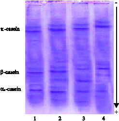 | |
| Fig. 1: | Electrophoretic pattern in casein of goat`s milk after 96 h of oral dioxin doses; Lane 1: Untreated goat`s milk (control); Lane 2 and 3: Treated goat`s milk with low dose of dioxin; Lane 4: Treated goat`s milk with high dose of dioxin |
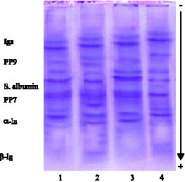 | |
| Fig. 2: | Electrophoretic pattern in whey proteins of goat`s milk after 96 h of oral dioxin doses; Lane 1: Untreated goat`s milk (control); Lane 2 and 3: Treated goat`s milk with low dose of dioxin; Lane 4: Treated goat`s milk with high dose of dioxin |
CONCLUSIONS
It is clear that the gross chemical composition of goat`s milk was affected with dioxin doses; this may be due to the physical and chemical properties of dioxin compounds which have adverse effects to body tissues and consequently transferred to milk. The percentages of dioxins transfer into milk and mammary gland after oral dose indicate the selectivity of the intestinal or mammary epithelial barrier for dioxins in lactating goats and their capacity to metabolize them (depending on their physical and chemical properties). This may explain the differential behavior within the compounds.
Goats exposed to dioxins revealed significant decrease in total WBCs count associated with neutropenia, also, the same significant decrease in total RBCs count and MCV indicating anemia. Doses of dioxin facilitated the incidence of mastitis. So that, the presence of pathogenic microorganisms, act as a hazard for milk consumers increasing the incidence of disease and food poisoning as well as milk deterioration. The oral ingestion of goats with dioxins resulted in different patterns of milk protein fractions in casein and whey proteins. Several bands were observed for dioxin low doses than dioxin higher doses in most all fractions of casein and whey proteins.
REFERENCES
- Barnett, A.J.G. and G.A. Tawab, 1957. A rapid method for the determination of lactose in milk and cheese. J. Sci. Food Agric., 8: 437-441.
CrossRefDirect Link - Cheng, P.S., M.S. Hsu, E. Ma, U. Chou and Y.C. Ling, 2003. Levels of PCDD/Fs in ambient air and soil in the vicinity of a municipal solid waste incinerator in HsinChu. Chemosphere, 52: 1389-1396.
Direct Link - Fouzy, A.S.M. and U. Rouff, 2006. Distribution of PCDDs/PCDFs into milk and organs of Egyptian Baladi goats after oral supplementation of dioxins. Kieler Milchwirtschaftliche Forschungsberichte, 58: 5-15.
Direct Link - Fouzy, A.S.M., H.M. Desouky, Y.G. Ghazi and A.M. Hammam, 2007. Some clinico and histopathological changes in female goats experimentally exposed to dioxin. Pak. J. Biol. Sci., 8: 1213-1220.
CrossRefDirect Link - Frazier, Jr., D.E., A.E. Silverstone and T.A. Gasiewicz, 1994. 2, 3, 7, 8-Tetrachlorodibenzo-p-dioxin-induced thymic atrophy and lymphocyte stem cell alterations by mechanisms independent of the estrogen receptor. Biochem. Pharmacol., 47: 2039-2048.
CrossRefDirect Link - Ganesh, K.C. and R.K. Jayaraj, 1995. Structural, clinical and transmission aspects of dioxins: Potential environmental pollutants. Curr. Sci., 69: 237-240.
Direct Link - Hall, S.M. and A.N. Rycroft, 2007. Causative organisms and somatic cell counts in subclinical intramammary infections in milking goats in the UK. Vet. Reco., 160: 19-22.
Direct Link - Kerkvliet, N.I., D.M. Shepherd and L. Baecher-Steppan, 2002. T lymphocytes are direct, aryl hydrocarbon receptor (AhR)-dependent targets of 2, 3, 7, 8-tetrachlorodibenzo-p-dioxin (TCDD): AhR expression in both CD4+ and CD8+ T cells is necessary for full suppression of a cytotoxic T lymphocyte response by TCDD. Toxicol. Applied Pharmacol., 185: 146-152.
CrossRefDirect Link - Kholif, A.M. and S.A.H. Abo El-Nor, 1998. Effect of replacing corn with powdered date seeds in diets of lactating goats on its productive performance. Egypt. J. Dairy Sci., 26: 25-38.
Direct Link - Laemmli, U.K., 1970. Cleavage of structural proteins during the assembly of the head of bacteriophage T4. Nature, 227: 680-685.
CrossRefDirect Link - Madhavan, N.D. and K.A. Naidu, 1995. Polycyclic aromatic hydrocarbons in placenta, maternal blood, umbilical cord blood and milk of Indian women. Hum. Exposure Toxicol., 14: 503-506.
CrossRefDirect Link - De Vos, R.H., W. van Dokkum, , A. Schouten and P. de Jong-Berkhout, 1990. Polycyclic aromatic hydrocarbons in Dutch total diet samples (1984-1986). Food Chem. Toxicol., 28: 263-268.
CrossRefDirect Link - Murante, F.G. and T.A. Gasiewicz, 2000. Hemopoietic progenitor cells are sensitive targets of 2,3,7,8-tetrachlorodibenzo-p-dioxin in C57BL/6J mice. Toxicol. Sci., 54: 374-383.
Direct Link - Sakai, R., T. Kajiume, H. Inoue, R. Kanno, M. Miyazaki, Y. Ninomiya and M. Kanno, 2003. TCDD treatment eliminates the long-term reconstitution activity of hematopoietic stem cells. Toxicol. Sci., 72: 84-91.
Direct Link - Schaeren, W. and J. Maurer, 2006. Prevalence of subclinical udder infections and individual somatic cell counts in three dairy goat herds during a full lactation. Schweiz Arch. Tierheilkd, 148: 641-648.
Direct Link - Scialli, A.R., 2001. Tampons, dioxins and endometriosis. Reprod. Toxicol., 15: 231-238.
CrossRefPubMedDirect Link - Travis, C.C. and H.A. Hattemer-Frey, 1987. Human exposure to 2,3,7,8-TCDD. Chemosphere, 16: 2331-2342.
CrossRefDirect Link