
Mohammed A. Ibrahim
Royal Scientific Society, Amman, Jordan
Noha Saleh
Al-Nharain University, Baghdad, Iraq ;National Commission for Biotechnology; Damascus, Syria
Esmael Archoukieh
Al-Nharain University, Baghdad, Iraq ;National Commission for Biotechnology; Damascus, Syria
Haithum W. Al-Obaide
Faith Biomedical Engineering, MI 48310, USA
Mohannad M. Al-Obaidi
Faith Biomedical Engineering, MI 48310, USA
Harun M. Said
Department of Radiation Oncology, University of Wurzburg, Germany
International Journal of Cancer Research
Year: 2010 | Volume: 6 | Issue: 1 | Page No.: 19-26


ABSTRACT
Molecular genetic studies suggested that genetic instabilities of genomic DNA are implicated in the pathogenesis of cancers. In this study the molecular genomic polymorphism of acute lymphoblastic leukemia was analyzed by 23 arbitrary primers of decamer oligonucleotides to investigate the genetic correlation by cluster tree analysis. Ten primers were able to amplify genomic DNA of leukemia patients, but varied in their ability to detect genomic DNA instability. One of amplifying primers showed novel pattern of amplified DNA fragments. It amplified DNA fragment (3.162 kbp) in normal individuals but was absent in 77.8% of patients, whereas other amplified DNA fragment (0.98 kbp) was absent in normal individuals but it was present in 55.6% of patients. Moreover, dissimilarity matrix analysis of amplified DNA fragments provided genetic relatedness among genomic DNA of patients.
PDF Abstract XML References Citation
How to cite this article
Mohammed A. Ibrahim, Noha Saleh, Esmael Archoukieh, Haithum W. Al-Obaide, Mohannad M. Al-Obaidi and Harun M. Said, 2010. Detection of Novel Genomic Polymorphism in Acute Lymphoblastic Leukemia by Random Amplified Polymorphic DNA Analysis. International Journal of Cancer Research, 6: 19-26.
DOI: 10.3923/ijcr.2010.19.26
URL: https://scialert.net/abstract/?doi=ijcr.2010.19.26
DOI: 10.3923/ijcr.2010.19.26
URL: https://scialert.net/abstract/?doi=ijcr.2010.19.26
INTRODUCTION
Cancer is a disorder of cell growth that leads to invasion and destruction of healthy tissue by abnormal cells. Although, cancer has affected human since early time, it was a rare disease until the twentieth century. Cancer now ranks second only to heart disease as a major cause of death in the world (Jemal et al., 2007). One of the common cancers in human is leukemia which is considered as neoplastic proliferation of leukocyte precursor in the bone marrow and could be classified by the type of white blood cell affected (either lymphoid cells or myeloid cells) and by the rate of cell growth (either acute or chronic). Acute leukemia involves an overgrowth of very immature blood cells and get worse quickly, whereas, chronic leukemia involves an overgrowth of mature blood cells and get worse slowly (Hoffbrand et al., 2001). There are four types of leukemia (Bolufer et al., 2006): Acute Lymphoblastic Leukemia (ALL), Acute Myeloid Leukemia (AML), Chronic lymphoid Leukemia (CLL) and Chronic Myeloid Leukemia (CML). Acute Lymphoblastic Leukemia (ALL) is a severe blood disorder in which abnormal leukocytes are identified as immature forms of lymphocytes. The disease accounts for about 3-4 new cases/100,000 per year and it is considered the most common type of leukemia in young children and is responsible for 25-30% of pediatric malignancies (Bolufer et al., 2006). The common manifestations of ALL are anemia, neutropenia, thrombocytopenia and pain of the bones (Xinias et al., 2005).
Molecular genetic studies suggested that genetic instabilities of genomic DNA are implicated in the pathogenesis of cancers. Molecular approaches have been used to detect genomic alterations of various types of cancers; one of these approaches is Random Amplified Polymorphic DNA (RAPD) analysis. In this method arbitrary primers of decamer oligonucleotides are used with variable success. The RAPD method can simply and rapidly detect genetic alterations in the entire genome without knowledge of specific DNA sequence information (Papadopoulos et al., 2002; Awamleh et al., 2009). Recently, the RAPD-PCR analysis was used as a mean for identifying the genetic alterations in human tumors and revealed that genetic alterations are frequently observed in various types of tumors, e.g., lung cancer (Ong et al., 1998); squamous cell carcinoma of the head and neck (Maeda et al., 1999); brain tumor (Misra et al., 1998), breast cancer (Papadopoulos et al., 2002; Singh and Roy, 2001); hepatocellular carcinoma (Xian et al., 2005; Zhang et al., 2004); lymphoma (Scarra et al., 2001) and leukemia (Odero et al., 2001).
The results reported in this study showed possible investigation of molecular genomic polymorphism of acute lymphoblastic leukemia by RAPD-PCR analysis and the potential use of RAPD primers for detection of ALL.
MATERIALS AND METHODS
Blood Samples Collection and DNA Extraction
The collection of blood samples was carried out during the period from 20/3/2007 to 30/12/2007. The patients were attending Al-Bairuni University Hospital and Al-Assad University Hospital in Damascus from different regions of Syria. Five-millimeters of blood were obtained from each patients and controls (normal individuals), placed in tubes containing anti-coagulant (K3EDTA) and kept at -20°C till further use. The extraction of genomic DNA from whole blood was carried out following the instructions of the DNA purification kit obtained from Promega Company. In this study we used DNA pooling procedure for normal male and female individuals.
DNA Amplification
The reaction mixture (25 μL) consisted of 2 μL template DNA (25 ng μL-1), 2.5 μL primer (10 pmol mL-1), 8 μL miliQ water and 12.5 μL PCR Master mix {0.05 units μL-1 Taq polymerase, 4 mM MgCl2 and dNTPs (0.4 mM of each of dATP, dCTP, dGTP, dTTP)}. The mixture was incubated in the Appollo thermocycler (with heating lid) programmed for 40 cycles, each one consisting of a denaturation step (30 seconds at 94°C), one annealing step (60 sec at 38°C) and an extension step (2 min at 72°C), an extra extension step was performed for 10 min at 72°C (Xian et al., 2005)). Twenty three primers: ( OPA- 01, OPA- 09, OPA- 11, OPA-12, OPA-13, OPA-14, OPB -18, OPB -17, OPB -11, OPB -12, OPB -15, OPC-01, OPE-05, OPF-18, OPI -18, OPJ -01, OPJ-04, OPJ -05, OPK -08, OPW -17, OPY-10, OPZ-02, OPZ-19) were used in the amplifications. The primers were obtained from Operon Technologies, Alameda, AL, USA.
DNA Bands Gel Electrophoresis and Size Determination
The reaction products were separated by electrophoresis on an Agarose gel (1.5%) containing ethiduim bromide with final concentration of 0.5 μg mL-1 were prepared in 1X TBE buffer. The DNA ladder size marker (Fermentas) used in this study contained 10 discrete fragments (in base pair): 1031, 900, 800, 700, 600, 500, 400, 300, 200 and 100 bp from nearest distance to the well to the far one from the well, respectively. This Ladder was used as a molecular size indicator in the experiments of this study. The DNA bands obtained were visualized under ultraviolet light and the molecular sizes of DNA fragments ( bands) were estimated according to standard curve represented the relationship between band molecular sizes of the Ladder measured by base pairs and distant of migration measured by millimeter (Sambrook and Russell, 2001).
Data Analysis
Twenty three different arbitrary primers were used for RAPD-PCR analysis. Loss, gain, shift and number of amplified bands were scored and subjected to statistical analysis. Cluster analysis methods were used for grouping the RAPD-PCR bands into respective categories. Multiple sequence alignments analysis were employed for investigation of RAPD primers sequences which can and cannot amplify genomic DNA of ALL patients. The bioinformatics toolkit in MATLAB was used for the statistical analysis for grouping the RAPD-PCR bands into respective categories and classifying primers sequence composition for creating phylogenetic tree to investigate the relationships among RAPD primers sequences.
RESULTS
Patients
During the period of study 14 male and female individuals attended the hospital were diagnosed as ALL patients. The results presented in Table 1 classified ALL patients in to in three age groups (2-11 years, 12-21 years and 22-32 years) for both female and male. The ALL incidence was higher for younger age groups (2-11 years) and (12-21) as compared with age group (22-32). The incidence in childhood of ALL in males is approximately equal to females.
Genomic DNA Isolation from Blood Cells
Variable quantities of DNA approximately (50-400 μg) were obtained from 300 μL of whole blood of the ALL patients, whereas the amount of extracted DNA from presumed normal individuals were in the range 100-500 μg 300 μL-1. The purity of isolated DNA ranged from 1.7-1.9.
Relatedness of RAPD Primers
In this study, twenty three different arbitrary primers of decamer oligonucleotides were screened for RAPD-PCR analysis using genomic DNA isolated from nine ALL patients and normal individuals as a control. Ten of twenty three RAPD primers used in the study were able to amplify genomic sequences of ALL leukemia patients (Table 2). The dendogram of the RAPD primers sequence analysis showed a clear distinction between the ten primers which can amplify the DNA and the thirteen primers which could not amplify the genomic DNA of ALL patients (Fig. 1). More over the cluster analysis gave genetic correlation as measured by the genetic distance.
| Table 1: | Distribution of Acute Lymphoid Leukemia (ALL) patients into sub-groups according to their age and sex |
 | |
| Table 2: | Sequences of amplifying and non amplifying oligonucleotide primers used for RAPD-PCR analysis |
 | |
| +: Amplification, -: No amplification | |
 | |
| Fig. 1: | Dendogram of the ten RAPD primers which can amplify the genomic DNA of ALL patients |
It is possible to classify the ten amplifying primers into two main clusters with sub groups (Fig. 1). The genetic relatedness among amplifying primers in each subgroup of two main clusters of amplifying primers is high (1.7-2) as compared with primers in subgroups of two main clusters of non-amplifying primers which is approximately 0.3 (Fig. 2).
RAPD-PCR Analysis of Genomic DNA
The ten amplifying primers varied in their ability to detect genomic DNA instability of ALL patients. The results showed that primer OPA-09 gave small number of amplified bands (31 DNA fragments) as compared with results of other primers. Moreover the primer OPA-09 illustrated novel DNA bands pattern in ALL patients in comparison with the results obtained with other patterns of nine amplifying primers.
 | |
| Fig. 2: | Dendogram of the 13 RAPD primers which could not amplify the genomic DNA of ALL patients |
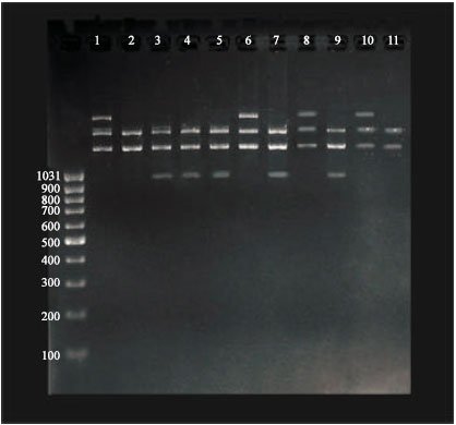 | |
| Fig. 3: | RAPD-PCR Patterns of ALL patients obtained with OPA-09 primer. Electrophoresis was performed on (1.5%) agarose gel and run with 3 volt cm-1. The lane 1 represent normal females, lanes from 2 to 7 represent the ALL female patient, lane 8 represent normal males; lanes from 9 to 11 represent ALL males’ patients. Lane L indicates the λ DNA as a ladder |
RAPD primer OPA-09 gave one band of 3.162 kbp which was present in normal male and female individuals but was absent in 77.8% of tested ALL patients. On the other hand, band which represents DNA fragment of 0.980 kbp was absent in normal individuals but it was present in 55.6% of ALL patients (Fig. 3).
 | |
| Fig. 4: | Dendogram generated by cluster analysis of the DNA fragments (bands) shown in Fig. 3. Key: P1-P10 indicate ALL patients, NM and NF indicate normal males and females, respectively |
Dissimilarity matrix analysis (dendogram) was developed using squared Euclidean distance and hierarchal clustering of 31 DNA fragments generated from RAPD data of primer OPA-09 (Fig. 3) the obtained results provided interesting information on genetic polymorphism and genetic relatedness among genomic DNA of ALL patients and controls (Fig. 4). Normal male and female individuals were found clustered in one subgroup with two patients, whereas, other seven patients were classified in three subgroups.
DISCUSSION
The RAPD-PCR results identified ten primers out of twenty three primers used in this study being able to amplify the genomic DNA of patients and controls. The failure of thirteen primers to amplify the genomic DNA of normal persons and leukemia patients may be attributed to the absence of suitable priming sites for these primers in the genomic DNA i.e., there are no complementary sequences for these primers in their genomes. Similar results have been reported by Papadopoulos et al. (2002).
Present results showed using the ten amplifying primers, a reasonable degree of DNA poly morphism was detected among normal persons and leukemia patients. The obtained data showed that DNA polymorphism in ALL patients might due to nucleotides sequence of primer in question and on the genotype of leukemia patients. In other words, the number of compatible sites of primer in genomic DNA of leukemia patients which is effected by different types of mutations and translocations, this will affect the primer-template interaction sites and will result in the loss or gain of a bands. Mutations such as (insertion or deletion or substitution) and translocations will result in adding or deleting primers binding sites, as a result this will lead to differences in number of amplified bands. The polymorphism also includes the differences in molecular weights of amplified bands which resulted from various types of mutations and translocation occurred, thus causing mobility shift of bands and might cause addition of new band(s) (Misra et al., 1998; Ong et al., 1998; Papadopoulos et al., 2002; Xian et al., 2005). It is worth mentioned that these investigators were able ascertain genomic DNA polymorphism by RAPD-PCR analysis in genomic DNA of cancer patients; however their results could not identify a RAPD primer comparable to OPA-09 which showed good detection power of ALL.
The presented results showed that although primer OPA-09 gave small number of amplified band as compared with results of other primers, it demonstrated high efficiency to detect ALL at genomic level. The primer efficiency could be demonstrated by its ability to give highest ratio of polymorphic bands as compared to total number of amplified bands. Another important characteristic of efficient primer is not by giving highest number of amplified bands, but it is shown by primer's ability to illustrate the polymorphism between normal individuals and leukemia patients.
Computational analysis of the ten amplifying RAPD primers revealed interesting sequence correlation ships which could be used for future studies in designing more reliable RAPD primers in detection of genomic instabilities of ALL genome and possible development of RAPD primers for diagnostic purposes.
CONCLUSION
Genetic instabilities are frequent in the genomic DNA of ALL of patients who were analyzed in this study and could be detected by using suitable RAPD primer. Specific bands could be generated as a result of RAPD-PCR analysis which might be used for diagnosis and prognosis of ALL. Multiple sequence alignment analysis provided interesting information on possible future development in design of RAPD primers for detection of ALL genomic DNA sequences. Further examination of other groups of leukemia patients with different phenotypic characteristics can give us a broader view about the polymorphic differences in genomic DNA of patients suffering from this type of cancer and provide an optimal base for the design of optimal therapeutic approaches against this disease.
ACKNOWLEDGMENTS
The authors would like to thank the Royal Jordanian Scientific Society, Al-Nahrain University, National Syrian Biotechnology Commission and Dept. of Radiation Oncology, University of Wurzburg for their support of this research.
REFERENCES
- Awamleh, H., D. Hassawi, H. Migdadi and M. Brake, 2009. Molecular characterization of pomegranate Punica granatum L. landraces grown in jordan using amplified fragment length polymorphism markers. Biotechnology, 8: 316-322.
CrossRef - Bolufer, P., E. Barragan, M. Collado, J. Cervera, J.A. Lopez and M.A. Sanz, 2006. Influence of genetic polymorphisms on the risk of developing leukemia and on disease progression. Leukemia Res., 30: 1471-1491.
CrossRef - Misra, D.A.A., I.M. Sulaiman, S. Sinha, C. Sarkar, A.K. Mahapatra and S.E. Hasnain, 1998. Genetic alterations in brain tumors identified by RAPD analysis. Gene, 206: 45-48.
CrossRef - Jemal, A., R. Siegel, E. Ward, T. Murray, J. Xu and M.J. Thun, 2007. Cancer statistics, 2007. CA: Cancer J. Clinicians, 57: 43-66.
CrossRefPubMedDirect Link - Maeda, T., A. Jikko, H. Hiranuma and H. Fuchihata, 1999. Analysis of genomic instability in squamous cell carcinoma of the head and neck using the random amplified polymorphic DNA method. Cancer Lett., 138: 183-188.
CrossRefDirect Link - Odero, M.D., J.L. Soto, E. Matutes, J.I.M. Subero and I. Zudaire et al., 2001. Comparative genomic hybridization and amplotyping by arbitrarily primed PCR in stage A B-CLL. Cancer Genet. Cytogenetic, 130: 8-13.
CrossRefDirect Link - Ong, T.M., B. Song, H.W. Qian, T.W. Qian, Z.L. Wu and W.Z. Whong, 1998. Detection of genomic instability in lung cancer tissues by random amplified polymorphic DNA analysis. Carcinogenesis, 19: 233-235.
CrossRefDirect Link - Papadopoulos, S., T. Benter, G. Anastassiou, M. Pape and S. Gerhard et al., 2002. Assessment of genomic instability in breast cancer and uveal melanoma by random amplified polymorphic DNA analysis. Int. J. Cancer, 99: 193-200.
PubMedDirect Link - Sambrook, J. and D.W. Russell, 2001. Molecular Cloning: A Laboratory Manual. 3rd Edn., Cold Spring Harbor Laboratory Press, New York, USA., ISBN-13: 9780879695774, Pages: 2344.
Direct Link - Scarra, A., P.S. Moore, G. Rigaud and F. Menestrina, 2001. Genetic alterations in primary mediastinal B-cell lymphoma: An update. Leukemia Lymphoma, 41: 47-53.
CrossRefDirect Link - Singh, K.P. and D. Roy, 2001. Identification of novel breast tumor-specific mutation(s) in the q11. 2 region of chromosome 17 by RAPD/AP-PCR fingerprinting. Gene, 269: 33-43.
CrossRefDirect Link - Xian, Z.H., W.M. Cong, S.H. Zhang and H.C. Wu, 2005. Genetic alteration of hepatocellular carcinoma by random amplified polymorphic DNA analysis and cloning sequencing of tumor differential DNA fragment. World J. Gastroenterol., 11: 4102-4107.
PubMedDirect Link - Zhang, S.H., W.M. Cong, Z.H. Xian, H. Dong and M.C. Wu, 2004. Genomic instability in hepatocelluar carcinoma revealed by using random amplified polymorphic DNA method. J. Cancer Res. Clin. Oncol., 130: 757-761.
Direct Link