
E.P. Sabina
School of Biotechnology, Chemical and Biomedical engineering,
VII University, Vellore-632 014, Tamil Nadu, India
M. Rasool
School of Biotechnology, Chemical and Biomedical engineering,
VII University, Vellore-632 014, Tamil Nadu, India
International Journal of Biological Chemistry
Year: 2007 | Volume: 1 | Issue: 3 | Page No.: 149-155


ABSTRACT
The aim of this research is to determine the protective efficacy of Triphala in adjuvant-induced arthritis with respect to lipid peroxidation, antioxidant status and inflammatory mediator tumour necrosis factor-alpha (TNF-α). Arthritis was induced by intradermal injection of Complete Freund’s Adjuvant (0.1 mL) into the right hind paw of Swiss albino mice. Triphala (1000 mg kg-1 day-1) and Indomethacin (3 mg kg-1 day-1) were orally administered for 8 days (from 11th to 18th day) after adjuvant injection. The levels of lipid peroxides, enzymic and non-enzymic antioxidants and tumour necrosis factor-α were measured in blood and spleen of control and arthritic animals. In arthritic animals, the lipid peroxidation and tumour necrosis factor-α were found increased, whereas a decrease in antioxidant levels was observed. These observed alterations were significantly modulated to near normal levels by oral administration of Triphala (1000 mg kg-1 day-1) in arthritic animals. The present study shows the antiarthritic effect of Triphala against adjuvant-induced arthritic mice.
PDF Abstract XML References
How to cite this article
E.P. Sabina and M. Rasool, 2007. Therapeutic Efficacy of Indian Ayurvedic Herbal Formulation Triphala on Lipid Peroxidation, Antioxidant Status and Inflammatory Mediator TNF-α in Adjuvant-induced Arthritic Mice. International Journal of Biological Chemistry, 1: 149-155.
URL: https://scialert.net/abstract/?doi=ijbc.2007.149.155
URL: https://scialert.net/abstract/?doi=ijbc.2007.149.155
INTRODUCTION
Rheumatoid Arthritis (RA) is a systemic autoimmune disease that primarily presents as a chronic symmetric polyarthritis associated with inflammation and cartilage destruction (Costello and Halverson, 2003). It is the most common inflammatory arthritis affecting approximately 1-2% of the general population worldwide (Harris, 1994). Incidence increases with age, with women being affected three times more than men. The pathogenesis of rheumatoid arthritis is multifactorial and recent research has implicated oxygen free radicals and inflammatory cytokine tumor necrosis factor (TNF)-α which have been found to play an important role as mediators of tissue damage (Fleischmann et al., 2004). Reactive oxygen species are highly reactive transient chemical species (nitric oxide, superoxide anion, hydrogen peroxide and hydroxyl radical) with the potential to initiate cellular damage (to protein, lipids, etc.) in joint tissues, especially in rheumatoid arthritis. TNF-α is produced abundantly by activated macrophages in synovial tissues from rheumatoid arthritis patients and by stimulating mesenchymal cells such as osteoclasts, fibroblasts and chondrocytes, which release joint-destructive matrix proteinase. Although a number of drugs (non-steroidal or steroidal anti-inflammatory agents and immunosuppressant) used in the treatment of rheumatoid arthritis have been developed over the past few decades, there is still an urgent need for more effective drugs with lower side effects. Thus, a desirable drug for treatment of rheumatoid arthritis, which possesses the properties of controlling joint inflammation, relieving joint pain and protecting the arthritic joints from damage without any side effects is required.
Triphala is the most commonly used Indian ayurveda herbal formulation, comprising the fruits of three trees, Indian goose berry (Emblica officinalis Gaertn, family-Euphobiaceae), Belleric myrobalan (Terminalia belerica Linn, family-Combretaceae) and Chebulic myrobalan (Terminalia chebula Retzr, family-Combretaceae). Triphala has been reported to be a rich source of Vitamin C, ellagic acid, gallic acid, chebulinic acid, bellericanin, β-sitosterol and flavanoids (Kaur et al., 2005). Its components Emblica officinalis, Terminalia belerica, Terminalia chebula are reported to possess anti-inflammatory, antimutagenic, antioxidant, cytoprotective, gastroprotective activity, myocardial necrosis, hepatoprotective, antibacterial, hypolipdemic and anticancer activity (Pulok Mukherjee et al., 2006; Srikumar et al., 2005; Sandhyaa et al., 2006; Saravanan et al., 2007). preliminary studies confirm its anti-inflammatory and lysosomal membrane stabilizing effect on adjuvant-induced arthritis in mice (Rasool and Sabina, 2007). In the current study, the anti-arthritic effect of Triphala was investigated using adjuvant-induced arthritis, a well known experimental model for rheumatoid arthritis in mice. The anti-arthritic effect of Triphala was assessed by measuring changes in lipid peroxidation, antioxidant status and inflammatory mediator Tumour Necrosis Factor (TNF)-α in plasma and spleen of arthritic animals. The standard non-steroidal anti-inflammatory drug, Indomethacin, was used as a reference drug for purposes of comparison.
MATERIALS AND METHODS
Animals
The study was performed with Swiss albino mice, 25-30 g, of either sex. The mice were brought from Tamil Nadu Veterinary College, Chennai, India. The mice were acclimatized for a week in a light and temperature-controlled room with a 12 h dark-light cycle. The mice were fed commercial pelleted feed from Hindustan Lever Ltd. (Mumbai, India) and water was made freely available.
Drug
The commercially available Triphala powder ( mixture of dried and powdered fruits of three plants, T. chebula, E. officinalis and T. bellerica in equal proportions (1:1:1) ) was obtained from Indian medical Practitioners Co-operative Stores and Society (IMCOPS), Adyar, Chennai, India and its aqueous suspension in 2% gum acacia was used at a dose 1 g kg-1 b.wt. orally. The Triphala powder used in this study was found to contain approximately 50% polyphenols as investigated by High Performance Thin Layer Chromatography (HPTLC) densitometer analysis (Rasool and Sabina, 2007). Indomethacin (Tamilnadu Dadha Pharmaceuticals, Chennai, India) was dissolved in 2% gum acacia solution and 3 g kg-1 b.wt. was administered orally (Rasool and Varalakshmi, 2007). All other reagents used were standard laboratory reagents of analytical grade and were purchased locally.
Dosage
Preliminary studies with different dosages (250 mg, 500 mg, 1 g) of this drug revealed that 1 g kg-1 b.wt. dosage produced significant anti-inflammatory effect by reducing paw swelling in adjuvant-induced arthritic animals. Hence 1 g kg-1 b.wt. dosage was considered for this study.
Experimental Protocol
The mice were divided into six groups each comprising six animals. Group 1 served as controls. In Group II, arthritis was induced by intra dermal injection of Complete Freund’s Adjuvant (CFA) (0.1 mL) into the right hind paw (Rasool and Sabina, 2007). The adjuvant (Tuberculosis Research Center, Chennai, India) contained heat-killed Mycobacterium tuberculosis (H37 RA) (10 mg) in paraffin oil (1 mL). Group III and IV were treated with Triphala and Indomethacin, respectively, for 8 days from the 11th to the 18th day of the experimental period. Groups V and VI comprised arthritic mice were treated with Triphala and Indomethacin, respectively, from day 11th to 18th after the administration of Complete Freund’s Adjuvant (CFA).
On the 19th day, at the end of the experimental period, the animals were sacrificed by cervical decapitation and the blood was collected. The spleen was immediately dissected out and homogenized in ice-cold 0.01 M, Tris HCL buffer, pH 7.4 to give a 10% homogenate. Spleen tissue homogenate and plasma were used for assaying the following biochemical investigations.
Biochemical Estimations
Lipid peroxidation in plasma was estimated by the method of Ledwozyw et al. (1986). Spleen lipid peroxidation was carried out by the procedure of Hogberg et al. (1974) using thiobarbutric acid (TBA) as the colouring agent. Malonaldehyde (MDA) produced during peroxidation of lipids served as an index of lipid peroxidation. malonaldehyde reacts with thiobarbutric acid to generate a colour product, which absorbs at 532 nm.
Spleen lipid peroxidation with inducer system namely, 10 mM FeSO4/0.2 ascorbate/10 mM H2O2 was measured by the method of Devasagayam and Tarachand (1987). The Malonaldehyde contents of the samples were expressed as nmoles of MDA formed/mg/protein.
Superoxide dismutase (SOD) activity in spleen was determined by the method of Marklund and Marklund (1974). The degree of inhibition of the auto-oxidation of pyrogallol at an alkaline pH by SOD was used as a measure of the enzyme activity. Catalase and glutathione peroxidase activities in spleen were estimated by the method of Sinha (1972) and Rotruk et al. (1973).The activity of catalase was expressed as μg of H2O2 consumed/mn/mg protein. Glutathione peroxidase was expressed as μg of glutathione utilized/minute/mg/protein. Non enzymic antioxidants, glutathione (Moron et al., 1979), ascorbic acid (Omaye et al., 1979), total suphydryl (TSH) and non protein sulphydryl (NPSH) (Sedlack and Lindsay, 1968) were estimated in the spleen.
TNF-α levels in plasma and spleen of control and arthritic mice were determined by enzyme-linked immunosorbent assay (ELISA, Cayman Chemicals Company, USA), according to the manufacturer's instruction.
Statistical Analysis
Results were expressed as mean±SD and statistical analysis was performed using ANOVA to determine significant differences between groups, followed by Student’s Newman-Keul’s test. p<0.05 implied significance.
RESULTS
In group II arthritic mice, basal lipid peroxide levels were elevated in both plasma and spleen. In presence of inducers like ascorbate, FeSO4 and H2O2, spleen lipid peroxide levels were found to be increased significantly compared to control group, whereas the administration of Triphala to arthritic mice altered the above changes by regulating the lipid peroxide level to nearly that of normal levels (Table 1).
The enzymic and non-enzymic antioxidants were significantly decreased in arthritic mice compared to control mice. These changes highlight the deteriorating antioxidant status in the arthritic mice. Furthermore, administration of Triphala increased the enzymic and non-enzymic antioxidant levels in arthritic mice considerably, which indicates its antiperoxidative action (Table 2).
| Table 1: | Effect of Triphala and Indomethacin on basal and induced lipid peroxidation in plasma and spleen of control and experimental animals |
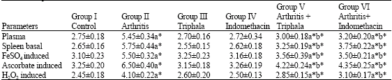 | |
| Treatment of groups: Group I-Control; Group II-Arthritic mice; Group III-Control mice treated with Triphala (1000 mg kg-1 b.wt) for 8 days from 11 th to 18th day; Group IV-Control mice treated with Indomethacin (3 mg kg-1 b.wt) for 8 days from 11 th to 18th day; Group V-Arthritic mice treated with Triphala (1000 mg kg-1 b.wt) from 11 th to 18th day post adjuvant; Group VI-Arthritic mice treated with Indomethacin (3 mg kg-1 b.wt) from 11 th to 18th day post adjuvant. Comparisons: A-Group I vs groups II, III, IV, V and VI. b-Group II vs group V and VI. Each value represent mean±SD (n = 6). Values are expressed as: nanomoles of malonaldehyde formed/mg protein; plasma mg dL-1. The symbols represent statistical significance at: * p<0.05 | |
| Table 2: | Effect of Triphala and Indomethacin on the enzymic and nonenzymic antioxidant levels in spleen of control and experimental animals |
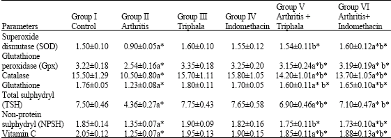 | |
| Treatment of groups: Group I: Control; Group II: Arthritic mice; Group III: Control mice treated with Triphala (1000 mg kg-1 b.wt) for 8 days from 11 th to 18th day; Group IV-Control mice treated with Indomethacin (3 mg kg-1 b.wt) for 8 days from 11 th to 18th day; Group V-Arthritic mice treated with Triphala (1000 mg kg-1 bw) from 11 th to 18th day; Group VI-Arthritic mice treated with Indomethacin (3 mg kg-1 b.wt) from 11 th to 18th day. Comparisons: A-Group I vs groups II, III, IV, V and VI and b: Group II vs group V and VI. Values are expressed as mean±SD (n = 6). Enzyme units are expressed as; SOD-units/mg protein (unit-Amount of enzyme required to inhibit the auto-oxidation reaction by 50%); Gpx-μg of GSH utilized/min/mg protein; Catalase-μmol of H2O2 consumed min-1 mg-1 protein. Glutathione, Total sulphydryl (TSH), Non-protein sulphydryl (NPSH), Vitamin C- μg mg-1 protein. The symbols represent statistical significance at: * p<0.05 | |
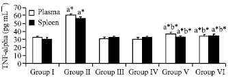 | |
| Fig. 1: | Effect of Triphala and Indomethacin on TNF-alpha production in adjuvant-induced arthritis in mice. Treatment of groups: Group 1-Control; Group II-Arthritic mice; Group III-Control mice treated with Triphala (1000 mg kg-1 b.wt) for 8 days from 11 th to 18th day; Group IV-Control mice treated with Indomethacin (3 mg kg-1 b.wt) for 8 days from 11 th to 18th day; Group V-Arthritic mice treated with Triphala (1000 mg kg-1 b.wt) from 11 th to 18th day post adjuvant; Group VI-Arthritic mice treated with Indomethacin (3 mg kg-1 b.wt) from 11 th to 18th day post adjuvant. Comparisons: a: Group I vs groups II, III, IV, V and VI and b-Group II vs group V and VI. The symbols represent statistical significance at: *p<0.05 |
Levels of tumour necrosis factor-α in the arthritic mice were systemically overproduced in the serum and spleen, while the elevated levels of tumour necrosis factor-α were found to be decreased in Triphala treated arthritic mice (Fig. 1).
DISCUSSION
Triphala an Indian ayurvedic herbal formulation is believed to promote health, immunity and longevity (Sandhyaa et al., 2006). Triphala, rich in antioxidants, plays an essential role in the treatment of a wide variety of conditions like infections, obesity, anemia, fatigue, constipation and in infectious diseases like tuberculosis, pneumonia and AIDs (El-Mekkawey and Merelhy, 1995). Here, we have evaluated the anti-arthritic effect of Triphala in adjuvant-induced arthritis in mice; an experimental model of arthritis, which resembles several aspects of human rheumatoid arthritis (Pearson, 1964).
In rheumatoid arthritis, the polymorphonuclear leukocytes are activated and reactive oxygen species are generated in excessive amounts. These are reactive ephemeral molecules which are known to play an important role in the progression of inflammation. In this study, the lipid peroxide level was found to be increased in arthritic mice compared to control animals. This is in agreement with other studies in which higher lipid peroxide levels have been reported in adjuvant-induced arthritis (Geetha et al., 1998; Rasool and Varalakshmi, 2007). The increased lipid peroxide observed in arthritic animals may be due to its release from neutrophils and monocytes during inflammation. It is an accepted fact that rheumatoid arthritis is accompanied by abnormalities in body iron metabolism. At the onset of inflammation, it was observed that there was a rapid fall in the total iron content of blood plasma followed by an increased deposition of iron proteins in the synovial fluid. The drop in plasma iron correlates closely with the activity of the inflammatory process (Gutteridge, 1986). In the synovial fluid of inflamed joints, the iron released during necrosis, might catalyze the formation of hydroxyl radicals from H2O2, thus contributing to an increase in inflammation.
In the present study, the concentration of enzymic and non-enzymic antioxidants was significantly decreased in arthritic mice (Group II), as compared to normal control mice. Similar results have been reported by Geetha et al. (1998) and Ramprasad et al. (2005). The increased rate of free radical production frequently results in a decrease in the level of antioxidant enzymes and the enzyme activities are reduced thereby leading to autocatalysis of oxidative damage process. Super oxide dismutase, glutathione peroxidase and catalase are considered primary antioxidant enzymes, since they are involved in direct elimination of free radicals. The decreased activity of enzyme antioxidants in adjuvant-induced arthritis may be due to the high concentration of reactive oxygen species formed. Due to increased lipid peroxidation, the levels of free radicals overcome the saturation level. The high concentrations of free radicals inhibit the activity of antioxidants and hence the activities of these enzymes appear to be reduced. Enzymic antioxidants are inactivated by the free radicals and hence the presence of non-enzymic antioxidants is presumably essential for the removal of these radicals (Allen, 1991). Vitamin C, glutathione and other thiols are water soluble antioxidants that remove free radical from cytosol by reacting directly with them (Allen, 1991). In the present observation, non-enzymic antioxidants were found to be significantly decreased in arthritic animals. The observed non-enzymic antioxidants depression in adjuvant-induced arthritis is associated with the protracted inflammatory phase of the disease.
In present study, it was observed that Triphala protects the arthritic animal from lipid peroxidation and increases the enzyme activities of superoxide dismutase, glutathione peroxidase, catalase and non-enzymic antioxidant levels, which suggests that Triphala prevents the oxidative damage due to reactive oxygen species overproduction from arthritis. The increased activities/levels of antioxidants observed in treated rats could be due to phenolic compounds (flavonoids) present in Triphala. It has been reported earlier that phenolic compounds has potential antioxidant activity and it could scavenge free radicals effectively and has also been found to inhibit the lipid peroxidation (Ramprasad et al., 2005). Hydrogen-donating hydroxyl groups on the aromatic ring of phenolic compounds present in Triphala might be responsible for the free radical scavenging and antioxidant activity of phenolics, which correlates with earlier report (Anand, 1997). Emblica officinalis, Terminalia belerica and Terminalia chebula, the constituents of Triphala extract have been reported to be a rich source of Vitamin C, ellagic acid, gallic acid, chebulinic acid, bellericanin, β-sitosterol and flavanoids. Most of these compounds have been reported to be a potent inhibitor of lipid peroxide formation, a scavenger of hydroxyl and superoxide radicals and to increase the antioxidant enzymes (Jagetia et al., 2002). Thus the modifying role of Triphala extract observed in our study may be due to the antiperoxidative action of its components that was reported earlier (Lee et al., 2005; Naik et al., 2005).
TNF-α, a proinflammatory cytokine, is produced by activated macrophages and other cell types and these cell types are abundant in the arthritic joints as has been shown in both the animal models and in rheumatoid patients. This abundance of TNF-α in arthritic joint provides evidence of its involvement in the disease pathology, which is supported by studies demonstrating that neutralization of TNF-α leads to decreased production of other inflammatory cytokines (Haworth et al., 1991; Brennan et al., 1989). Present results confirm that Triphala suppressed the inflammatory process by reducing the production of tumour necrosis factor-α in adjuvant-induced arthritic mice.
To conclude, the results of the present study have empirically indicate that Triphala is effective in the treatment of rheumatoid arthritis and that it supports the common belief prevailing in traditional medicines, worldwide.
REFERENCES
- Anand, K.K., B. Singh, A.K. Saxena, B.K. Chandan, V.N. Gupta and V. Bhardwaj, 1997. 3, 4, 5-Trihydroxybenzoic acid (gallic acid), the hepatoprotective principle in the fruits of Terminalia belerica bioassay guided activity. Pharmacol. Res., 36: 315-321.
PubMedDirect Link - Costello, J.C. and P.B. Halverson, 2003. A new era in rheumatoid arthritis treatment. Wisc. Med. J., 102: 29-33.
Direct Link - Fleischmann, R., R. Stern and I. Iqbal, 2004. Anakinra: An inhibitor of IL-1 for the treatment of rheumatoid arthritis. Exp. Opin. Biol. Therapy, 4: 1333-1344.
Direct Link - Hogberg, J., R.E. Larson, A. Kristoferson and S. Orrenius, 1974. NADPH-dependent reductase solubilised from microsomes of peroxidation and its activity. Biochem. Biophys. Res. Commun., 56: 836-842.
CrossRef - Jagetia, G.C., M.S. Baliga, K.J. Malagi and M.S. Kamath, 2002. The evaluation of the radioprotective effect of Triphala (an ayurvedic rejuvenating drug) in the mice exposed to γ-radiation. Phytomedicine, 9: 99-108.
CrossRefDirect Link - Kaur, S., H. Michael, S. Arora, P.L. Harkonen and S. Kumar, 2005. The in vitro cytotoxic and apoptotic activity of Triphala--an Indian herbal drug. J. Ethanopharmacol., 10: 15-20.
CrossRefPubMedDirect Link - Ledwozyw, A., J. Michalak, A. Stepien and A. Kadziolka, 1986. The relationship between plasma triglycerides, cholesterol, total lipid and lipid peroxidation product during human arterosclerosis. Clin. Chem. Acta, 155: 275-283.
CrossRefPubMedDirect Link - Lee, H.S., N.H. Won, K.H. Kim, H. Lee, W. Jun and K.W. Lee, 2005. Antioxidant effects of aqueous extract of Terminalia chebula in vivo and in vitro. Biol. Pharmaceut. Bull., 28: 1639-1644.
CrossRefPubMedDirect Link - Marklund, S. and G. Marklund, 1974. Involvement of the superoxide anion radical in the autoxidation of pyrogallol and a convenient assay for superoxide dismutase. Eur. J. Biochem., 47: 469-474.
CrossRefPubMedDirect Link - Moron, M.S., J.W. Depierre and B. Mannervik, 1979. Levels of glutathione, glutathione reductase and glutathione S-transferase activities in rat lung and liver. Biochim. Biophys. Gen. Subj., 582: 67-78.
CrossRefPubMedDirect Link - Naik, G.H., K.I. Priyadarsini, R.G. Bhagirathi, B. Mishra, K.P. Mishra, M.M. Banavalikar and H. Mohan, 2005. In vitro antioxidant studies and free radical reactions of triphala, an ayurvedic formulation and its constiuents. Pytother. Res., 19: 582-586.
Direct Link - Omaye, S.T., J.D. Turnbull and H.E. Sauberlich, 1979. Selected methods for the determination of ascorbic acid in animal cells, tissues and fluids. Methods Enzymol., 62: 3-11.
CrossRefPubMedDirect Link - Pulok, M.K., S. Rai, S. Bhattacharyya, P.K. Debnath and T.K. Biswas et al., 2006. Clinical study of Triphala: A well known phytomedicine from India. Iran. J. Pharmacol. Therapeut., 5: 51-54.
Direct Link - Ramprasad, V.R., P. Shanthi and P. Sachdanandam, 2005. Evaluation of antioxidant effect of Semecarpus anacardium Linn.nut extract on the components of immune system in adjuvants arthritis. Vascular Pharmacl., 42: 179-186.
CrossRef - Rasool, M. and E.P. Sabina, 2007. Anti-iflammatory effect of Indian ayurvedic herbal formulation Triphala on adjuvant-induced arthritic mice. Phytotherapy Res., 21: 889-894.
Direct Link - Rasool, M. and P. Varalakshmi, 2007. Protective effect of Withania somnifera root powder in relation to lipid peroxidation, antioxidant status, glycoproteins and bone collagen on adjuvant-induced arthritis. Fundamental Clin. Pharmacol., 21: 157-164.
Direct Link - Rotruck, J.T., A.L. Pope, H.E. Ganther, A.B. Swanson, D.G. Hafeman and W.G. Hoekstra, 1973. Selenium: Biochemical role as a component of glutathione peroxidase. Science, 179: 588-590.
CrossRefPubMedDirect Link - Sandhya, T., K.M. Lathika, B.N. Pandey and K.P. Mishra, 2006. Potential of traditional ayurvedic formulation, Triphala, as a novel anticancer drug. Cancer Lett., 231: 206-214.
CrossRefDirect Link - Saravanan, S., R. Srikumar, S. Manikandan, N.J. Parthasarathy and R.S. Devi, 2007. Hypolipidemic effect of triphala in experimentally induced hypercholesteremic rats. Yakugaku Zasshi, 127: 385-388.
PubMedDirect Link - Sedlak, J. and R.H. Lindsay, 1968. Estimation of total, protein-bound, and nonprotein sulfhydryl groups in tissue with Ellman's reagent. Anal. Biochem., 25: 192-205.
CrossRefPubMedDirect Link - Sinha, A.K., 1972. Colorimetric assay of catalase. Anal. Biochem., 47: 389-394.
CrossRefPubMedDirect Link - Srikumar, R., N.J. Parthasarathy and R.S. Devi, 2005. Immunomodulatory activity of triphala on neutrophil functions. Biol. Pharm. Bull., 28: 1398-1403.
CrossRefPubMedDirect Link