
Roghayeh Abbasalipourkabir
Department of Biochemistry, Faculty of Medicine, Hamadan University of Medical Science, Hamadan, Iran
Aref Salehzadeh
Department of Parasitology, Faculty of Medicine, Hamadan University of Medical Science, Hamadan, Iran
Rasedee Abdullah
Department of Clinical Pathology and Hematology, Faculty of Veterinary Medicine, Universiti Putra Malaysia, Malaysia
Biotechnology
Year: 2011 | Volume: 10 | Issue: 6 | Page No.: 528-533


ABSTRACT
Recently more focus has been put to the development of innovative drug-delivery systems that includes liposomes and solid lipid nanoparticles (SLNs). An in vitro study was conducted to determine the effect of solid lipid nanoparticle on the human breast cancer cell lines (MCF-7 and MDA-MB231). The SLNs based on palm oil were prepared using the high pressure homogenization method and were characterized by the particle size and polydispersity index (particle sizer), zeta potential (zetasizer), ultrastructure [transmission electron microscopy (TEM)] and MTT assay and natural red assay. Homogenization of solid lipid nanoparticles at 1000 bar for 20 cycles produced particles with 145.00±3.39 nm in size and zeta potential of -19.50±1.80 mv. The SLNs were generally round and uniform in shape. The cytotoxicity of the components of the SLN formulation was low: SLN with 1% oleyl alcohol displayed no significant cytotoxicity effect on breast cancer cells. In the light of these findings, SLN stabilized with 30% lecithin and 1% oleyl alcohol as nonionic co-surfactant in aqueous phase was found to be safe for cells and the acceptable for the incorporation lipophilic drugs.
PDF Abstract XML References Citation
Received: August 03, 2011;
Accepted: November 02, 2011;
Published: December 12, 2011
How to cite this article
Roghayeh Abbasalipourkabir, Aref Salehzadeh and Rasedee Abdullah, 2011. Cytotoxicity Effect of Solid Lipid Nanoparticles on Human Breast Cancer Cell Lines. Biotechnology, 10: 528-533.
DOI: 10.3923/biotech.2011.528.533
URL: https://scialert.net/abstract/?doi=biotech.2011.528.533
DOI: 10.3923/biotech.2011.528.533
URL: https://scialert.net/abstract/?doi=biotech.2011.528.533
INTRODUCTION
Recently solid lipid nanoparticles (SLN) as colloidal particulate drug delivery system have received much attention from drug development researchers (Fontana et al., 2005; Mehnert and Mader, 2001). The solid lipid nanoparticles contain a lipid matrix composed of physiologically tolerable lipids that is not toxic. The use of SLNs has several advantages including avoidance organic solvent for production of SLN, capacity of large scale production, widespread application, high bioavailability, provides protection of drug from degradation agent like water and light and improved controlled-drug release (Jores et al., 2005; Yuan et al., 2007). Solid lipid nanoparticles (SLN) containing stabilized lipid dispersions have been designed as an alternative colloidal drug delivery system to polymer nanoparticles, emulsions and liposomes. Among their disadvantages are that their undesired toxicities have remained largely unaddressed (Zhanga and Fenga, 2006). With SLN the potential toxicity may be due to its unique size and other physico-chemical characteristics. Stabilizers offer stability to the dispersions, avoiding aggregation and allow the design of SLN of favorable size. Generally surfactants like phospholipids contributing to steric and electrostatic stabilization are also used in SLN formulations (Mehnert and Mader, 2001). The stabilizers are adsorbed on to the particle surface and hence affect the properties of SLN. Therefore they may increase the risk of undesired or toxic effects (Unfried et al., 2007). Until now only a small number of basic in vitro cell culture studies evaluated the potential toxicity and biocompatibility of SLN formulation and stabilizers (Kristl et al., 2008).
The objective of this study was to examine the influence of component of solid lipid nanoparticles on the viability of breast cancer cells using the MTT assay.
MATERIALS AND METHODS
Softisan® 154 (S154), or hydrogenated palm oil, was a gift from CONDEA (Witten, Germany). Lipoid S100 (soy lecithin) was a gift from Lipoid KG (Ludwigshafen, Germany). Thimerosal, Sorbitol and were purchased from Sigma. Oleyl alcohol, also called octadecenol, or cis-9- octadecen-1-ol, is a fatty alcohol and nonionic surfactant or emulsifier was purchased from Fluka and water was used in bidistilled quality.
Preparation of solid lipid nanoparticles: Solid Lipid Nanoparticle (SLN) was prepared using the high-pressure homogenization technique (Abbasalipurkabir et al., 2011). Briefly a mixture of S154 and Lipoid S100 at a ratio of 70:30 was ground in a ceramic crucible. The mixture was then heated to 65-70°C while being stirred with a PTFE-coated magnet until a clear-yellowish lipid matrix (LM) solution was obtained. A solution containing 1 mL oleyl alcohol, 0.005 g thimerosal, 4.75 g Sorbitol and 89.25 mL bidistilled water (all w/w) at the same temperature was added to 5 g of LM. A pre-emulsion of SLN was obtained using the homogenizer (Ultra Turrax, Ika, Staufen, Germany) at 13000 rpm for 10 min and high-pressure homogenizer (EmulsiFlex-C50 CSA10, Avestin, Ottawa, Canada) at 1000 bar, 20 cycles and 60°C. The obtained SLN was exposed to air to solidify.
Physical characterization of solid lipid nanoparticles: Mean particle size (diameter, nm±SD) and size distribution (polydispersity index, PI) of solid lipid nanoparticles were determined using the high performance particle sizer (HPP5001, Malvern Inst. UK.). Measurement was at 25°C performed in triplicates. Prior to measurement, each sample was dispersed in filtrated bidistilled water by sonic water bath. The zeta potential (electrophoretical movement) of the nanoparticle formulations was then measured by Zeta potential analyzer (Zeta sizer; ZEN-2600; Malvern, UK.), in triplicates. The morphology of SLN was viewed using a transmission electron microscopy (Hitachi H-7100, Japan). After dispersion with water, the samples were negatively stained with 1.5% (w/v) phosphotungstic acid for viewing.
Treatment of the cells: The SLN was dispersed in PBS. Breast cancer cells (MCF-7 and MDA-MB231-ATCC) were maintained in culture medium RPMI supplemented with 10% Fetal Bovine Serum (FBS) at 37°C in a humidified incubator containing 5% CO2/95% air. The cells were allowed to grow in 25 cm3 cell culture flask until confluent. The old media was removed and the confluent cells washed with PBS and detached with trypsin. A haemocytometer was used to estimate the concentration of the cells in the flask. At a concentration, One hundred microlitres of breast cancer cells (105 cell mL-1) in culture medium were seeded into a 96-well plate. The confluent cells were treated with SLN at concentration ranging 37.5, 75, 150 and 300 μg mL-1 for 24, 48 and 72 h. The control wells received PBS as the vehicle.
MTT assay: The viability and rate of proliferation of the mammalian cells can be determined by several biological assays including the [3-(4,5-dimethylthiazol-2-yl)-2,5-diphenyl tetrazolium bromide] (MTT) assay. In the MTT assay viable cells are able to reduce the water-soluble yellow tetrazolium salt into a water-insoluble purple formazan compound (Mosmann, 1983). The color reaction determined spectrophotometically is used as a measure of cell viability and proliferation (Yang et al., 2001) .
The viability of the breast cancer cells in presence of SLN was assessed by the MTT assay. After the incubation for defined time (24, 48 and 72 h), 10 μL of MTT solution (5 mg mL-1 in PBS) was added to each well and the plates incubated at 37°C for 2 additional hours. Fifty microlitres of DMSO was added to each well and shaken for 15 min. The optical density of the cell suspention was read on an ELISA reader (Tecan, Magellan, Austria) at 570 nm after background correction at 690 nm. Average cell viability of treated cells was expressed as percentage of the absorbance of control-treated cells.
Natural red assay: Breast cancer cell lines (MCF-7 and MDA-231) were maintained in RPMI as described in the MTT assay. One hundred microlitres of confluent cell monolayer were plated in 24-well plates (105 cells/well). After incubation overnight 500 μL of SLNs (SLN-03a, SLN-03b, SLN-03c and SLN-03d) dispersed in PBS, pH = 7.4 at concentrations of 60 and 80 μg mL-1, were added to the MCF-7 and MDA-231 cell, respectively. Control wells contained PBS only. After incubation for 24 h, adherent cells were washed once with 500 μL PBS (37°C), the buffer then replaced with 500 μL NR solution (50 μg mL-1). The cells were incubated for an additional 3 h allowing the lysosomes of viable cells to take up the dye. Then the cells were again washed three times with PBS to remove extracellular NR. The incorporated dye was extracted by adding of 1200 μL of 50% ethanol with 1% glacial acetic acid solution and the plates shaken vigorously for 10-15 min. The absorbance was read at 540 nm using the ELISA reader (Tecan, Magellan, Austria) against primary medium as the blank and the results determined as % of control (100% viability).
Statistical analysis: All the data were subjected to one way analysis of variance (ANOVA) followed by Post Hoc multiple comparison and Duncan test after verification of the normal distribution of the data.
RESULTS AND DISCUSSION
Physical analyses: The particle sizes, particle size distribution, specific surface area and zeta potential of different SLN formulations are presented in Table 1. The low PI for SLNs indicates an acceptable particle size distribution. Zeta potential is an important factor to the determination of stability of colloidal formulation (Mountasser et al., 2002).
| Table 1: | Effect of different amount of lecithin on SLN characteristics |
| All values are represented as Means±SD, (n = 5) | |
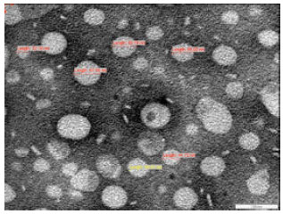 | |
| Fig. 1: | SLNat 7 days after preparation. The particles were negatively stained with aqueous solution of phosphotungstic acid (PTA) 10 min prior to imaging. Particles exhibited spherical and uniform shapes |
In formulations where the zeta potential is high, aggregation of particles is unlikely to occur, because there is electrical repulsion (Mehnert and Mader, 2001). The TEM image of SLN after 7 days preparation is shown in Fig. 1. The particles were nonporous, spherical and uniform in shape.
Effect of SLN on the viability of breast cancer cell lines: Before applying SLN as a drug delivery system, its potential cytotoxicity needs to be determined. The MTT assay was conducted to determine the potency and cytotoxicity effect of SLN on human breast cancer cell lines, MCF-7 and MDA-MB231. The 50% inhibitory concentration (IC50) of SLN on two breast cancer cell lines, MCF-7 and MDA-MB231 for 24, 48 and 72 h are presented in Table 2. The IC50 of SLN was time-dependent and different between the tested breast cancer cell lines. The IC50 values suggest that SLN has low cytotoxicity to the MDA-MB231 and the MCF-7 cells. The cytotoxicity effect of blank SLN on MCF-7 cell and MDA-MB231 cell lines are presented in Fig. 2 and 3, respectively. Greatest cytotoxicity of SLN was observed at 72 and then of 48 and 24 h. The low cytotoxicity of the SLN can be attributed to the lecithin (Schubert and Muller-Goymann, 2005) and components of the aqueous phase used, especially the nonionic emulsifier (Scholer et al., 2000; Scholer et al., 2001).
| Table 2: | The ICS0 of breast cancer cell lines treated with SLN formulation |
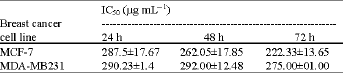 | |
| All values are represented as Means±SD, (n = 5) | |
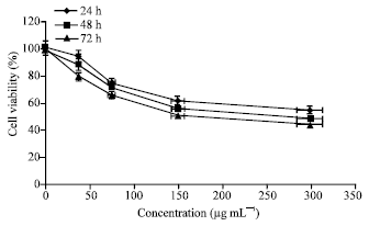 | |
| Fig. 2: | Cell viability of MCF-7 cells treated with SLN-03. The cells were incubated with SLN-03 formulations for 24, 48 and 72 h. The percentage of cell viability was expressed as a ratio of treated cells to untreated control cells. (Each point represents the mean and standard deviation), (n = 5) |
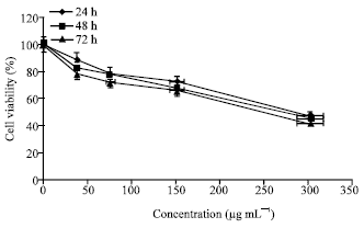 | |
| Fig. 3: | Cell viability of MDA-MB231 cells treated with SLN-03. The cells were incubated with SLN-03 formulations for 24, 48 and 72 h. The percentage of cell viability was expressed as a ratio of treated cells to untreated control cells. (Each point represents the mean and standard deviation), (n = 5) |
However the cytotoxicity of the SLN in this study is comparable to the nanoparticulate systems consisting of polylactic acid/glycolic acid or polycyanocrylate nanoparticles (Muller et al., 1997).
The cytotoxicity effect of particles is due to their adherence to the cell membrane, particle internalization and degradation of products in the cell culture medium or inside the cells.
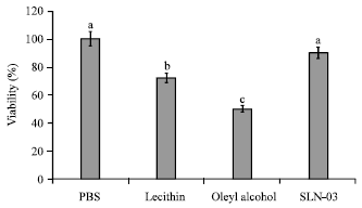 | |
| Fig. 4: | Effect of lecithin, oleyl alcohol and SLN dispersed in phosphate- buffer saline on the viability of MCF-7 cells. Means with superscripts a-c are significantly different (p<0.05), (n = 5) |
However, different cells have different susceptibilities to different particulate carriers (Lherm et al., 1992). Since the SLN contain natural lipids, it is well-tolerated by the living organisms. Most formulations of SLNs were designed with glycerides consisting of fatty acids which are safe. However, the emulsifiers used may be potentially toxic. Since the prerequisite for human use is an acceptable low toxicity (Muller et al., 1997), the nanoparticles must be assessed first in vitro cell cultures, before testing in vivo.
In this study the cytotoxicity effects of 30% lecithin and 1% oleyl alcohol used for stabilization of SLN was assessed on breast cancer cell lines and their viability also determined by MTT assay. The results of MTT assay for MCF-7 and MDA-MB231 treated with SLN are presented in Fig. 4 and 5, respectively. Lecithin and oleyl alcohol reduced the viability of MCF-7 cells by 30 and 50%, respectively. With the MDA-MB231 cells, lecithin by 25% and oleyl alcohol by 30% reduced viability of the cells. Although SLN had some effect on the viability of breast cancers, the effect is negligible.
The cytotoxicity of 1% oleyl alcohol on the breast cancer cells in present study was comparable to the findings of Maaben et al. (1993). In their study, poloxamer at concentration of 1% (w/w) stabilized polylactide/polyglycolide particles and reduced the cell viability by 50%. According to Muller et al. (1997) surfactant toxicity is significantly reduced in SLN formulations. Similarly, in the current study the cytotoxicity of lecithin and oleyl alcohol was significantly reduced once they are incorporated in the SLN formulation. The result suggests that lecithin and oleyl alcohol are suitable for the preparation of SLNs in the application as drug carriers.
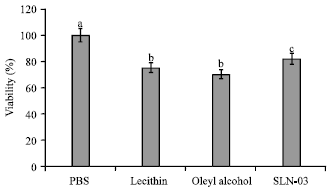 | |
| Fig. 5: | Effect of lecithin, oleyl alcohol and SLN dispersed in phosphate- buffered saline on the viability of MDA-MB231 cells. Means with superscripts a-c are significantly different (p<0.05), (n = 5) |
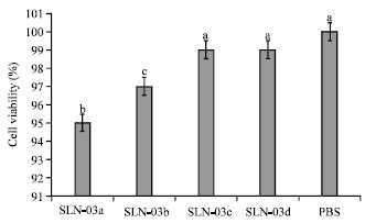 | |
| Fig. 6: | Effect of SLN stabilized with oleyl alcohol as cosurfactant on viability of MCF-7 cells. Means with superscripts a-c are significantly different (p<0.05), (n = 5) |
This information is particularly useful, because it allows the use of high concentrations of these surfactants to physically stabilize the SLNs.
Effect of surfactant concentration on the cytotoxicity of SLN: Toxicity of four formulations of SLN (SLN-03a, SLN-03b, SLN-03c and SLN-03d) stabilized with several concentrations of oleyl alcohol (0.5, 1, 1.5 and 2% w/w, respectively) was further evaluated on breast cancer cell line using the NR assay. The NR protocol is a quick, reliable and useful method for determination of cell viability. The neutral red enters the lysosomes of viable cells and its concentration measured at 540 nm (Fautz et al., 1991). The results for NR analysis of MCF-7 and MDA-MB231 cell lines are presented in Fig. 6 and 7, respectively. Increased concentration of oleyl alcohol inside the SLN formulations did not produce significant cytotoxic effect on the cells. It seems that the reduced cell viability as the result of SLN treatment is likely to be independent of its surfactant concentration.
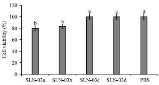 | |
| Fig. 7: | Effect of SLN stabilized with oleyl alcohol as cosurfactant on viability of MDA-MB231 cells. Means with superscripts a-c are significantly different (p<0.05), (n = 5) |
It can be concluded that the toxicity of SLN is probably dependent on the other factors such as size, charge and formulation (Heydenreich et al., 2003).
CONCLUSION
The use of Solid Lipid Nanoparticle (SLN) opens up new perspectives for the formulation of poorly soluble drugs. The SLN is a very complex system with some advantages and disadvantages over other colloidal carrier systems. This study showed that the cytotoxicity of the component of the SLN formulation was low. Solid lipid nanoparticle with 1% oleyl alcohol displayed no significant cytotoxicity effect on breast cancer cells. In the light of these findings SLN stabilized with 1.5% lecithin and 1% nonionic cosurfactant in the aqueous phase was found to be safe for cells and the acceptable formulation for incorporation of lipophilic drugs.
REFERENCES
- Abbasalipourkabir, R., A. Salehzadeh and R. Abdullah, 2011. Delivering tamoxifen within solid lipid nanoparticles. Pharmaceut. Technol., 35: 74-80.
Direct Link - Fontana, G., L. Maniscalco, D. Schillaci, G. Cavallaro and G. Gimmona, 2005. Solid lipid nanoparticles containing tamoxifen characterization and in vitro antitumoral activity. Drug Deliv., 12: 385-392.
CrossRefPubMedDirect Link - Fautz, R., B. Husein and C. Hechenberger, 1991. Application of the neutral red assay (NR assay) to monolayer cultures of primary hepatocytes: Rapid colorimetric viability determination for the unscheduled DNA synthesis test (UDS). Mutat. Res., 253: 173-179.
PubMed - Heydenreich, A.V., R. Westmeier, N. Pedersen, H.S. Poulsen and H.G. Kristensen, 2003. Preparation and purification of cationic solid lipid nanospheres-effects on article size, physical stability and cell toxicity. Int. J. Pharm., 254: 83-87.
PubMed - Jores, K., A. Haberland, S. Wartewig, K. Mader and W. Mehnert, 2005. Solid Lipid Nanoparticles (SLN) and oil-loaded SLN studied by spectrofluorometry and raman spectroscopy. Pharm. Res., 22: 1887-1897.
CrossRefPubMedDirect Link - Kristl, J., K. Teskac, M. Milek and I. Milinaric-Rascan, 2008. Surface active Stabilizer tyloxapol in colloidal dispersions exerts cytostatic effects and apoptotic dismissal of cells. Toxicol. Applied Pharmacol., 232: 218-225.
PubMed - Lherm, C., R.H. Muller, F. Puisieux and P. Couvreur, 1992. Alkylcyanoacrylate drug carriers: II. Cytotoxicity of cyanoacrylate nanoparticles with different alkyl chain length. Int. J. Pharm., 84: 13-22.
CrossRef - Mehnert, W. and K. Mader, 2001. Solid lipid nanoparticles: Production, characterization and applications. Adv. Drug Deliv. Rev., 47: 165-196.
CrossRef - Mosmann, T., 1983. Rapid colorimetric assay for cellular growth and survival: Application to proliferation and cytotoxicity assays. J. Immunol. Methods, 65: 55-63.
CrossRefPubMedDirect Link - Mountasser, I., H. Fessi and A.W. Coleman, 2002. Atomic force microscopy imaging of novel type of polymeric colloidal nanostructures. Eur. J. Pharm. Biopharm., 54: 281-284.
CrossRefPubMedDirect Link - Scholer, N., C. Olbrich, K. Tabatt, R.H. Muller, H. Hahn and O. Liesenfeld, 2001. Surfactant but not the size of solid lipid nanoparticles (SLN) influences viability and cytokine production of macrophages. Int. J. Pharm., 221: 57-67.
PubMed - Schubert, M.A. and C.C. Muller-Goymann, 2005. Characterisation of surface-modified solid lipid nanoparticles (SLN): Influence of lecithin and nonionic emulsifier. Eur. J. Pharm. Biopharm., 61: 77-86.
CrossRefPubMedDirect Link - Unfried, K., C. Albrecht, L.O. Klotz, A.V. Mikecz, S. Grether-Beck and R.P.F. Schins, 2007. Cellular responses to nanoparticles: Target structures and mechanisms. Nanotoxicology, 1: 52-71.
CrossRef - Yang, X.H., T.L. Sladek, X. Liu, B.R. Butler, C.J. Froelich and A.D. Thor, 2001. Reconstitution of caspase 3 sensitize MCF-7 breast cancer cells to doxorubicine and etopside-induced apoptosis. Cancer Res., 61: 348-354.
Direct Link - Yuan, H., L.F. Huang, Y.Z. Du, X.Y. Ying, J. You, F.Q. Hu and S. Zeng, 2008. Solid lipid nanoparticles prepared by solvent diffusion method in a nanoreactor system. Colloids Surfaces B: Biointerf., 61: 132-137.
CrossRefPubMedDirect Link - Zhanga, Z. and S.S. Fenga, 2006. The drug encapsulation efficiency, in vitro drug release, cellular uptake and cytotoxicity of paclitaxel-loaded poly(lactide)-tocopheryl polyethylene glycol succinate nanoparticles. Biomaterials, 27: 4025-4033.
PubMed