
M. Rahaie
Biotechnology Research Institute, School of Agriculture, Shiraz University, Shiraz, Iran
S.S. Kazemi
Department of Plant Breeding, Faculty of Agricultural Sciences and Engineering, College of Agriculture and Natural Resources, University of Tehran, Karaj, Iran
Biotechnology
Year: 2010 | Volume: 9 | Issue: 4 | Page No.: 428-443


ABSTRACT
The reliable, quick and cost-effective method for detection and quantification of different analytes such as pathogens is crucial in many analytical applications. Biosensors have revolutionized diagnostics and been emerged as a highly promising tool for rapid identification of food and environmental contaminants, biological warfare agents, illicit drugs and human/animal disease markers and other required fields. It is foresighted that the future of detection analytical methods for biological agents shall be guided by biosensor, which has already contributed enormously in sensing and identification technology. However, the increasing numbers of analytes requiring monitoring and others that require control and the need for high sensitivity, speed and accuracy of analytical measurements have stimulated considerable efforts in developing different sensors as diagnostics tools. A range of molecules with biorecognition powers are available naturally such as antibodies, enzymes, cell receptors and nucleic acids which are used as the sensing receptors in biosensors. Currently, Lectins as new bioreceptors has being started to play a pivotal role in many biosensing devices due to their exquisite specificity for their cognate carbohydrates. In this review, biosensor technology and different bio-receptor systems and methods of transduction are described briefly and then lectins characterizations with lectin based biosensors and their application in different fields are discussed. It is expected that the views presented in this article will provide both new ideas and a toolbox in a single reference work for both the beginner and experienced scientists who work on lectins and biosensors.
PDF Abstract XML References Citation
Received: June 18, 2010;
Accepted: July 25, 2010;
Published: October 21, 2010
How to cite this article
M. Rahaie and S.S. Kazemi, 2010. Lectin-based Biosensors: As Powerful Tools in Bioanalytical Applications. Biotechnology, 9: 428-443.
DOI: 10.3923/biotech.2010.428.443
URL: https://scialert.net/abstract/?doi=biotech.2010.428.443
DOI: 10.3923/biotech.2010.428.443
URL: https://scialert.net/abstract/?doi=biotech.2010.428.443
INTRODUCTION
Recent integration of biology, electronic vision and computer technology has opened the research horizons for greater accuracy in adverse biological agents diagnostics, product quality and process control in the agricultural and food industry, environment monitoring and health care. The term biosensor has been variously applied to a number of devices either used for monitoring living systems or incorporating biotic elements. Biosensor is a progressing interdisciplinary research between analytical chemistry, biology and microelectronics (Neethirajan et al., 2005). Historically, the enzyme electrode was demonstrated Clark and Lyons in 1962 as the first biosensor. The biosensor coupled glucose oxidase to an amperometric electrode for monitoring oxygen in blood (D’Orazio, 2003). Vo-Dinh et al. (1987) showed that antibodies could be utilized in situ for the detection of a chemical carcinogen in a fiber optic-based immunosensor. Nowadays, biosensors are widely used for the identification, detection and characterization of biological matters which are critical in medical (Jha and Sharma, 2010; Dillon et al., 2005; Li et al., 2008; Turkarslan et al., 2009; Kamikawa et al., 2010), industrial (Dhall et al., 2008; Kim et al., 2009), environmental (Castillo et al., 2004; Vianello et al., 2007; Oh et al., 2010; Bromage et al., 2007; Vamvakaki and Chaniotakis, 2007 ) and food (Choi et al., 2005; Muller-Renaud et al., 2005; Hildebrandt et al., 2008; Huet et al., 2010; Luo et al., 2009) analysis. Generally, analytes detection and quantification has always been a subject of particular concern in important fields, such as clinical diagnostics, food technology and environmental monitoring. Due to the rapid development of biosensors, bioanalysis has progressed at exciting rates. Unlike complicated analytical techniques, biosensors provide easy, fast and low-cost methods to detect and quantify analytes in real time (Campas et al., 2008). This way prepares approaches for the development of a wide range of sensors to detect biological compounds using a variety of enzymes, examples being urea detection using Urease, NADH using NAD+, Glutamate dehydrogenase and Lactate dehydrogenase (Turner, 2007). Although, these sensors are based on relatively simple principle, the next generation of sensors incorporated more sensitive recognition elements and complex methods of detection. These recognition elements include antigens, antibodies, nucleic acids, whole cells and proteins such as lectins. The changes in these elements upon sensing a signal are detected via optical, electrochemical, calorimetric, acoustic, piezoelectric, magnetic and micromechanical transducers (Nayak et al., 2009). Hence, it can be described biosensor as analytical devices incorporating biological materials such as enzymes, tissues, microorganisms, antibodies, cell receptors or biologically derived materials or a biomimic component intimately associated with or integrated within a physicochemical transducer or transducing microsystem which may be either optical, electrochemical, thermometric, piezoelectric or magnetic (Sharma et al., 2003). Also, currently, application of nanoparticles as solid support in sensors, specially biosensors, cause to open a new window for using of them in different fields mentioned above (Khodayari and Rahaie, 2007).
In this review, we describe the biosensors, their components and applications generally. In follow we explain about lectin proteins and their characterization for using in biosensor synthesis and then focus on lectin based biosnesors and their applications in different fields.
AN OVERVIEW ON BIOSENSORS APPLICATIONS
Biosensors are moving from the laboratory level to field testing and commercialization in US, Europe and Japan (Sharma et al., 2003). Due to its simplicity, high sensitivity and potential ability for real-time and on-site analysis, biosensors have been widely applied in various fields including medicine, food, environment, process industries, security, defense and diagnostics, etc. (Shaojun and Chen, 2002; Sharma et al., 2003). In general, ideal characteristics for a biosensor are higher accuracy, quick assay time, better sensitivity, higher specificity, robust and user friendliness. Different fields have different necessities and biosensors have the flexibilities for application in various bioanalysis. For example, in agriculture and food industries, the quality of a product is evaluated through periodic chemical and microbiological analyses which are expensive, slow and need to well trained operators and in some cases, require to steps of extraction or sample pretreatment, increasing the time of analysis. Biosensors can provide rapid, non-destructive and affordable methods for monitoring quality of a product. Biosensors reduce assay time and cost or increase the product safety and they have been adapted to identify or quantify analytes in real-time. Biosensors have the potential to improve analytical methods to resolve the problems in different bioindustries. For example in agriculture and food industry as mentioned above, for quality assessment, grading and sorting of biological products, in medicine, for quantification of body liquid analytes such as blood sugar and harmful microorganisms, in environmental monitoring for detecting biological and chemical pollutant agents such as heavy metals, several types of electronic sensors are being developed in order to provide rapid and non-destructive determination. New markets for biosensors and other bioelectronic devices are developing (Neethirajan et al., 2005).
PRINCIPLE OF BIOSENSOR ACTION AND ITS COMPONENTS
The principle of detection is the specific binding of the interest analyte or group of analytes (Fig. 1) to the biorecognition element immobilized on a suitable support medium (Sharma et al., 2003; Neethirajan et al., 2005). This interaction is converted to a measurable signal by the transduction system and then the signal is converted into a readout or display (Conroy et al., 2009). In fact, the specific interaction results in a change in one or more physico-chemical properties (pH change, electron transfer, mass change, heat transfer, uptake or release of gases or specific ions) which are detected and may be measured by the transducer (Neethirajan et al., 2005).
A biosensor generally consists of three main components as shown in Fig. 1. The biorecognition element, the transducer and the signal display or readout (Vo-Dinh and Cullum, 2000). The biological component of biosensor can be divided into two distinct groups, i.e., catalytic and non-catalytic. The catalytic group includes enzymes, microorganisms and tissues, while the non-catalytic consists of antibodies, receptors, nucleic acids and some proteins such as lectins etc.
Various types of transducers are available for detection of analytes such as electrochemical (amperometric, potentiometric and conductometric), optical, colorimetric and acoustic etc. The biological materials especially enzymes, multi enzyme complex, tissues, microorganisms, organelles, cell receptors, antibodies, nucleic acids or whole cells (bacterial, fungal, animal or plant) are responsible for recognition of the analyte (Luong et al., 1988).
Usually, Biosensors are categorized according to the biorecognition component and, as it mentioned before, they can be:
| • | Enzymes, which recognize specific substrates and catalyze the corresponding reactions |
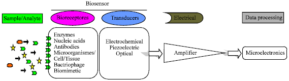 | |
| Fig. 1: | Principle of operation of a typical biosensor with its components |
| • | Antibodies, which interact with the corresponding antigens by affinity |
| • | Oligonucleotides, which recognize complementary sequences also by affinity |
| • | Cells and whole organisms, whose respiration, growth, fluorescence or bioluminescence can be indicative of the presence of certain chemicals |
| • | Tissues, which usually act as enzyme source |
| • | Biomimetic materials, also called synthetic bioreceptors |
It should be added proteins such as lectin based biosensors to these categories. Each of these techniques has advantages and disadvantages and, in fact, they are usually complementary (Campas et al., 2008).
BIORECOGNITION ELEMENTS
Bioreceptors or the biological recognition elements are the key points of specificity for biosensor technologies. A bioreceptor is a molecular species that utilizes a biochemical mechanism for recognition. They are responsible for binding the analyte of interest to the sensor for the measurement. Bioreceptors can generally be classified into five different major categories. These categories include antibody/antigen, enzymes, nucleic acids/DNA, cellular structures/cells, biomimetic and bacteriophage (phage). The enzymes, antibodies and nucleic acids are the main classes of bioreceptors which are widely used in biosensor applications. Though the enzymes are one of the biorecognition elements, they are mostly used to function as labels than the actual bioreceptor (Velusamy et al., 2010).
Enzyme: Biosensors that make use of enzymes as the biorecognition elements are a well developed and widely studied area. The use of enzymes as the biological recognition element was very popular in the first generation of biosensor development due to their commercial availability or ease of isolation and purification from different sources. Among various oxidoreductases, glucose oxidase, horseradish peroxidase and alkaline phosphatase have been employed in most biosensor studies (Wang, 2000; Laschi et al., 2000; Luong et al., 2008). Enzymes offer the advantages of high sensitivity, possibility of direct visualization and stability for years. But there are some disadvantages found when using enzymes as labels, which include multiple assay steps and the possibility of interference from endogenous enzymes. Many enzyme detection methods are visual, eliminating the need for expensive, complicated equipments (Velusamy et al., 2010). But the enzyme stability is still problematic and the ability to maintain enzyme activity for a long period of time still remains a formidable task (Tothill, 2001; D'Orazio, 2003).
Enzyme-based biosensors have been popular with over 2000 (by 2001) articles published in the literature and this is plausibly due to the need for monitoring glucose in blood (Tothill, 2001; D'Orazio, 2003) and the ease of construction of such biosensors (Luong et al., 2008). Many papers have been reported since the past 20 years, where immunosensor systems used enzymes as labels, for the detection of pathogens in food. For example, a report discussing the applications of highly dispersed carbon particles which utilized sandwich immunoassay format used Horseradish Peroxidase (HRP) enzyme to label the antibody for the detection of pathogenic bacteria's such as L. monocytogenes, E. coli and C. jejuni (Chemburu et al., 2005).
Nucleic acid: Recent advances in nucleic acid recognition have enhanced the power of DNA (deoxyribonucleic acid) biosensors and biochips (Velusamy et al., 2010). The development of nucleic acid biosensors alone has resulted in over 700 papers published since 1997 to 2008 (Luong et al., 2008). In the case of nucleic acid bioreceptors for pathogen detection, the identification of a target analyte's nucleic acid is achieved by matching the complementary base pairs that are often the genetic components of an organism. Since each organism has unique DNA sequences, any self-replicating microorganism can be easily identified (Velusamy et al., 2010). The preferred methods of measurement include optical (SPR, Surface Plasmon Resonance), electrochemical or piezoelectric detection systems. The detection of specific DNA sequences has been advocated for detecting microbial and viral pathogens (Yang et al., 1997) as viruses are almost uniquely DNA or RNA composed within an outer coat or capsid of protein (Hall, 1990). Due to their wide range of physical, chemical and biological activities, simple, rapid and inexpensive detection, nucleic acid based biosensors have been reported by many researchers for the detection of food pathogen like E. coli O157:H7 (Chen et al., 2008a), Salmonella sp. (Lermo et al., 2007), Cumpylohacter jejuni (Uyttendaele et al., 1997) etc.
Recent advances in nucleic acid recognition, like the introduction of Peptide Nucleic Acid (PNA) has opened up exciting opportunities for DNA biosensors. PNA is a synthesized DNA in which the sugar-phosphate backbone is replaced with a pseudopeptide. PNA as a probe molecule has several advantages like superior hybridization characteristics, detection of single-base mismatches and improved chemical and enzymatic stability relative to nucleic acids. PNA based nucleic acid recognition have been reported by many researchers (Briones et al., 2004; Fan et al., 2007; Steichen et al., 2007). Rahaie et al. (2010a) currently also reported the new DNA based microdumbells and nanodumbells which have dimensions that are close to biomolecules, thus making them viable templates for single molecule barcoding in nucleic acids analyzes.
Antibody: Antibodies are common bioreceptors used in biosensors. Antibodies may be polyclonal, monoclonal or recombinant, depending on their selective properties and the way they are synthesized (Lazcka et al., 2007). The way in which an antigen and an antigen-specific antibody interact is similar to a lock and key fit (Vo-Dinh and Cullum, 2000). An antigen-specific antibody fits its unique antigen in a highly specific manner, so that the three-dimensional structures of antigen and antibody molecules are matching. This unique property of antibodies is the key point that makes the immunosensors as powerful analytical tool and their ability to recognize molecular structures allows one to develop antibodies that bind specifically to chemicals, biomolecules, microorganisms, etc (Velusamy et al., 2010).
Few examples of biosensors fabricated using antibodies as the sensing element for the detection foodborne pathogens includes, Surface Plasmon Resonance (SPR) (Taylor et al., 2006; Waswa et al., 2007), evanescent wave fiber-optic biosensors (Lim, 2003), nanowire labeled direct-charge transfer biosensor (Pal et al., 2007), self-excited PZT-glass microcantilevers (Campbell and Mutharasan, 2005), mag-netoelastic resonance sensors (Guntupalli et al., 2007) and immuno-sensors (Tokarskyy and Marshall, 2008).
Cell: In cellular structures/cells based biorecognition is either based on whole cell/microorganism or a specific cellular component that is capable of specific binding to certain species. The cellular systems, enzymes and non-enzymatic proteins are the three major subclasses of this category (Velusamy et al., 2010).
Bacteriophages: Recently, bacteriophages are employed as biorecognition elements for the identification of various pathogenic microorganisms. These powerful bacteriophages (phages) are viruses that bind to specific receptors on the bacterial surface in order to inject their genetic material inside the bacteria. These entities are typically of 20-200 nm in size (Singh et al., 2009). Phages recognize the bacterial receptors through its tail spike proteins. Since the recognition is highly specific it can be used for the typing of bacteria and hence opened the path for the development of specific pathogen detection technologies. Researchers have reported the application of phage as a biorecognition element for the detection of various pathogens such as E. coli (Singh et al., 2009), S. aureus (Balasubramanian et al., 2007) and B. anthracis spores (Huang et al., 2008; Xie et al., 2009) by using different sensing platforms.
Biomimetic: A receptor that is fabricated and designed to mimic a bioreceptor (antibody, enzyme, cell or nucleic acids) is often termed a biomimetic receptor. Though there are several methods, such as genetically engineered molecules and artificial membrane fabrication. The molecular imprinting technique has emerged as an attractive and highly accepted tool for the development of artificial recognition agents. Molecular imprinting is one of the techniques of producing artificial recognition sites by forming a polymer around a molecule which can be used as a template.
Biochromic Conjugated Polymer (BCP) sensors are another type of biomimetic based biosensor for pathogen detection, demonstrated by Song et al. (2002). Biologically active cell membrane components were incorporated into conjugated polymers with desirable optical properties (Velusamy et al., 2010).
TRANSDUCTION PLATFORMS
Although, a variety of transduction methods have been feasible toward the development of biosensor technology, the most common methods are electrochemical and optical followed by piezoelectric (Wang, 2000; Collings and Caruso, 1997).
Electrochemical methods: Electrochemical transducers are the oldest and most commonly used (Conroy et al., 2009). Electrochemical sensors measure the electrochemical changes that occur when chemicals interact with a sensing surface of the detecting electrode. The electrical changes can be based on a change in the measured voltage between the electrodes (potentiometric transducer), a change in the measured current at a given applied voltage (amperometric transducer), or a change in the ability of the sensing material to detect changes in the electric field (Impedance including capacitance/conductance) (Jiang et al., 2008). Electrochemical biosensors appear more suited for field monitoring applications (e.g., hand-held) and miniaturization towards the fabrication of an implantable biosensor (Luong et al., 2008). They offer high specificity, low-detection limits, relative freedom from matrix interference and low cost. However, some challenges remain including high performance and cost-effectiveness (Jiang et al., 2008).
Piezoelectric methods: Piezoelectric sensors employ materials that resonate on the application of an external alternating electrical field (Jiang et al., 2008; Marco et al., 1995). Typically, Acoustic wave devices, made of piezoelectric materials such as quartz crystals are utilized and the frequency of oscillation in the field is a function of the crystal mass (Patel, 2002). Shifts in the frequency of the oscillation occur as a result of mass changes at the crystal surface. Therefore, the interaction of an analyte with a biorecognition element immobilized on the quartz crystal leads to a mass change and, thus, an oscillation frequency changes (Patel, 2002). Piezoelectric crystal devices are advantageous as they facilitate direct measurement without the need for labeling (Mutlu et al., 2008).
Piezoelectric sensors can be further categorized into bulk and surface acoustic wave instruments. Bulk Wave (BW) instruments are applied in gravimetric devices known as Quartz-Crystal Microbalances (QCM), which are related to mass sensitivity of the crystals, or Thickness Shear Mode (TSM) resonators, which describe the motion of the crystals vibration (Leonard et al., 2003).
Optical methods: Optical immunosensors consist of a light source, components to generate light with specific characteristics, a modulating agent, a sensing area and a photodetector. Optical sensors are advantageous due to their low signal-to-noise ratio and low-reagent volume requirements (Jiang et al., 2008). Direct optical sensors are most useful for biosensor applications due to the lack of requirements for labelling of either the analyte or the biorecognition element and the ability to work effectively in complex matrices. However, indirect optical sensors are also very valuable and are generated by incorporation of a suitable label, such as a fluorophore (D’Orazio, 2003).
Techniques commonly employed in optical biosensors include Reflectometric Interference Spectroscopy (RIfS), interferometry, Optical Waveguide Lightmode Spectroscopy (OWLS), Total Internal Reflection Fluorescence (TIRF), Surface Plasmon Resonance (SPR), resonant mirrors, fibre optics, ellipsometry, fluorescence and ultra violet/visible (UV/vis) spectroscopy (Conroy et al., 2009).
Nowadays, the integration of nanotechnology approaches into biosensors has provided new analytical methods for different diagnostic systems (Sanvicens and Marco, 2008; Choi et al., 2007; Jain, 2007). In this context, Nanoparticles (NPs) have raised attractive research field with respect to have several distinctive physical and chemical characterizations such as the large surface-to-volume ratio, unique optical, electronic and magnetic properties depending on their core materials (Sanvicens et al., 2009; Agasti et al., 2010). In addition, nanoparticles can be functionalized with a wide range of small organic ligands and large biomacromolecules by using tools and techniques of surface modification. These make them promising synthetic scaffold for the creation of novel chemical and biological detection systems (Rosi and Mirkin, 2005) and suitable labels in optical biosensors (or bioassays) to address the significant chemical and spectra limitations of organic fluorophores (Liu et al., 2008). Each of these capabilities has allowed researchers to design novel diagnostic systems that offer significant advantages in terms of sensitivity, selectivity, reliability and practicality. Currently, nanostructured materials, such as noble metal nanoparticles, quantum dots, magnetic nanoparticles and nucleic acid-nanoparticle hybrid nanostructures have been employed in a broad spectrum of highly innovative approaches for diagnostic and quantification bioassays of biologic molecules (Alivisatos, 2004; Niemeyer, 2001; West and Halas, 2000; Parak et al., 2003; Rahaie et al., 2010a). some of examples of application of NPs into biodevices and nanobiosensors (Merkoci, 2007; Ligler, 2009; Balasubramanian and Burghard, 2006; Sadik et al., 2009; Palecek and Fojta, 2007) are detection of specific biological molecules such as proteins (Hu et al., 2009), enzymes (Hong et al., 2009), nucleic acids (Litos et al., 2009), detection of infectious agents (Liu et al., 2007) and sometimes for detection and quantification of gene transcript simultaneously (Rahaie et al., 2010b).
LECTIN BASED BIOSENSING
For analytical purpose, although analytes can be detected by a wide range of immobilized ligands, such as antibodies, enzymes, cell receptors and oligonucleotide probes, the increasing numbers of analytes requiring monitoring and control and also, the need for high sensitivity, speed and accuracy of analytical measurements have stimulated considerable efforts in developing different sensors as diagnostics tools. Currently, carbohydrate binding proteins (CBFs, Lectins) as new bioreceptors has being started to play a pivotal role in many biosensing devices due to their exquisite specificity for their cognate carbohydrates.
Lectins description: Lectins are proteins of non-immune origin that specifically interact with carbohydrates without modifying those (Monzo et al., 2007). Historically, The name of lectin is derived from the Latin word legere, meaning, among other things to select. There are different definitions of lectins but the one which is represented by Goldstein et al. (1980) is more accepted. According to his description, a lectin is a carbohydrate-binding protein or glycoprotein of non-immune origin which agglutinates cells or precipitates glycoconjugates or both.
Most lectins specifically recognize sugar units (e.g., N-acetylneuraminic acid, N-acetylglucosamine, N-acetylgalactosamine, galactose, mannose, or fucose) (Scherz and Bonn, 1998; Monzo et al., 2007). Stillmark in 1888, as first descriptor of lectins, isolated ricin, an extremely toxic hemagglutinin from seeds of the castor plant (Ricinus communis). In 1919, the great biochemist J.B. Sumner crystallized the protein now known as Concanavalin A (Fig. 2) from the common Jackbean (canavalia ensiformis) and later in 1936 he discovered that ConA is able to bind and precipitate some polysaccharides such as glycogen and starch (Slifkin and Doyle, 1990).
Due to their multifaceted biological properties, lectins were later developed by cell biologists as probes to investigate cell surface structures and functions. Interaction with lectins can be used to obtain independent information about the presence of specific carbohydrates, the configuration of anomeric linkages and the location or position of carbohydrate residues polysaccharide molecules (Slifkin and Doyle, 1990; Damjanov, 1987).
Most reports suggest that plant lectins primarily bind monosaccharides, but it is also known that they bind complex determinants with higher affinity (Cummings, 1999).
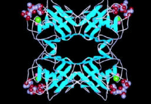 | |
| Fig. 2: | Structure of tetrameric a common used lectin, ConA, at 2.35 Å |
Apparently, the affinity of lectins to bind complex carbohydrates is often an order of magnitude higher than that for monosaccharides. In addition, the haptenic sugars used to inhibit lectins may not reflect the structural nature of the glycans (oligosaccharide chains attached to proteins) recognized with high affinity, as the binding of two competing ligands can be deduced from the law of mass action and depends on two parameters: the concentration of the ligands and their affinity to the lectins. Another aspect to consider in lectin affinity is that metal-carbohydrate interactions play an important role during the binding process of some lectins. For example, crystallographic studies on mannose-binding lectins proved that carbohydrate binding is only possible in the presence of Ca+2, while others interact with their saccharide counterpart in the absence of metal ions (Weis et al., 1992; Zheng et al., 1997).
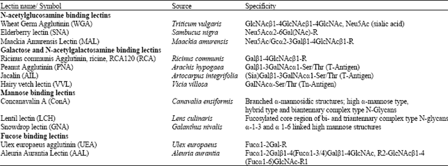
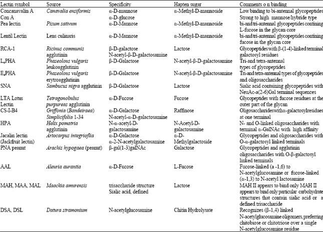
Lection sources and characterizations: Lectins may be derived from plant, microbial, or animal resources and may be soluble or membrane bound. Plant and animal lectins may be classified into groups on the basis of their carbohydrate-binding specificities. Generally, lectins are classified in five groups on the basis of their affinity for (1) N-acetylglucosamine, (2) galactose and N-acetyl-D-galactosamine, (3) glucose/mannose, (4) L-fucose and (5) salic acids (Slifkin and Doyle, 1990; Damjanov, 1987). Table 1 has shown the major lectins.
Roles of lectins in organisms: The biological role of lectins is quite speculative. lectins may be involved in sugar transport or carbohydrate storage. Some lectins may be associated with the binding of symbiotic rhizobia to root nodules. Because of their role in adhesion and agglutination, lectins have been considered to be important in both symbiotic and pathogenic interactions between some microorganisms and host. The presence of lectins in food products such as tomato, for example, is common.
| Table 1: | The main classes of lectins with their specifity (adapted form ref. No. 52) |
 | |
Lectin-cell binding can elicit a wide variety of biological phenomena. Some lectins induce mitosis in resting lymphocytes and some agglutinate neoplastic transformed cells, but not their healthy nonmalignant counterparts, whereas others agglutinate healthy nonmalignant cells (Slifkin and Doyle, 1990; Damjanov, 1987; Etzler, 1986).
Animal lectins: As mentioned above, lectins are not exclusively found in plants. Some animal lectins that we know today were discovered even before plant lectins, but they were not recognized as carbohydrate-binding proteins for quite a while (Kilpatrick, 2000). Further, lectins serve many different biological functions in animals from cell-adhesion regulation, through glycoprotein synthesis, to control of certain blood-protein levels (Danguy et al., 1998). Lectins are also known to play important roles in the immune system by recognizing carbohydrates that are exclusive to pathogens (Karlsson, 1999). Carbohydrate recognition of lectins often requires a certain anomeric configuration and specific adjacent sugar residues (Monzo et al., 2007).
Lectins have also been used to fractionate animal cells, including B and T lymphocytes and to demonstrate changes in cell surface architecture following virus infection or parasite infection.
Some lectins are found on the surface of mammalian liver cells which specifically recognize galactose residues. It is believed that these cell-surface receptors are responsible for the removal of certain glycoproteins from the circulatory system. Another lectin is a receptor that recognizes hydrolytic enzymes containing mannose-6-phosphate and targets these proteins for delivery to the lysosomes. Lectins are also known to play pivotal roles in the immune system by recognizing carbohydrats that are found exclusively on pathogens, or that are inaccessible on host cells. Examples are the lectin complement activation pathway and mannose-binding lectin (Slifkin and Doyle, 1990; Damjanov, 1987; Etzler, 1986; Dietz et al., 1988).
Microbial lectins: The microbial lectins may play an essential role in mediating adhesion to surfaces colonized by the microorganism. Many bacteria contain surface-associated lectins that enable these organisms to adhere to surfaces. Bacterial lectins resemble plant lectins in carbohydrate specificity, relative thermostability, divalent cation requirements and other properties and may be used for some of the same purposes as other lectins that yield coaggregation responses with certain yeast cells (Slifkin and Doyle, 1990; Mirelman, 1986).
Plants lectins: The function of lectins in plants is still uncertain. The best characterized family of plant lectins is the leguminosae (lectins such as ConA, soybean agglutinin and lentil lectin). Two other families of plants whose lectins have been characterized are the Graminea (cereals such as wheat germ) and Solanaceae (potatoes and tomatoes). Most leguminous lectins are metalloproteins with tightly bound Ca+2 and Mn+2 which are essential for carbohydrate-binding activity. Some plant lectins, more properly classified as toxins, however, are among the most poisonous proteins on our planet and can readily result in death not only to cells in cultures but also to animals. This kind of lectins contains one subunit for binding to carbohydrate ligands (B chain) and a second subunit that has no carbohydrate-binding activity but is instead an enzyme (A chain) with adenosine-N-glycosidase activity that can catalytically inactivate ribosomes. Such enzymes are termed ribosome-inactivating proteins or RIPs. These toxic plant lectin subunits conjugated to specific antibodies and other targeting ligands are now being tested as treatments for cancer and other disorders of cellular proliferation (Slifkin and Doyle, 1990; Jordinson et al., 1997; Takeya et al., 1998).
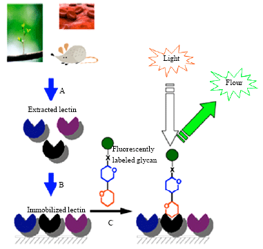 | |
| Fig. 3: | A typical method for the production of a lectin based optically detection approach including (A) lectin extraction, (B) immobilization on a solid surface and (C) biosensing of fluorescently labeled glycan |
Lectins are found in different parts of plants including seeds, bark, tubers, stem and leaves. lectins probably have many different and important functions in plants such as seed storage proteins, to help maintaining seed dormancy, Defense against fungal, viral and bacterial pathogens, Defense against animal predators, Symbiosis in legumes (rhizobia), Transport of carbohydrates, Mitogenic stimulation of embryonic plant cells, Elongation of cell walls and Recognition of pollen (Slifkin and Doyle, 1990; Damjanov, 1987; Rudiger, 1998; Sharon, 1998).
Lectins for analytical applications: As of today, lectins are normally chosen in bioseparation applications where glycoconjugates are involved. Figure 3 shows a typical method for the production of a lectin based optically detection approach. Different lectins are recommended to isolate glycoproteins and glycopeptides with distinct types of carbohydrate structures, e.g., just to list a few: galectins are specific for N-acetyl-lactosamine-containing carbohydrates found in both N-linked and O-linked glycans (Kasai and Hirabayashi, 1996); Concanavalin A (Con A ) recognizes oligomannosyl motifs in N-linked-glycans (Ohyama et al., 1985); peanut agglutinin is specific for O-linked-glycans (Neurohr et al., 1980) and Aleuria aurantia lectin shows broad specificity for fucose-containing oligosaccharides (Kochibe and Furukawa, 1980; Monzo et al., 2007). Table 2 provides a summary of the most frequently used lectins in bioseparations. In follow, it is mentioned the different opportunities for using Lectins for biosensing purposes and biosensor technology.
Medical samples analysis: The most important applications of lectin-based biosensors are in the field of medicine and therapeutics. Many different researches have already been done in this field and a lot of samples of this kind of sensors have been produced. Also, the large amount of published articles about the applications of lectin in the assessment process of clinical samples, laboratories and diagnostic instruments can prove the importance of this issue. Actually, the applicability of biosensing systems for precise determination of analytes of biomedical interest from diverse physiological clinical samples such as blood, urine, saliva, cell tissues, etc. is highly desirable to establish quick and reliable analytical tools avoiding time-and sample-consuming pretreatment methods (Shankaran et al., 2007).
Safina et al. (2008) proposed a novel lectin-based Quartz Crystal Microbalance (QCM) biosensor to identify the presence of different (bacteria) using the lectins immobilized on the surface of QCM crystal which bind specifically to the certain oligosaccharides present on the cell wall of the injected bacteria. The proposed biosensor is able to detect 103 cells and the flow-injection assay of the bacterial cells takes about 30 min.
| Table 2: | The common lectins used for detection and isolation of glycopeptides and oligosaccharides (Adapted from Ref No. 67) |
 | |
Dayarathna et al. (2008) introduced a proteomic platform for analysis of plasma samples. Their method utilizes a double fractionation approach which combines the MARS immunodepletion column with multi-lectin affinity chromatography, M-LAC, to deplete the most abundant proteins in plasma, the majority of which are glycosylated such as angiotensinogen and apolipoprotein CI which are present at high levels in patients with obesity plus diabetes and hypertension. The methodology was shown to be effective for profiling changes in the plasma proteome of subjects with obesity and its associated complications such as the mentioned diseases.
Currently, one of the main issues in pharmaceutical studies is the intelligent drug delivery to a specific tissue or cell such as cancer cells. To achieve this purpose, it’s essential to design a special pharmaceutical vector like liposomes which have target molecules such as a lectin for binding to the surface of a specific cell and then releasing the drug molecule. Different studies have been done and lots of articles related to this issue have been published.
Yamazaki et al. (2005) reported a method to prepare two types of sugar-chain-remodeled glyco-protein-liposome conjugates with different levels of complexity of sugar-chain structure, their lectin-binding affinity using in vitro experiments in order to assess their tissue-specific distribution. This attempt can be used to produce active targeting DDS nanoparticles for so-called missile drugs.
Bies et al. (2004) reviewed using lectins to target and deliver drugs to their site of action. It should be mentioned that besides the targeting to specific cells, the lectin-sugar interaction can also been used to trigger vesicular transport into or across epithelial cells.
In general, glycosylation is the key step in a number of processes at the cellular level such as cell attachment, migration and invasion (Xiea et al., 2009). Also, Glycosylation is a prevalent post-translational modification on most cell-surface bound and soluble proteins and lipids. The alteration in glycosignatures associated with malignant transformation, tumor progression and metastasis is very well documented. The relative abundances and branch structures of glycans on the membrane surface of cancer cells are often altered compared with normal cells. These alterations in glycosylation may be indicative of the different stages of the disease, thus being useful for diagnosis. Furthermore, different cancer cells express with different types and quantities of glycoconjugates on its surface. The information coded on cell surface glycans is frequently deciphered by lectins that recognize specific carbohydrate residues. Therefore, lectin may be used for disease diagnosis and encoding of different cancer cells. In addition, lectin has been widely used for drug delivery system (Xiea et al., 2009).
Existing methods for probing lectin-carbohydrate interactions are tedious, required to extensive instrumental setup and technical expertise. Accordingly, there are critical needs for developing effective new glycotechnologies and biosensors which are sensitive, rapid, simple, reliable and cost-effective.
Wang et al. (2006) reported a novel nanoparticle-based bioassay based on glycan interaction with surface-immobilized lectins. The lectin acts as the glycans recognition element for the competition between a nanocrystal-labeled glycans and the target glycans for the carbohydrate binding sites on lectin. The extent of competition is monitored by highly sensitive electrochemical stripping voltammetry of the captured nano-crystals. Electrochemical readout offers attractive advantages of miniaturization and low-cost (for meeting the demands of point-of-care diagnostics) and elegant ways for interfacing biorecognition events and signal transduction.
The utility of nanoparticle tracers for monitoring lectin-sugar interactions and for bioassays of disease-related glycan markers has been demonstrated. The glyconanosensors for electrochemical biosensing of free glycans can be readily expanded for analogous measurements of glycoconjugates and for other nanoparticle-based transductions (e.g., fluorescence) of glycan-lectin interactions. The assay has been optimized and tested using a model system involving a surface-bound pure Arachis hypogaea (peanut agglutinin, PNA) lectin and various analytes, including the cancer associated T antigen (β-D-Gal-[1f3]-D-GalNAc disaccharide) and it has been shown excellent discrimination between target and nontarget sugars (Dai et al., 2006).
Xue et al. (2010) have reported to construct a novel electrochemical lectin-probe by conjugation of FcCOOH to ConA for in situ homogeneous cytosensing and facile evaluation of cell surface mannose moieties. The proposed ferrocene-lectin based electrochemical method shows good performance for detection of cancer cells with broad detection range, low detection limit, good reproducibility and simple detection procedure. The designed electrochemical lectin-probe can be conveniently used to evaluate quantitatively surface carbohydrates on K562 cells in response to drugs with acceptable sensitivity and reliability, thus could be employed to evaluate quantitatively the mannosyl moieties on the cell surface.
Nagaraj et al. (2010) have developed an ultrasensitive diagnostic platform called NanoMonitor to enable rapid label-free analysis of highly promising class of glycans biomarkers with high sensitivity and selectivity. The glycosylation of fetuin, a serum protein and extracts from a human pancreatic cancer line were analyzed to demonstrate the capabilities of the NanoMonitor. The NanoMonitor device consists of a silicon chip with an array of gold electrodes forming multiple sensor sites and works on the principle of electrochemical impedance spectroscopy. Lectins are conjugated to the surface of the electrode. When, specific glycans from a test sample are bonded to lectins on the base of each nanowell, a perturbation of electrical double-layer occurs, which results in a change in the impedance. Based on its performance metrics, the NanoMonitor has excellent potential for development as a point-of-care handheld electronic biosensor device for routine detection of glycan biomarkers from clinical samples.
Agricultural and food industry: Simple, rapid and automated analytical systems for qualification and safety certainty of products are essential in food industry at various stages, from food material collection, processing, storage, transportation to deliver to consumers. It is need to be examined specific analytes, a wide group of materials ranging from small molecular organic compounds, fungal metabolites, microbes to whole cell microbes in order to ascertain the quality, hygienic condition and purity of food products. Also, biosensors are expanding in food-related fields for detection of various individual analytes which include pesticides or herbicides in farm products, veterinary drugs in animal products like dairy products, meat and fish, food preservatives, nutritional additives and so on. The analytical techniques can be classified into two major categories depending on the nature of their field applications. Primarily, cost-effective and high throughput automated analytical systems are required to routine periodical analysis at industrial establishments. However, rapid, miniaturized field-portable sensor systems are in high consideration at the times of pandemic outburst, enforcing food regulations regarding preservatives and nutrients, prevention of food adulteration, etc (Shankaran et al., 2007).
According to the lectins characteristics and the existence of glycosidic analytes in food samples, various biosensors based on these proteins have been fabricated. There are many different examples in this field. For instance, Carlsson et al. (2005) used simple microcontact printed gold-wafers to make a lectin panel for investigation and discrimination of different meat juices from different fresh meat. The results showed that the different meat juices from different species could be discriminated from each other.
Microorganism detection is a key issue in fields like food and drinking water quality assurance, biological terrorism threat control, applied medicine and fermentation technology. In most cases, early detection of very low concentrations of microorganisms is essential for an effective action; therefore there is a demand for rapid, inexpensive and highly sensitive analytical tools such as biosensors for their detection (Serra et al., 2008; Invitski et al., 1999).
Ertl and Mikkelsen (2001) created a novel method of microorganism species identification. They investigated an electrochemical biosensor array, in which transduction is based on respiratory cycle activity measurements, where the microorganism’s native respiratory chain is interrupted with non-native external oxidants. The selective biochemical recognition agents employed in this study were lectins that, once immobilized, recognize and bind to cell surface lipopolysaccharides. In this research, they supplied lectins from the species Artocarpus integrifolia, Arachis hypogaea, Galanthusnivalis, Phytolacca americana, Lens culinaris, Helix pomatia, Triticum vulgaris and Codium fragile as well as Con A and biotinylated Con A.
Serra et al. (2008) provided a new method which is proposed for the easy and early detection, identification and quantification of microorganisms based on specific recognition through wall cell glycocalyx components of bacteria by immobilizing lectin proteins on a gold-plated quartz crystal surface. The piezoelectric transduction allows label-free real-time monitoring of the binding event. Nonspecific adsorption of non-targeted organisms is minimized. Further development of the method would consist of building up a battery of lectin-quartz crystals as sensors to obtain a response profile for different microorganisms which would permit the identification via statistical analysis.
Environmental monitoring: A great variety of toxic chemicals are continuously released in the environment due to human and various technological activities including domestic and industrial wastages, use of drugs and pesticides, terrorist attacks and biological weapons and etc. Among the number of analytical methods developed for extensive environmental monitoring programs, lectin based biosensing is more attractive and effective owing to its advantages.
One of the pesticide that has attracted most research interest and that is used as a model pesticide in many investigations is 2, 4-dichlorophenoxyacetic acid (2, 4-D). A competitive immunoassay based on Surface Plasmon Resonance (SPR) for the detection of the pesticide 2, 4-dichlorophenoxyacetic acid (2, 4-D) was reported by (Svitel et al., 2000). The novelty of the assay was based on the regeneration of the chip surface by the reversible interaction between monosaccharide (D-glucose) and lectin (Concanavalin A). Concanavalin A-2, 4-D conjugate was chemically synthesized, purified and used for binding to the SPR chip modified with covalently bound α-D glucose. The interaction between anti-2, 4-D antibody and the surface-bound Concanavalin A-2, 4-D conjugate was monitored by surface plasmon resonance and the response was used for the quantification of 2, 4-D.
Phenolic compounds and aromatic amines have been widely distributed in nature and possessed relevance in several industrial processes. Owing to the toxicity and persistency, sensitive and rapid monitoring of phenolic compounds and aromatic amines has gained significance in environmental control and human health (Silva Luz et al., 2006; Yang et al., 2006; Morales et al., 2005). Bienzyme electrode with Horseradish Peroxidase (HRP) and Glucose Oxidase (GOD) multilayers has been constructed based on sugar-lectin biospecific interactions for amperometric determination of phenolic compounds and aromatic amines (Chen et al., 2008b). Substituted phenolic compounds and aromatic amines could be determined with in situ generation of H2O2 by GOD-catalyzed oxidation of glucose. The biosensor possessed high sensitivity and fast response for phenolic compounds and 95% of the maximum response could be reached in about 3S. The biosensor presented high stability due to the design for in situ generation of H2O2 with bienzyme system.
CONCLUSION AND PROSPECTIVE
The diagnostics market is expanding rapidly and covering a wide range of disciplines including agricultural and food industry, medicine and environment monitoring. It’s expected to produce a significant effect on quality improvement and cost-reduction in that area in order to establish appropriate technologies for biosensing. Recent advancements in nanotechnology have enabled a paradigm shift in biosensing. It seems that biosensors could play a pivotal role in providing powerful analytical tools to the required diagnosis sectors, particularly where rapid, low cost, high sensitive and specific measurements in field situations are required. Due to their unique characteristics and flexibility, biosensors show great promise for food safety and environmental monitoring applications. Advances in areas such as toxicity, bioavailability and multi pollutant screening could widen the potential market and allow these techniques to be competitive. There are different approaches to combine biology, chemistry and electronics in order to develop new biosensors with different applications in diagnostic filed. A wide range of transducers is available to engineer new biosensing devices. Also, a range of molecules with biorecognition properties can be used as the sensing element in biosensors. Lectin proteins are one of the most attractive biorecognition elements for providing biosensors. In this review, we summarized on-going developments in this field and made an attempt to create points of view of promising lectin-based biosensors. It is anticipated that lectin-affinity-based separation and detection methods and instruments, especially biosensors, will maintain their importance in diagnostic researches. The future of researches in this field appears exceedingly bright, judging by the surge in publications related to the creation of lectin-based biosensors and protein-carbohydrate based diagnostic technologies. New techniques also continue to be developed in order to study lectins and to utilize their specificities in functional assays. Based on the recent achievements in this field, we expect to see more and more innovative strategies in the near future to reduce the complexity of lectin-based detection and quantification techniques. At last, it is hoped that the review will provide new ideas and prospective for researchers and technologists who interest in creating new methods for diagnostic purposes.
REFERENCES
- Agasti, S.S., S. Rana, M.H. Park, C.K. Kim, C.C. You and V.M. Rotello, 2010. Nanoparticles for detection and diagnosis. Adv. Drug Deliv. Rev., 62: 316-328.
CrossRef - Alivisatos, P., 2004. The use of nanocrystals in biological detection. Nat. Biotechnol., 22: 47-52.
PubMed - Balasubramanian, K. and M. Burghard, 2006. Biosensors based on carbon nanotubes. Anal. Bioanal. Chem., 385: 452-468.
CrossRef - Bies, C., C.M. Lehr and J.F. Woodley, 2004. Lectin-mediated drug targeting: History and applications. Adv. Drug Delivery Rev., 56: 425-435.
CrossRef - Briones, C., E. Mateo-Marti, C. Gomez-Navarro, V. Parro, E. Roman and J.A. Martin-Gago, 2004. Ordered self-assembled monolayers of peptide nucleic acids with DNA recognition capability. Phys. Rev. Lett., 93: 208103-208103.
CrossRef - Bromage, E.S., T. Lackie, M.A. Unger, J. Ye and S.L. Kaattari, 2007. The development of a real-time biosensor for the detection of trace levels of trinitrotoluene (TNT) in aquatic environments. Biosens. Bioelectron., 22: 2532-2538.
PubMed - Campas, M., R. Carpentier and R. Rouillon, 2008. Plant tissue-and photosynthesis-based biosensors. Biotechnol. Adv., 26: 370-378.
CrossRef - Campbell, G.A. and R. Mutharasan, 2005. Detection of pathogen Escherichia coli O157: H7 using selfexcited PZT-glass microcantilevers. Biosens. Bioelectron., 21: 462-473.
CrossRef - Carlsson, J., F. Winquist, B. Danielsson and I. Lundstrom, 2005. Biosensor discrimination of meat juice from various animals using a lectin panel and ellipsometry. Anal. Chimica Acta, 547: 229-236.
CrossRef - Castillo, J., S. Gaspar, S. Leth, M. Niculescu and A. Mortari et al., 2004. Biosensors for life quality-Design, development and applications. Sens. Actuators B. Chemical, 102: 179-194.
Direct Link - Chemburu, S., E. Wilkins and I. Abdel-Hamid, 2005. Detection of pathogenic bacteria in food samples using highly-dispersed carbon particles. Biosens. Bioelectron., 21: 491-499.
CrossRef - Chen, S.H., V.C.H. Wu, Y.C. Chuang and C.S. Lin, 2008. Using oligonucleotide-functionalized Au nanoparticles to rapidly detect foodborne pathogens on a piezoelectric biosensor. J. Microbiol. Methods, 73: 7-17.
CrossRef - Choi, J.W., B.K. Oh, Y.K. Kim and J. Min, 2007. Nanotechnology in biodevices. J. Microbiol. Biotechnol., 17: 5-14.
PubMed - Choi, S.H., J.W. Lee and S.J. Sim, 2005. Enhanced performance of a surface plasmon resonance immunosensor for detecting Ab-GAD antibody based on the modified self-assembled monolayers. Biosens. Bioelectron., 21: 378-383.
PubMed - Collings, A.F. and F. Caruso, 1997. Biosensors: Recent advances. Rep. Prog. Phys., 60: 1397-1445.
CrossRef - Conroy, P.J., S. Hearty, P. Leonard, R.J. O`Kennedy, 2009. Antibody production, design and use for biosensor-based applications. Semin. Cell Dev. Biol., 20: 10-26.
PubMed - Dai, Z., A.N. Kawde, Y. Xiang, J.T. La Belle, J. Gerlach,V.P. Bhavanandan, L. Joshi and J. Wang, 2006. Nanoparticle-Based sensing of glycan-lectin interactions. J. Am. Chemical Soc., 128: 10018-10019.
CrossRef - Danguy, A., C. Decaestecker, F. Genten, I. Salmon and R. Kiss, 1998. Applications of lectins and neoglycoconjugates in histology and pathology. Acta Anatom., 161: 206-218.
PubMed - Dayarathna, M.K.D.R., W.S. Hancock and M. Hincapie, 2008. A two step fractionation approach for plasma proteomics using immunodepletion of abundant proteins and multi-lectin affinity chromatography: Application to the analysis of obesity, diabetes and hypertension diseases. J. Sep. Sci., 31: 1156-1166.
PubMed - Dhall, P., A. Kumar, A. Joshi, T. Kumar Saxsena, A. Manoharan, S.D. Makhijani and R. Kumar, 2008. Quick and reliable estimation of BOD load of beverage industrial wastewater by developing BOD biosensor. Sens. Actuators B Chemical, 133: 478-483.
CrossRef - Dillon, P.P., A.J. Killard, S.J. Daly, P. Leonard and R. O`Kennedy, 2005. Novel assay format permitting the prolonged use of regeneration-based sensor chip technology. J. Immunol. Methods, 296: 77-82.
PubMed - Ertl, P. and S.R. Mikkelsen, 2001. Electrochemical biosensor array for the identification of microorganisms based on lectin-lipopolysaccharide recognition. Anal. Chem., 71: 4241-4248.
PubMed - Fan, Y., X.T. Chen, J.M. Kong, C.H. Tung and Z.Q. Gao, 2007. Direct detection of nucleic acids by tagging phosphates on their backbones with conductive nanoparticles. Angew. Chem. Int. Ed., 46: 2051-2054.
CrossRef - Goldstein, I.J., R.C. Hughes, M. Monsigny, T. Osawa and N. Sharon, 1980. What should be called a lectin? Nature, 285: 66-66.
CrossRef - Guntupalli, R., J. Hu, R.S. Lakshmanan, T.S. Huang, J.M. Barbaree and B.A. Chin, 2007. A magnetoelastic resonance biosensor immobilized with polyclonal antibody for the detection of Salmonella typhimurium. Biosens. Bioelectron., 22: 1474-1479.
PubMed - Hildebrandt, A., R. Bragos, S. Lacorte and J.L. Marty, 2008. Performance of a portable biosensor for the analysis of organophosphorus and carbamate insecticides in water and food. Sens Actuators B Chemical, 133: 195-201.
CrossRef - Hong, S., I. Choi, S. Lee, Y.I. Yang, T. Kang and J. Yi, 2009. Sensitive and colorimetric detection of the structural evolution of superoxide dismutase with gold nanoparticles. Anal. Chem., 81: 1378-1382.
CrossRef - Hu, J., P.C. Zheng, J.H. Jiang, G.L. Shen, R.Q. Yu and G.K. Liu, 2009. Electrostatic interaction based approach to thrombin detection by surface-enhanced raman spectroscopy. Anal. Chem., 81: 87-93.
CrossRef - Huang, S, S.Q. Li, H. Yang, M. Johnson and J. Wan et al., 2008. Optimization of phage-based magnetoelastic biosensor performance. Sens. Transducers, 3: 87-96.
Direct Link - Huet, A.C., T. Fodey, S.A. Haughey, S. Weigel, C. Elliott and P. Delahaut, 2010. Advances in biosensor-based analysis for antimicrobial residues in foods. TrAC Trends Anal. Chem.
CrossRef - Jain, K.K., 2007. Applications of nanobiotechnology in clinical diagnostics. Clin. Chem., 53: 2002-2009.
PubMedDirect Link - Jha, R. and A.K. Sharma, 2010. Design of a silicon-based plasmonic biosensor chip for human blood-group identification. Sensors Actuators B: Chem., 145: 200-204.
CrossRef - Jiang, X., D. Li, X. Xu, Y. Ying, Y. Li, Z. Ye and J. Wang, 2008. Immunosensors for detection of pesticide residues. Biosens. Bioelectron., 23: 1577-1587.
PubMed - Jordinson, M., R.J. Playford and J. Calam, 1997. Effects of a panel of dietary lectins on cholecystokinin release in rats. Am. J. Physiol., 273: G946-G950.
Direct Link - Karlsson, K.A., 1999. Bacterium-host protein-carbohydrate interactions and pathogenicity. Biochem. Soc. Trans., 27: 471-474.
PubMed - Kasai, K.I. and J. Hirabayashi, 1996. Galectins: A family of animal lectins that decipher glycocodes. J. Biochem., 119: 1-8.
PubMed - Kim, Y.H., J.S. Park and H.I. Jung, 2009. An impedimetric biosensor for real-time monitoring of bacterial growth in a microbial fermentor. Sens. Actuators B Chemical, 138: 270-277.
Direct Link - Laschi, S., M. Franek and M. Mascini, 2000. Screen-printed electrochemical immunosensors for PCB detection. Electroanal, 12: 1293-1298.
3.0.CO;2-5/abstract target='_blank' class='btn btn-sm btn-outline-primary mr-3 mt-3'>Direct Link - Lazcka, O., F.J. Del Campo and F.X. Munoz, 2007. Pathogen detection: A perspective of traditional methods and biosensors. Biosen. Bioelectronic, 22: 1205-1217.
PubMed - Leonard, P., S. Hearty, J. Brennan, L. Dunne, J. Quinn, T. Chakraborty and R. O`Kennedy, 2003. Advances in biosensors for detection of pathogen in food and water. Enzyme Microbial Technol., 32: 3-13.
CrossRef - Lermo, A., S. Campoy, J. Barbe, S. Hernandez, S. Alegret and M. Pividori, 2007. In situ DNA amplification with magnetic primers for the electrochemical detection of food pathogens. Biosens. Bioelectron., 22: 2010-2017.
PubMed - Li, Y.S., W.P. Liu, X.F. Gao, D.D. Chen and W.G. Li, 2008. Immobilized enzymatic fluorescence capillary biosensor for determination of sulfated bile acid in urine. Biosens. Bioelectron., 24: 538-544.
PubMed - Ligler, F.S., 2009. Perspective on optical biosensors and integrated sensor systems. Anal. Chem., 81: 519-526.
CrossRef - Lim, D.V., 2003. Detection of microorganisms and toxins with evanescent wave fiber-optic biosensors. Proc. IEEE, 91: 902-907.
CrossRef - Litos, I.K., P.C. Ioannou, T.K. Christopoulos, J. Traeger-Synodinos and E. Kanavakis 2009. Multianalyte, dipstick-type, nanoparticle-based DNA biosensor for visual genotyping of single-nucleotide polymorphisms. Biosens. Bioelectron., 24: 3135-3139.
CrossRef - Liu, Y., R. Brandon, M. Cate, X. Peng, R. Stony and M. Johnson, 2007. Detection of pathogens using luminescent cdse/zns dendron nanocrystals and a porous membrane immunofilter. Anal. Chem., 79: 8796-8802.
CrossRef - Luo, J., X. Liu, Q. Tian, W. Yue, J. Zeng, G. Chen and X. Cai, 2009. Disposable bioluminescence-based biosensor for detection of bacterial count in food. Anal. Biochem., 394: 1-6.
CrossRef - Luong, J.H.T., A. Mulchandani and G.G. Guilbaul, 1988. Developments and applications of biosensors. Trends Biotechnol., 6: 310-316.
CrossRefDirect Link - Luong, J.H.T., K.B. Male and J.D. Glennon, 2008. Biosensor technology: Technology push versus market pull. Biotechnol. Adv., 26: 492-500.
Direct Link - Marco, M.P., S. Gee and B.B. Hammock, 1995. Immunochemical techniques for environ- mental analysis. Trend Anal. Chem., 4: 341-350.
CrossRef - Monzo, A., G.K. Bonn and A. Guttman, 2007. Lectin-immobilization strategies for affinity purification and separation of glycoconjugates. Trends Anal. Chem., 26: 423-432.
Direct Link - Morales, M.D., M.C. Gonzalez, B. Serra, J.B. Zhang, A.J. Reviejo and J.M. Pingarron, 2005. Biosensing of aromatic amines in reversed micelles with self-generation of hydrogen peroxide at glucose oxidase-peroxidase bienzyme electrodes. Electroanalysis, 17: 1780-1788.
CrossRef - Muller-Renaud, S., D. Dupont and P. Dulieu, 2005. Development of a biosensor immunoassay for the quantification of αS1-casein in milk. J. Dairy Res., 72: 57-64.
Direct Link - Nayak, M., A. Kotian, S. Marathe and D. Chakravortty, 2009. Detection of microorganisms using biosensors-A smarter way towards detection techniques. Biosens. Bioelectron., 25: 661-667.
PubMed - Neurohr, K.J., N.M. Young and H.H. Mantsch, 1980. Determination of the carbohydrate-binding properties of peanut agglutinin by ultraviolet difference spectroscopy. J. Biol. Chem., 255: 9205-9209.
Direct Link - Niemeyer, C.M. and P. Doz, 2001. Nanoparticles, proteins and nucleic acids: Biotechnology meets materials science. Angew. Chem. Int. Ed., 40: 4128-4158.
3.0.CO;2-S/abstract target='_blank' class='btn btn-sm btn-outline-primary mr-3 mt-3'>Direct Link - Ohyama, Y., K. Kasai, H. Nomoto and Y. Inoue, 1985. Frontal affinity chromatography of ovalbumin glycoasparagines on aconcanavalinA-Sepharose column. J. Biol. Chem., 260: 6882-6887.
Direct Link - Oh, S.E., S.H.A. Hassan and S.W. van Ginkel, 2010. A novel biosensor for detecting toxicity in water using sulfur-oxidizing bacteria. Sens. Actuators B Chemical.
CrossRef - Pal, S., E.C. Alocilja and F.P. Downes, 2007. Nanowire labeled direct-charge transfer biosensor for detecting Bacillus species. Biosens. Bioelectron., 22: 2329-2336.
PubMed - Palecek, E. and M. Fojta, 2007. Magnetic beads as versatile tools for electrochemical DNA and protein biosensing. Talanta, 74: 276-290.
PubMed - Parak, W.J., D. Gerion, T. Pellegrino, D. Zanchet and C. Micheel et al., 2003. Biological applications of colloidal nanocrystals. Nanotechnology, 14: 15-27.
Direct Link - Patel, P.D., 2002. (Bio) sensors for m easurement of analytes implicated in food safety: A review. Trend Anal. Chem., 21: 96-115.
CrossRef - Rahaie, M., R. Ghai, B. Babic and K. Dimitrov, 2010. Synthesis and characterization of DNA-based micro- and nanodumbbell structures. J. Bionanosci., 3: 73-79.
CrossRef - Rosi, N.L. and C.A. Mirkin, 2005. Nanostructures in biodiagnostics. Chem. Rev., 105: 1547-1562.
CrossRefPubMedDirect Link - Rudiger, H., 1998. Plant lectins-More than just tools for glycoscientists: Occurrence, structure and possible functions of plant lectins. Acta Anat. (Basel)., 161: 130-152.
PubMed - Sadik, O.A., A.O. Aluoch and A. Zhou, 2009. Status of biomolecular recognition using electrochemical techniques. Biosens. Bioelectron., 24: 2749-2765.
CrossRef - Safina, G., M. van Lier and B. Danielsson, 2008. Flow-injection assay o f the pathogenic bacteria using lectin-based quartz crystal microbalance biosensor. Talanta, 77: 468-472.
CrossRef - Sanvicens, N. and M.P. Marco, 2008. Multifunctional nanoparticles-properties and prospects for their use in human medicine. Trends Biotechnol., 26: 425-433.
CrossRefPubMedDirect Link - Sanvicens, N., C. Pastells, N. Pascual and M.P. Marco, 2009. Nanoparticle-based biosensors for detection of pathogenic bacteria. Trends Anal. Chem., 28: 1243-1252.
CrossRef - Serra, B., M. Gamella, A.J. Reviejo and J.M. Pingarron, 2008. Lectin-modified piezoelectric biosensors for bacteria recognition and quantification. Anal. Bioanal. Chem., 391: 1853-1860.
CrossRef - Shankaran, D.R., K.V. Gobi and N. Miura, 2007. Recent advancements in surface plasmon resonance immunosensors for detection of small molecules of biomedical, food and environmental interest. Sens. Actuators B, 121: 158-177.
Direct Link - Shaojun, D. and X. Chen, 2002. Some new aspects in biosensors. Rev. Mol. Biotechnol., 82: 303-323.
PubMed - Sharma, S.K., N. Sehgal and A. Kumar, 2003. Biomolecules for development of biosensors and their applications. Curr. Appl. Physics, 3: 307-316.
CrossRef - Sharon, N., 1998. Glycoproteins now and then: A personal account. Acta Anat. (Basel)., 161: 7-17.
Direct Link - Silva Luz, R.D.C., F.S. Damos, A.B. de Oliveira, J. Beck and L.T. Kubota, 2006. Development of a voltammetric sensor for catechol in nanomolar levels using a modified electrode with Cu (phen)(2)(TCNQ)(2) and PLL. Sens. Actuators B Chem., 117: 274-281.
CrossRef - Singh, A., N. Glass, M. Tolba, L. Brovko, M. Griffiths and S. Evoy, 2009. Immobilization of bacteriophages on gold surfaces for the specific capture of pathogens. Biosens. Bioelectron., 24: 3645-3651.
CrossRef - Slifkin, M. and R.J. Doyle, 1990. Lectins and their application to clinical microbiology. Clin. Microbiol. Rev., 3: 197-218.
PubMedDirect Link - Song,.J, Q. Cheng, S.M. Zhu and R.C. Stevens, 2002. Smart materials for biosensing devices: Cellmimicking supramolecular assemblies and colorimetric detection of pathogenic agents. Biomed. Microdev., 4: 213-221.
CrossRef - Steichen, M., Y. Decrem, E. Godfroid and C. Buess-Herman, 2007. Electrochemical DNA hybridization detection using peptide nucleic acids and [Ru(NH3)(6)](3+) on gold electrodes. Biosens. Bioelectron., 22: 2237-2243.
CrossRef - Svitel, J., A. Dzgoev, K. Ramanathan and B. Danielsson, 2000. Surface plasmon resonance based pesticide assay on a renewable biosensing surface using the reversible concanavalin A monosaccharide interaction. Biosens. Bioelectron., 15: 411-415.
PubMed - Takeya, A., O. Hosomi and T. Kogure, 1998. Vicia villosa B4 lectin inhibits nucleotide pyrophosphatase activity toward UDP-GalNAc specifically. Biochim. Biophys. Acta., 1425: 215-223.
CrossRef - Taylor, A.D., J. Ladd, Q.M. Yu, S.F. Chen, J. Homola and S.Y. Jiang, 2006. Quantitative and simultaneous detection of four foodborne bacterial pathogens with a multi-channel SPR sensor. Biosens. Bioelectron., 22: 252-258.
CrossRef - Tothill, I.E., 2001. Biosensors developments and potential applications in the agricultural diagnosis sector. Comput. Electron. Agric., 30: 205-218.
CrossRef - Turkarslan, O., S.K. Kayahan and L. Toppare, 2009. A new amperometric cholesterol biosensor based on poly (3,4-ethylenedioxypyrrole). Sens. Actuators B Chem., 136: 484-488.
CrossRef - Uyttendaele, M., A. Bastiaansen and J. Debevere, 1997. Evaluation of the NASBA® nucleic acid amplification system for assessment of the viability of Campylobacter jejuni. Int. J. Food Microbiol., 37: 13-20.
PubMed - Vamvakaki, V. and N.A. Chaniotakis, 2007. Pesticide detection with a liposome-based nano-biosensor. Biosens. Bioelectron., 22: 2848-2853.
CrossRefDirect Link - Velusamy, V., K. Arshak, O. Korostynska, K. Oliwa and C. Adley, 2010. An overview of foodborne pathogen detection: In the perspective of biosensors. Biotechnol. Adv., 28: 232-254.
CrossRef - Vianello, F., R. Boscolo-Chio, S. Signorini and A. Rigo, 2007. On-line detection of atmospheric formaldehyde by a conductometric biosensor. Biosens. Bioelectron, 22: 920-925.
CrossRefDirect Link - Vo-Dinh, T., B.G. Tromberg, G.D. Griffin, K.R. Ambrose, M.J. Sepaniak and E.M. Gardenhire, 1987. Antibody-based fiber optics biosensor for the carcinogen benzo (a) pyrene. Appl. Spectro. Sci., 41: 735-738.
Direct Link - Vo-Dinh, T. and B. Cullum, 2000. Biosensors and biochips: Advances in biological and medical diagnostics. Fresen. J. Anal. Chem., 366: 540-551.
PubMed - Waswa, J., J. Irudayaraj and C. DebRoy, 2007. Direct detection of E. coli O157: H7 in selected food systems by a surface plasmon resonance biosensor. LWT-Food Sci. Technol., 40: 187-192.
CrossRef - West, J.L. and N.J. Halas, 2000. Applications of nanotechnology to biotechnology commentary. Curr. Opin. Biotechnol., 11: 215-217.
PubMed - Xie, F., H. Yang, S. Li, W. Shen and J. Wan et al., 2009. Amorphous magnetoelastic sensors for the detection of biological agents. Intermetallics, 17: 270-273.
CrossRef - Xiea, M., J. Hua, Y.M. Longa, Z.L. Zhanga, H.Y. Xieb and D.W. Panga, 2009. Lectin-modified trifunctional nanobiosensors for mapping cell surface glycoconjugates. Biosens. Bioelectron., 24: 1311-1317.
PubMed - Xue, Y., L. Ding, J. Lei and H. Ju, 2010. A simple electrochemical lectin-probe for in situ homogeneous cytosensing and facile evaluation of cell surface glycan. Biosens. Bioelectron., 26: 169-174.
CrossRef - Yamazaki, N., S. Kojima and H. Yokoyama, 2005. Biomedical nanotechnology for active drug delivery systems by applying sugar-chain molecular functions. Curr. Appl. Phys., 5: 112-117.
CrossRef - Yang, M., M.E. McGovern and M. Thompson, 1997. Genosensor technology and the detection of interfacial nucleic acid chemistry. Anal. Chim. Acta, 346: 259-375.
CrossRef - Yang, S.M., Y.M. Li, X.M. Jiang, Z.C. Chen and X.F. Lin, 2006. Horseradish peroxidase biosensor based on layer-by-layer technique for the determination of phenolic compounds. Sens. Actuators B Chem., 114: 774-780.
CrossRef - Zheng, Y.J., R.L. Ornstein and J.A. Leary, 1997. A density functional theory investigation of metal ion binding sites in monosaccharides. Theochem, 289: 233-240.
CrossRef - Wang, J., Z. Dai, A.N. Kawde, Y. Xiang and J.T. La Belle et al., 2006. Nanocrystal-based electrochemical biosensors of glycan-lectin interactions suitable for point-of-care use. Nanomedicine: Nanotechnology Biol. Med., 2: 314-318.
CrossRef