
Yahaya Tajudeen
Department of Cell Biology and Genetics, University of Lagos, Nigeria
Joy Okpuzor
Department of Cell Biology and Genetics, University of Lagos, Nigeria
Adedayo Titilola Fausat
Department of Cell Biology and Genetics, University of Lagos, Nigeria
Asian Journal of Scientific Research
Year: 2011 | Volume: 4 | Issue: 4 | Page No.: 315-325


ABSTRACT
The general health problems that may arise from prolonged exposure to cement dust were investigated using black rats (Rattus rattus L.) living around a Portland Cement Company in Sagamu, Ogun State, Nigeria. 24 black rats, comprising of 12 rats from the cement factory and 12 rats from an environment free from cement dust were used for the study. Elemental analysis, hematology examination, histopathology examination and UV spectroscopy of the DNA of the rats in the two locations were carried out in the laboratory. The elemental analysis of the lung tissues of the exposed rats showed significant (p<0.05) concentrations of calcium, silicon, aluminum, chromium and lead compared to the control rats. The hematology examinations of the exposed rats showed marked reduction in the PCV, HB, RBC, WBC and total protein compared to the control rats. The histopathology analysis of the lung tissues of the exposed rats showed abnormal alveolar architecture, damaged bronchioles, disrupted bronchus, weak respiratory connective tissues, degenerated epithelium linings and inflammations. The liver tissues had abnormal cellular pattern, damaged central veins, disruption of portal triad and inflammations. And the kidney tissues showed damaged epithelium linings, convoluted tubules, damaged renal corpuscles and inflammations. There was no significant difference between the UV spectroscopy of the DNA of the exposed and the control rats. The results further confirm that cement dust is both toxic and pathogenic to animals including man. There is no doubt that people working or living within the vicinity of the cement company stand the risk of being affected by different types of diseases arising from exposure to cement dust.
PDF Abstract XML References Citation
Received: May 22, 2011;
Accepted: June 08, 2011;
Published: September 08, 2011
How to cite this article
Yahaya Tajudeen, Joy Okpuzor and Adedayo Titilola Fausat, 2011. Investigation of General Effects of Cement Dust to Clear the Controversy Surrounding its Toxicity. Asian Journal of Scientific Research, 4: 315-325.
DOI: 10.3923/ajsr.2011.315.325
URL: https://scialert.net/abstract/?doi=ajsr.2011.315.325
DOI: 10.3923/ajsr.2011.315.325
URL: https://scialert.net/abstract/?doi=ajsr.2011.315.325
INTRODUCTION
Since the advent of industrial revolution, economic performances have been considered as the principal criteria for measuring progress. However, industrial revolution has been accompanied by environmental pollution and degradation. Activities such as processing, manufacturing, transportation and consumption not only deplete the stock of natural resources but also add stress to the environment by accumulating wastes (World Resources Institute, 1998).
In the past, companies often discharged wastes indiscriminately, leaving the government to bear the cost of environmental clean-up. But today, companies are responsible for the clean-up of polluted environments. As a result of increased awareness of the effects of environmental pollution on human and other organisms, different monitoring agencies are putting pressure on companies to put control strategies in place. Some companies are making efforts to decrease any negative impacts their activities may have on the environment, while some companies have continued to pollute the environment and professing to be environmentally conscious (Doyle, 2002). Cement companies are important sources of environmental pollution with detrimental effects on plant and animal (El-Abssay et al., 2011). The main impacts of cement production on the environment are the broadcast of dust and gases (Bilen, 2010). But cement industry is among the industries that have not complied with full disclosure of their activities and profess to be environmentally friendly.
The basic components of cement are calcium oxide, silicon dioxide, aluminum trioxide and iron oxide (Fell et al., 2010). However, the burning and calcination process of cement produces pollutants such as heavy metals, dioxin, particulates, chromium, sulfur dioxide, nitrogen dioxide and carbon dioxide (Akinola et al., 2008; Ade-Ademilua and Obalola, 2008). These toxic elements and gases have been fingered in a lot of diseases including respiratory diseases, genetic diseases, hematology problems, multi-organ damage and cancer (Mohammed and Sambo, 2008; Leem et al., 2008; Zeleke et al., 2010; Gbadebo and Amos, 2010; Ogunbileje and Akinosun, 2011). Furthermore, various residents of cement plants are protesting against health problems affecting them which they attribute to the quality of the air they breathe in (Fortune, 2011). Despite these claims, cement manufacturers have continued to deny the health effects of their activities. Their argument is being supported by the fact that most of the studies mentioned above were done on the basis of radiology or spirometry (Meo, 2004) which give rooms for controversy. Because the study organism, man, is mobile and could have developed the diseases from environments he has previously lived. Therefore, to clear this controversy, a research, describing the effects of cement dust on various organs and systems of an animal which has spent its entire life around Cement Company, is needed. The objective of this study was to settle the rift between cement producers and residents of cement plants by investigating the veracity of all the alleged health hazards of cement dust.
MATERIALS AND METHODS
Sources of test animals: The exposed rats (12 in number) were caught using traps at about 100 m from the cement factory while the control rats (12 in number) were caught at Ogijo town, about 6 km from the cement company. The weights of the rats in the two locations were taken and recorded.
Experimentation: The experiment commenced in mid May, 2010, a week after capturing the rats and lasted for about 4 weeks. The 12 rats from the cement factory were labeled the exposed rats while the 12 rats from Ogijo town were labeled the control rats. Ogijo town is free from cement dust pollution and is in the same climatic zone as the cement factory. The rats in the two groups were moved into the Environmental Biology Laboratory, University of Lagos, where they were dissected. Elemental analysis, hematology examinations, UV spectroscopy of the DNA and histopathology analysis of the rats were carried out.
Chemical analysis of the lung tissues: The chemical analysis of the lung tissues of the rats in the two groups was done by Atomic Absorption Spectroscopy using UNICAM model969 Spectrophotometer at the Environmental Biology Laboratory.
Hematological examination: The rats were sedated with chloroform in the laboratory. Total death was prevented to allow continuous flow of blood for proper blood collection. Each rat was pegged down on a work bench and held firmly with office pins. Surgical blades were used to cut through the chest region of the rats in a dorsal-ventral direction. The blood was then collected from a beating heart using a Na Heparinized capillary tube through capillary action into EDTA bottles. EDTA serves as anti-coagulant and Na heparin in the capillary tube. The blood parameters (PCV, HB, WBC and RBC) were determined using Sysmex auto-analyzer while the total protein was determined using VET 360 Veterinary Refractometer (Phoenix series).
Preparation of internal organs for histopathology examinations: The internal organs (heart, kidney, liver and reproductive organs) of the rats were prepared for histopathology examinations using the method of Taylor et al. (2003).
Ultra-violet spectroscopy of the DNA: Total DNA content of the blood samples of the exposed and control rats were isolated using the protocol described by Dellaporta et al. (1983). The purity of the DNAs was then estimated using Ultra -Violet Spectroscopy, because DNA and other nucleic acids absorb ultra-violet light in the 260 to 280 nm range. After isolating the DNAs, impurities such as protein and RNA were removed, because good estimation can only be made on clean preparation. A cuvette was filled with the DNA samples and TE buffer in ratio of 1:1000 microliters and mixed thoroughly. The spectrophotometer containing the mixture was set on and the Optical Density (OD) or Absorbance (AB) at 260 and 280 nm in respect of each DNA sample was taken. The concentration or purity of the DNAs was estimated by dividing OD at 260 nm with OD at 280 nm. The optical density of a pure and normal DNA is between 1.5-2.0.
Statistical analysis: A database file was created in a personal computer and all statistical analysis was carried out with the Statistical Package for Social Sciences (SPSS) version 17 for windows and Microsoft Office Excel 2007. Comparison of data among exposed and control groups were calculated using Student’s t-test. p<0.05 was considered statistically significant.
RESULTS
Table 1 showed that both control and exposed rats grew normally. The minimum and maximum weights of the control rats were 230 and 280 g, respectively while the minimum and maximum weights of the exposed rats were 208 and 274 g, respectively. Also, the mean weights of the control and the exposed rats were 255 and 251 g, respectively. There was no significant difference (p>0.05) between the mean weights of the two groups of rats.
The elemental analysis of the lungs of the control and the exposed rats showed that the concentrations of some elements associated with cement-kilns burning wastes as alternative fuel were significantly higher (p<0.05) in the lungs of the exposed rats than in the lungs of the control rats. For example, in Table 2, the minimum and maximum concentrations of calcium, silicon, aluminum, chromium, lead in the control rats were 8.63, 0.035, 0.015, 0.038, 0.031 mg kg-1, respectively and 10.10, 0.096, 0.044, 0.062, 0.059 mg kg-1, respectively while the minimum and maximum concentrations of calcium, silicon, aluminum, chromium, lead in the exposed rats were 47.81, 0.96, 1.20, 0.123, 0.133 mg kg-1, respectively and 60.30, 1.12, 1.56, 0.172, 0.315 mg kg-1, respectively.
| Table 1: | Comparison between the weights (g) of control and exposed rats |
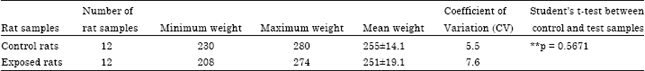 | |
| Data were expressed as Mean±SD. Since **p>0.05, no significant difference exist between the mean weights of the control and exposed rats | |
| Table 2: | Comparison between the concentrations in ( mg kg-1) of the elements detected in the lungs of the control and exposed rats |
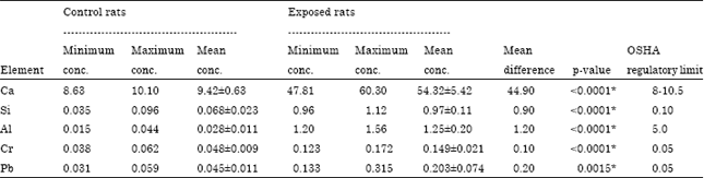 | |
| Values were expressed as Mean±SD. When *p<0.05 = significant from control and when **p>0.05 = not significant from control. OSHA: Occupational safety and health administration | |
Furthermore the mean concentrations in (mg kg-1) of calcium, silicon, aluminum, chromium and lead in the lungs of the exposed rats were 54.32, 0.97, 1.25, 0.149 and 0.203 mg kg-1, respectively while the mean concentrations in (mg kg-1) of calcium, silicon, aluminum, chromium and lead in the lungs the control rats were 9.42 mg kg-1, 0.068, 0.028, 0.048 and 0.045 mg kg-1, respectively. A significant difference (p<0.05) exists between the mean concentrations of all the chemical elements in the lungs of the control and exposed rats.
The results of the hematological examinations of the exposed rats showed marked reduction in the blood parameters compared to the blood parameters of the control rats. For instance, the minimum and maximum values of the packed cell volume, the hemoglobin, the red blood cells, the white blood cells, the total protein of the control rats were 36%, 12 g dL-1, 3.75x10¹², 3700 mm3, 6.40 g dL-1, respectively and 43%, 14.3 g dL-1, 4.51x10¹², 5500 mm3, 7.00 g dL-1, respectively while the minimum and maximum values of the packed cell volume, the hemoglobin, the red blood cells, the white blood cells, the total protein of the exposed rats were 18%, 6.00 g dL-1, 2.21x10¹², 1450 mm3, 4.96 g dL-1, respectively and 25%, 8.33 g dL-1, 2.95x10¹², 3300 mm3, 6.21 g dL-1, respectively. Moreover, the mean packed cell volume, the hemoglobin, the red blood cells, the white blood cells and the total protein of the control rats were 39.40%, 13.1 g dL-1, 4.07x10¹² and 6.66 g dL-1, respectively compared to 22.00%, 7.31 g dL-1, 2.59x10¹² and 5.36 g dL-1, respectively of the exposed rats (Table 3). There was a significant difference (p<0.05) between the mean values of all the blood parameters of the control and the exposed rats.
The Ultra-Violet Spectroscopy of the DNA of the exposed rats showed no noticeable contaminations when compared with the control rats. In Table 4, the absorbance at 260/280 of the control rats was 1.90 while the absorbance at 260/280 of the control rats was 1.80. These values are normal and need no further investigation. Furthermore, there was no significant difference (p>0.05) between the mean ultra-violet spectroscopy of the control and exposed rats.
The results of the histopathology examinations of the lung, liver and kidney tissues of the exposed rats showed marked histological changes compared to the histopathology of the lung, liver and kidney tissues of the control rats.
| Table 3: | Comparison between the blood parameters of the control and exposed rats |
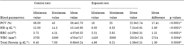 | |
| Values were expressed as Mean±SD. When *p<0.05 = Significant from control and when **p>0.05 = not significant from control | |
| Table 4: | Comparison between the ultra-violet spectroscopy of the control and exposed rats |
 | |
| Values were expressed as Mean±SD. When absorbance (A) at 260/280 is between 1.5-1.9 = Normal values, below 1.5 and above 2.0 = abnormal values. Since **p>0.05, no significant difference exist between the mean UV spectroscopy of the DNA of the control and exposed rats | |
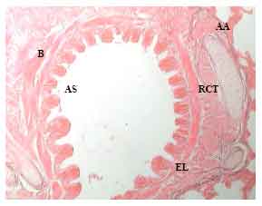 | |
| Fig. 1: | Histological section through the lung tissues showing areas of normal Alveolar Architecture (AA), normal Alveolar Space (AS), thin Respiratory Connective Tissues (RCT), normal Epithelium Lining (EL), normal Bronchioles (B) |
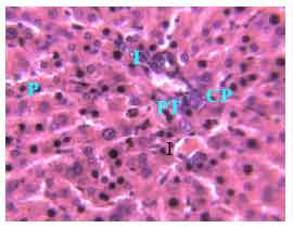
The lung tissues of the control rats showed normal Alveolar Architecture (AA), normal Alveolar Space (AS), normal Respiratory Connective Tissues (RCT), normal Epithelium Linings (EL) and normal Bronchioles (B) (Fig. 1),while the lung tissues of the exposed rats had abnormal Alveolar Architecture (AA), disrupted Bronchioles (B), damaged Bronchus (BR), weak Respiratory Connective Tissues (RCT), blue-black Pigments (P), Inflammations (I) and degenerated Epithelium Linings (EL) (Fig. 2). The liver tissues of the control rats showed normal Cellular Pattern (CP), normal Central Vein (CV), normal Hepatocytes (H), normal Portal Triad (PT), no Pigment (P) and no Inflammations (I) (Fig. 3) but the liver tissues of the exposed rats had abnormal Cellular Pattern (CP), damaged Central Veins (CV), disruption of Portal Triad (PT), Kuffer Cells (KC), blue-black Pigments (P) and Inflammations (I) (Fig. 4).
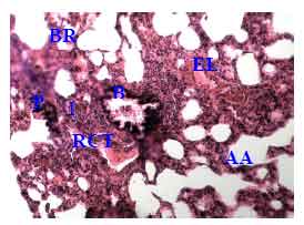 | |
| Fig. 2: | Histological section through the lung tissues showing areas of abnormal Alveolar Architecture (AA), disrupted Bronchioles (B), damaged Bronchus (BR), weak Respiratory Connective Tissues (RCT), blue-black Pigment (P), Inflammation (I) and degenerated Epithelium Lining(EL) |
 | |
| Fig. 3: | Histological section through the liver tissues showing areas of normal Cellular pattern (CP), normal Central Vein (CV), normal Hepatocytes (H), normal Portal Triad (PT), no Pigment (P) and no Inflammation (I) |
 | |
| Fig. 4: | Histological section through the liver tissues showing areas of abnormal Cellular Pattern (CP), damaged Central Vein (CV), disruption of Portal Triad (PT), Kuffer Cells (KC), area of blue-black Pigment (P) and areas of Inflammations (I) |
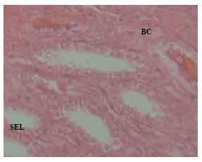 | |
| Fig. 5: | Histological section through the kidney tissues showing areas of normal Bowman Capsules (BC), normal Collecting Duct (CD), normal epithelium lining (SEL), no Pigment (P) and no Inflammation (I) |
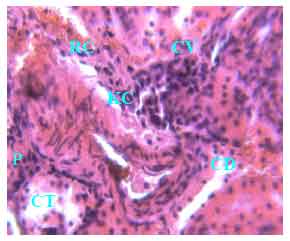 | |
| Fig. 6: | Histological section through the kidney tissues showing areas of abnormal convoluted tubules (CT) degenerated collecting duct (CD), abnormal renal corpuscles (RC), blue-black pigment (P) and inflammation (I) |
The kidney tissues of the control rats showed normal Bowman Capsules (BC), normal Collecting Duct (CD), normal Epithelium Linings (EL), no Pigments (P) and no inflammations (I) (Fig. 5), while the kidney tissues of the test rats had abnormal Convoluted Tubules (CT), degenerated Collecting Duct (CD), abnormal Renal Corpuscles (RC), blue-black Pigments (P) and Inflammation (I) (Fig. 6).
DISCUSSION
The discovery of silicon, aluminum, chromium and lead in the lungs of the exposed rats inhabiting the cement factory is a confirmation of the findings of previous studies such as Abdul-Wahab (2006), Gbadebo and Bankole (2007), Akinola et al. (2008), Ade-Ademilua and Obalola (2008), Akpan et al. (2011) and El-Abssay et al. (2011). It is also a confirmation of the fear and assertions of the residents of Huang Shan Cement, Ethiopia that the dust from the factory is the cause of their health problems (Fortune, 2011). Furthermore, it supports the findings of American Independent Pollution Monitoring Agency (IPC, 1996) that apart from the major non-toxic constituents of cement dust (e.g., calcium and iron), cement dust may contain heavy metals, poisonous gases (NO2, SO2, CO2), particulates and dioxins which may pose health risks to man and other animals in the environment. The presence of the toxic elements in the cement dust showed that the cement company is using hazardous wastes (e.g., tire) as alternative sources of energy to reduce the cost of cement production. This confirms the findings of IPC (1996) who found that the levels of heavy metals and dioxins in cement-kiln dust from Ribblesdale, U.K. were higher when cement fuel (alternative fuel) was burned. It also confirms the assertion of Doyle (2002) that some companies have continued to pollute the environment while professing to be environment conscious.
The normal weights of the exposed rats compared with the control rats is in line with the reports of Merenu et al. (2007) who found that the exposed factory workers in Sokoto Cement Company, Nigeria were heavier than the un-exposed group in the same environment. It also supports Shalabi et al. (2007) who observed increase in milk production, lactose, protein and glucose in lactating buffaloes fed with fugitive cement dust. Calcium has been fingered to be the cause of the observed increase in the weights of the exposed rats because it a basic nutrient of animal which specializes in blood and bone formations.
The decrease in the blood parameters of the exposed rats at the cement factory is an indication of microcytic anemia. This result confirms the findings of Mojimoniyi et al. (2007) who reported a fall in the red blood cell, packed cell volume and hemoglobin of cement factory workers in Sokoto, Nigeria. It also supports the findings of Mohammed and Sambo (2008) who observed a fall in the blood parameters of the Nile Tilapia in water treated with cement dust. Furthermore, it supports the findings of Ogunbileje and Akinosun (2011) that cement dust adversely affect the biochemical and hematological parameters of animals. The reduction in the blood parameters showed that cytotoxic interactions exist between the blood of the exposed rats and the toxic elements in the cement dust. Lead has been known to alter the hematological system of animals including man, inhibiting the activities of several enzymes involved in hem-biosynthesis (ATSDR, 2005). The reduction in the total protein of the exposed rats is an indication of the poor physiological conditions of the rats.
The UV spectroscopy of the DNA of the exposed rats did not show any form of contaminations and as such further analysis of the DNA of the rats was not necessary. A possible explanation for this is that the rats have been residing in the environment for a long period and has developed tolerance against the pollutants after a period of stress. The rats have possibly developed a pure line.
The marked histological changes in the lung, liver and kidney tissues of the exposed rats showed deleterious interactions between the tissues and the toxic elements in the cement dust. This observation supports the findings of Merenu et al. (2007), Mirzaee et al. (2008), Leem et al. (2008), Adbolh-Hossein et al. (2010), Zeleke et al. (2010) and Gbadebo and Amos (2010), all of them observed respiratory problems, laryngeal cancer, immune disorders and inflammation of cells in individuals exposed to cement dust. Some elements found in cement dust from kilns burning hazardous wastes have been fingered in pathogenesis of some diseases. Chromium VI compounds are known to cause respiratory problems, liver, kidney and circulatory damage (Martin and Griswold, 2009). Furthermore, lead has been fingered in the damage of the liver, kidney, heart, male gonads and immune system (ATSDR, 2005). Silicon has also been implicated in silicosis, increased risk of cancer (Hughes et al., 2001), chronic obstructive pulmonary diseases, lupus, rheumatoid, arthritis and renal disease (Hnizdo and Valyathan, 2003). Finally, excessive exposure to aluminum can cause respiratory problems (e.g., cough, shortness of breath), Alzheimer’s disease and kidney problem (ATSDR, 2010).
The levels at which the toxic elements were detected in the lungs of the exposed rats at the cement factory are frightening, because they have exceeded all regulatory standards. It showed that the cement plant is badly polluting the environment and it confirms the assertion of Bilen (2010) that cement production is of the main sources of environment pollution. It also showed that control strategies for prevention of dust release by cement plants have not been implemented in the cement company. Fell et al. (2003) said that in advanced countries, control-strategies for the prevention of dust release by machinery enclosure, local exhaust ventilation, work automation and greater diligence in maintenance of machinery have been implemented, presumably leading to less dust exposure. The cement factory is old, built in 1978 with old technologies and no major turn-around maintenance has been done since inception. In fact, at the factory site and settlements around the factory are heavy heaps of cement dust on roof-tops, automobiles, trees and every available space.
CONCLUSION AND RECOMMENDATION
This study has established that the cement plant is emitting toxic elements and poisonous gases. These toxic elements were injurious to the exposed rats inhabiting the cement factory environment. No doubt, all other animals including man in the vicinity of the cement factory will be suffering from the same problems. This then settled the argument on toxicity of cement dust between cement manufacturers and residents.
This is a wake-up call to various government monitoring agencies to be alive to their duties and put the welfare of its citizenry first. They should force the cement company and others in the country to implement prevention and control strategies and follow all Environment Protection Agency’s standards. A policy on minimum standard between a cement plant and residential areas should be formulated; cover trees could also be planted around cement factories to serve as dust breakers. Residents of cement companies should be enlightened on the use of detoxifiers, especially medicinal plants to eliminate toxic elements from the body. These will go a long way in preserving the health of people living around cement factories.
REFERENCES
- Abdul-Wahab, S.A., 2006. Impact of fugitive dust emissions from cement plants on nearby communities. Ecol. Modell., 195: 338-348.
CrossRef - Ade-Ademilua, O.E. and D.A. Obalola, 2008. The effect of cement dust pollution on Celosia argentea (Lagos Spinach) plant. J. Environ. Sci. Technol., 1: 47-55.
CrossRefDirect Link - Akinola, M.O., N.A. Okwok and T. Yahaya, 2008. The effects of cement dust on albino rats (Rattus norvegicus) around West African portland cement factory in Sagamu, Ogun state, Nigeria. Res. J. Environ. Toxicol., 2: 1-8.
CrossRefDirect Link - Akpan, I.O., A.E. Amodu and A.E. Akpan, 2011. An assessment of the major elemental composition and concentration in limestones samples from Yandev and Odukpani areas of Nigeria using nuclear techniques. J. Environ. Sci. Technol., 4: 332-339.
CrossRefDirect Link - Bilen, S., 2010. Effect of cement dust pollution on microbial properties and enzyme activities in cultivated and no-till soils. Afr. J. Microbiol. Res., 4: 2418-2425.
Direct Link - Dellaporta, S.L., J. Wood and J.B. Hicks, 1983. A plant DNA minipreparation: Version II. Plant Mol. Biol. Rep., 1: 19-21.
CrossRefDirect Link - Fell, A.K.M., T.R. Thomassen, P. Kristensen, T. Egeland and J. Kongerud, 2003. Respiratory symptoms and ventilatory function in workers exposed to Portland cement dust. J. Occup. Environ. Med., 45: 1008-1014.
PubMedDirect Link - Fell, K.M.A., L.I.B. Sikkeland, M.V. Svendson and J. Kongerad, 2010. Airway inflammations in cement production workers. Occup. Environ. Med., 67: 395-400.
CrossRefPubMedDirect Link - Gbadebo, A.M. and O.D. Bankole, 2007. Analysis of potentially toxic metals in airborne cement dust around sagamu, Southwestern Nigeria. J. Applied Sci., 7: 35-40.
CrossRefDirect Link - Gbadebo, A.M. and A.J. Amos, 2010. Assessment of radionuclide pollutants in bedrocks and soils from Ewekoro cement factory, Southwest Nigeria. Asian J. Appl. Sci., 3: 135-144.
CrossRefDirect Link - Hnizdo, E. and V. Valyathan, 2003. Chronic Obstructive Pulmonary disease due to Occupational exposure to Silica dust: A review of epidemiological and pathological evidence. Occup. Environ. Med., 60: 237-243.
CrossRef - Hughes, J.M., H. Weill, R.J. Rando, R. Shi, A.D. McDonald and J.C. McDonald, 2001. Cohort mortality study of North American industrial sand workers. II. Case-response analysis of lung cancer and silicosis deaths. Ann. Occup. Hyg., 45: 201-207.
CrossRef - Leem, J., E. Lee, H. Kim and M. Kim, 2008. The health effects of chromium containing cement dust assessed by combined methods of epidemiological and toxicological approach. Epidemiology, 19: s230-s230.
Direct Link - Merenu, I.A., F.B. Mojimoniyi, C.H. Njoku and M.T. Ibrahim, 2007. The effects of chronic cement dust exposure on lung function of cement factory workers in Sokoto, Nigeria. Afr. J. Biomed. Res., 10: 139-143.
Direct Link - Mirzaee, R., A. Kabriaei, S.R. Hashemi, M. Sadeghi and M. Shahrakipour, 2008. Effects of exposure to Portland cement dust on lung function in Portland cement factory workers in Khash, Iran. Iran. J. Environ. Health Sci. Eng., 5: 201-206.
Direct Link - Mojimoniyi, F.B.O, I.A. Merenu, M.T.O. Ibrahim and C.H. Njoku, 2007. The effects of cement dust exposure on hematological and liver function parameters of cement factory workers in Sokoto, Nigeria. Niger. J. Physiol. Sci., 23: 111-114.
PubMedDirect Link - Mohammed, A.K. and A.B. Sambo, 2008. Haematological assessment of the Nile tilapia Oreochromis niloticus exposed to sublethal concentrations of portland cement powder in solution. Int. J. Zool. Res., 4: 48-52.
CrossRefDirect Link - Shalabi, M.E.H., S.M. Kholif and M.M. Khorshed, 2007. Effect of by-pass cement dust supplementation level to diets on the productive performance of lactating buffaloes. Int. J. Dairy Sci., 2: 321-329.
CrossRefDirect Link - Zeleke , Z.K., B.E. Meon and M. Bratveit, 2010. . Cement dust exposure and acute lung function: A cross shift study. BMC. Pulmonary Med., 10: 19-19.
PubMedDirect Link