
A. Jafari
Department of Biology, Faculty of Science, Islamic Azad University of Mashhad, Iran
M. Nikian
Department of Biology, Faculty of Science, Islamic Azad University of Mashhad, Iran
Asian Journal of Plant Sciences
Year: 2008 | Volume: 7 | Issue: 8 | Page No.: 736-741


ABSTRACT
The present study tends to investigate the micromorphology, anatomy and palynology of four desert species of Salvia in center of Iran. For comparative micromorphology investigation, the shape of leaf, bracts and calyx were studied with SEM. To conduct the comparative study of anatomy characters, sections from stem were prepared using microtome and differential staining. In this part of investigation, arrangement of vessel and arrangement of tissues in stem were studied. For the palynology study, too, a comparative investigation on the species showed, the pollen was problate spheroidal, hexacolpate, bireticulate and semi- tectate or tectate. Finally, micromorphology study, is useful for identification of studied species.
PDF Abstract XML References Citation
How to cite this article
A. Jafari and M. Nikian, 2008. Micromorphological, Anatomical and Pollen Ornamentation Study on
Four Desert Species of Salvia in Center of Iran. Asian Journal of Plant Sciences, 7: 736-741.
DOI: 10.3923/ajps.2008.736.741
URL: https://scialert.net/abstract/?doi=ajps.2008.736.741
DOI: 10.3923/ajps.2008.736.741
URL: https://scialert.net/abstract/?doi=ajps.2008.736.741
INTRODUCTION
Salvia L. from Lamiaceae Family, Stachyioideae subfamily, Salvieae tribe having numerous variety of species, it is expanded from Italy to Iran, Iraq, Pakistan, Afghanistan (Parsa, 1949; Tutin, 1972; Boissier, 1975; Davis, 1982; Hedge, 1982, 1990). Salvia has 56 species in Iran about four of which are found in the deserts of center of Iran and 14 of them are endemic for Iran (Hedge, 1982). This genus has aromatic essential oils and antimicrobial effects. We collected S. eremophila, S. macilenta, S. tebessana, S. santolonifolia from their localities in Kerman and Yazd provinces. S. eremophila was endemic for Iran. Then micromorphologically, we studied shape of bracts, calyx and density of hairs. Also we prepared some cross section of stem of them. In terms of anatomic studies on Salvia, there has been a report about S. sclerea, S. trichoclada and S. napiflora (Ozdemir and Senel, 1999; Baran and Ozdemir, 2006). Another report for anatomy study of Salvia explained by Metcalf and Chalk (1983). In palynological studies, the comparison were made among the pollen grains of Salvia. In this part of study, pollen grains were extracted and acetolysed , to study shape and ornamentation of the pollen through SEM and LM. The previous palynology investigation had been done in Salvia, Origanum and Lycopus (Moon and Hong, 2003; Akyalcin, 2003). But, comparative palynology of studied species carried out for first time in Iran. The purpose of present study was to investigate variation of internal structure and identification of Salvia species on the basis of micromorphology because morphologically identification of this species is difficult.
MATERIALS AND METHODS
For micromorphological study, bracts, calyx and stem observed with SEM. As for the anatomic study, the examined species were collected from the localities in desert of center of Iran (Kerman and Yazd provinces) during May-June 2005-2006 (Table 1). For preparing of cross section of stem, the base of stem were selected from 8-9 specimens. The fresh specimens were fixed in FAA then, dehydrated with ethanol and later, some slices prepared with microtome. The section- 12 micron thick- stained with Safranine and Fast-green (Johnson, 1940; Chamberlain, 1990).
In the palynological study, the pollen was extracted from the anther and dehydrated by glacial acetic acid, then acetolised, coated with sputter finally studied by LM Olympus and SEM LEO1450VP (Erdtman, 1971; Moore et al., 1991). The pollen terminology was adapted from Punt et al. (1994).
| Table 1: | The localities of studied Salvia species |
 | |
RESULTS AND DISCUSSION
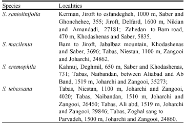
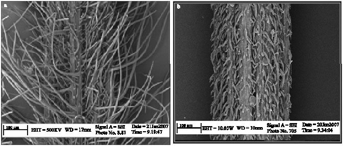
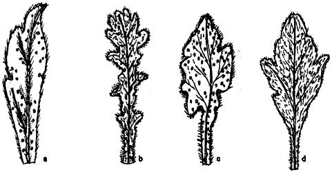
Micromorphological study: The results of stems hairs showed dense, long hairs (e.g., S. eremophila and S. santolonifolia and lax, short hairs (e.g., S. macilenta and S. tebessana) (Fig. 1a, b). The shape of leaves were obovate-oblong (e.g., S. santolonifolia), obovate (e.g., S. macilenta), triangular (e.g., S. tebessana) and obovate- oblong and retuse (S. eremophila) (Fig. 2a-d). The stems hairs of S. eremophila was dichotomous (Fig. 3). The shape of bracts were lanceolate- oblong with dense and long hairs (e.g., S. tebessana), rhomboid- obovate with and lax, short hairs (e.g., S. santolonifolia), rhomboid with long hairs (e.g., S. eremophila) and obovate, retuse with lax, short hairs (S. macilenta).
(Fig. 4a-d). The shape of calyx was tubular- campanulate except S. eremophila which was campanulate (Fig. 5a, b, Table 2).
Anatomic results: The results from the anatomic studies demonstrated the stem with following tissues:
| • | Epidermis layers with rectangular and ellipsoid cells. |
| • | Three to five layers of spongy parenchymatous under the epiderm |
| • | Some angular collenchymatous layers |
| • | Some sclerenchymatous layers between vascular bundles |
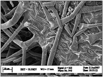
The arrangement of vessel was variable for example solitary in S. eremophila and S. tebessana , radial chain pore in S. santolinifolia and S. macilenta. Reticulate and pitted thickening vessel were observed in S. tebessana and S. eremophila and spiral in S. santolinifolia (Fig. 6, 7a, b).
 | |
| Fig. 1: | Electromicrograph of stems hairs (a) S. eremophila and (b) S. macilenta |
 | |
| Fig. 2: | Leaf shape (a), S. macilenta, (b) S. santolinifolia, (c) S. eremophila and (d) S. tebessana |
 | |
| Fig. 3: | Dichotomous hairs on the leaf of S. macilenta |
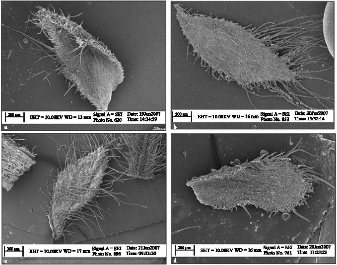 | |
| Fig. 4: | Electromicrograph of bracts shape (a) S. macilenta, (b) S. santolinifolia, (c) S. eremophila and (d) S. tebessana |
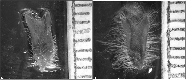 | |
| Fig. 5: | Calyx shape (a) campanulate in S. eremophila and (b) tubular-campanulate in S. tebessana |
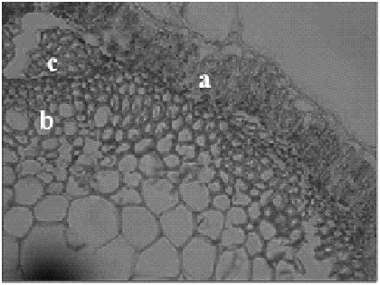 | |
| Fig. 6: | Cross section of S. santolinifolia stem (a) spongy parenchyma, (b) radial chain pore vessel and (c) angular collenchyma |
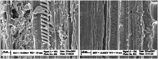 | |
| Fig. 7: | Electromicrograph of (a) spiral vessel in S. santolinifolia pitted and (b) reticulate vessel in S. eremophiola |
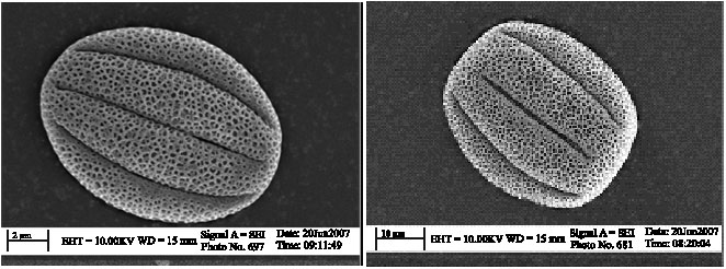 | |
| Fig. 8: | Electromicrograph of pollen (a) S. eremophila and (b) S. macilenta |
| Table 2: | The morphology and micromorphology characters of studied Salvia species |
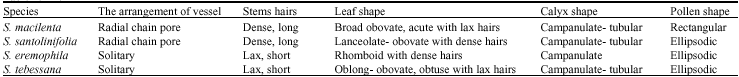 | |
Palynology analysis: The pollen was problate, elipsodic, bireticulate, hexacolpate and semi- tectate. Only S. macilenta had rectangular pollen (Fig. 8a, b, Table 2).
In present research to identifying species of Salvia used floral segments micromorphology study for first time. The studied species were similar as anatomy structure. Metcalf and Chalk (1983) carried out Labiatae anatomy structure. Also, anatomical study on S. sclerea, S. trichoclada and S. napiflora showed sclerenchymatous tissue above the phloem and around the vascular bundles (Ozdemir and Senel, 1999; Baran and Ozdemir, 2006). The results of palynology study on studied species showed similar structure. The other reports about Salvia sect. Audiertia, Hymenocrater and Lycopus showed similar pollen shape with studied species (Emboden, 1965; Moon and Hong, 2003). Finally we recommend using micromorphological study of floral and leaves segments help to identify exactly.
REFERENCES
- Akyalcin, H., 2003. Pollen morphology of Origanum L. (Labiatae) taxon in Turkey. Asian J. Plant Sci., 2: 28-41.
Direct Link - Baran, P. and C. Ozdemir, 2006. The morphological and tomical characters of Salvia napifolia Jaco. in Turkey. Bangladesh J. Bot., 35: 77-84.
Direct Link - Emboden, W.A., 1965. Pollen morphology of the genus Salvia section Audibertia. Pollen Spores, 6: 527-536.
Direct Link - Moon, H. and S. Hong, 2003. Pollen morphology of the genus Lycopus (Lamiaceae). Ann. Bot. Fennici., 40: 191-198.
Direct Link - Ozdemir, C. and G. Senel, 1999. The morpholoyical, anatomical and karyological properties of Salvia sclarea L. Turk. J. Bot., 23: 7-18.
Direct Link