
R.F. Dehkordi
Department of Anatomical Sciences, Faculty of Veterinary Medicine, University of Shahrekord, Shahrekord, Iran
A. Parchami
Department of Anatomical Sciences, Faculty of Veterinary Medicine, University of Shahrekord, Shahrekord, Iran
P. Kheibari
Department of Anatomical Sciences, Faculty of Veterinary Medicine, University of Shahrekord, Shahrekord, Iran
Asian Journal of Biological Sciences
Year: 2008 | Volume: 1 | Issue: 2 | Page No.: 100-102


ABSTRACT
The aim of this study was to determine sheep fetal testis developmental aspects. Twenty clinically healthy prenatal Lori Bakhtiyari sheep foeti, ranging from 41 to 86 days of intra-uterine life were studied. Foeti were collected from Shahrekord abattoir, Iran and allocated into 4 age groups according to measured CRL. Tissue sections of the organs showed that the TA and tubular diameter and the No. of testicular tubules per any microscopic field increase progressively with age especially between the first and the second group than the other groups. A trend of absolute growth of the foetal testes on biometry regarding length, width and thickness was noticed as per the advancement of gestation period. The biometry of the right and left testis were different in all four age groups between right and left testes.
PDF Abstract XML References Citation
How to cite this article
R.F. Dehkordi, A. Parchami and P. Kheibari, 2008. Studies on Foetal Testicular Development in Sheep. Asian Journal of Biological Sciences, 1: 100-102.
URL: https://scialert.net/abstract/?doi=ajbs.2008.100.102
URL: https://scialert.net/abstract/?doi=ajbs.2008.100.102
INTRODUCTION
Sheep occupy an important place in the Iranian livestock industry but he biometric values of prenatal testicular development in this species are still not well understood. Nasr et al. (1966) and Khalil (1969) studied the microscopic picture of the buffalo testis. Baishya and Vyas (1991) studied the various quantitative gross parameters of the testis of prenatal Surti buffalo at different phases of development. Testicular development of sheep fetus was studied by Hochereau-de et al. (1995). Proper development of testis is critical to establish the male phenotype and attain maximal reproductive capacity (Dufour et al., 2002). Disturbance in prenatal development and differentiation of the testis can thus be responsible for an array of undermusculinisation syndromes, ranging from XY females to males with subnormal fertility (Tohonen et al., 2003). Only few reports on testicular development in sheep can be traced in the available literature. The target of this study was to give exact biometric values of ovine embryonic testis of Lori Bakhtiyari breed in Iran.
MATERIALS AND METHODS
Testes from 21 apparently healthy prenatal Lori Bakhtiyari sheep ageing approximately from 41 to 86 days of intrauterine life, were collected from the Shahrekord slaughterhouse for this investigation in September 2007. Immediately after collection, samples were taken to Shahrekord campus laboratory and the foeti were measured for their Crown-Rump Length (CRL) in mm with the help of a graduated nylon tape. The foeti were then subjected to proper dissection of testes from the body. The different gross observations including length, width and thickness of the testes (right and left), were recorded by means of digital dial calipers and mettler balance, respectively. The testes were cut longitudinally into two halves to allow rapid fixation in 10% buffered formalin; they were then dehydrated and embedded in paraffin. Thin sections of 5 μ were cut and stained with H and E stain. Thirty clearly cut tubules were chosen at random from each testis and examined for the number and diameter of the seminiferous tubules in each viewing field (40x magnification) using an eyepiece micrometer. Data were analysed statistically, using one-way ANOVA test. p<0.05 was considered as significant.
The estimation of the fetal age was based on the CRL (McGeady et al., 2006).
RESULTS AND DISCUSSION
The material was distributed according to the approximated ages in 4 groups (Table 1).
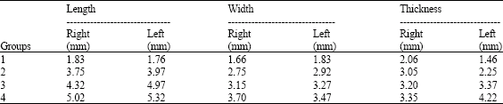
In the present investigation on the development of the foetal testis in the Lori Bakhyiyari sheep, the testicular growth in relation to foetal growth, has been shown in (Table 2, 3). Results of this study revealed that in all four age groups the length of the right testis was numerically greater than that of the left, but these differences are not statistically significant. In the first three age groups the width of the left testis is numerically greater than that of the left, but the reverse is true in the fourth group. The differences of the weight between right and left testis in all four age groups were not statistically significant. This study also showed that in the first two age groups the thickness of the right testis was greater than that of left and the reverse is true in the third and fourth age groups. The differences between right and left testis with regard to thickness are statistically significant in all four age groups.
As early as the day 41 the sex cords are clearly organized, solid and possesses no lumina. The cavities of the cords are filled with mesenchymal transparent mass and the cords are widely separated from each other. This simulates the findings of Abdel-Raouf (1960) and Abd El-Maksoud (2005) in cattle and Abdel-Raouf et al. (1974) in buffalo. The number of the seminiferous tubules in the first group averaged 7.86 μ and reached 22.45 μ in the fourth group. The diameter of the cords averaged 11.65 μ in the first group and reached 48.96 μ in the fourth group. The rapid increase in the tubular No. with age from the first to the second group may be due to the rapid increase in the length of the sex cords in the early stages of intra-uterine development. The diameter of the tunica albuginea (TA) averaged 7.21 μ in the first group and reached 50.61 μ in the fourth group. Each initial gonad is peripherally delineated by a relatively thick layer of mesenchymal tissue characterizing the first appearance of tunica albuginea, a distinct event in the pathway of testicular differentiation (Schrag, 1983).
| Table 1: | Distribution of the material among groups |
 | |
| Table 2: | Testicular measurements among four T groups |
 | |
| Table 3: | Tubular No., diameter and TA diameter among groups |
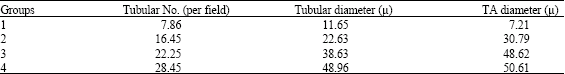 | |
| TA = Tunica Albuginea | |
Firstly, TA is shown to be homologous layer consisting mainly of undifferentiated mesenchymal cells and is covered externally by a layer of coelomic epithelium. Later on, the TA increases considerably in thickness and Separates into two well-defined layers: outer fibrous and inner cellular. The fibrous and cellular contents are found to increase progressively with age (Abd El-Maksoud, 2005; Schrag 1983), thus progressive increase in TA diameter reported in this study is due to progressive increase of both fibrous and cellular contents of the TA with age. This is in agreement with the observation of Abd El-Maksoud (2005) in bovine foetal testis. The increasing rate of the tubular number, tubular diameter and TA diameter was considerably greater between the first and the second group than the other groups. In the light of non-availability of any report on biometry of foetal testes in small ruminants, the present observations remain uncompared.
REFERENCES
- Abdel-Raouf, M., 1960. The postnatal development of the reproductive organs in bulls with special reference to puberty (incluing growth of the hypophysis and the adrenals). Acta Endocr., 49: 1-109.
PubMedDirect Link - Abdel-Raouf, M., M.A. El-Naggar and M.R. Fateh El-Bab, 1974. The development of the fetal testis in the buffalo. Z. Anat. Entwikcl. Gesch, 144: 227-236.
CrossRefDirect Link - Dufour, J.M., R.V. Rajotte and G.S. Korbutt, 2002. Development of an in vivo model to study testicular morphogenesis. J. Androl., 23: 635-644.
Direct Link - Hochereau-de Reviers, M.T., C. Perreau, C. Pisselet, A. Locatelli and M. Bosc, 1995. Ontogenesis of somatic and germ cells in sheep fetal testis. J. Reprod. Fertil., 103: 41-46.
Direct Link - McGeady, T.A., P.J. Quinn, E.S.F. Patrick and M.T. Ryan, 2006. Veterinary Embryology. 1st Edn., Blackwell Publishing Ltd., UK.
Direct Link - Tohonen, V., E.M. Ritzen, K. Nordqvist and A. Wedell, 2003. Male sex determination and prenatal differentiation of the testis. Endocrinol. Dev., 5: 1-23.
CrossRefDirect Link