
H. Rostami
Young Researchers Club, Tehran Medical Branch, Islamic Azad University, Tehran, Iran
M. Kazemi
Young Researchers Club, Karaj Branch, Islamic Azad University, Karaj, Iran
S. Shafiei
Department of Soil Science, Science and Research Branch, Islamic Azad University, Tehran, Iran
Asian Journal of Biochemistry
Year: 2012 | Volume: 7 | Issue: 3 | Page No.: 133-142


ABSTRACT
Aromatic and medicinal plants are widespread throughout world. Essential oils obtained from different aromatic and medicinal plants parts have been shown antibacterial, antifungal, antiviral and antioxidant properties. The experiment was started in season 2010-2011. In this study we studied the chemical compositions of essential oils of Lavandula officinalis and Melissa officinalis and also tested antibacterial and anticandidal activities of essential oils. The essential oils of Lavandula officinalis and Melissa officinalis collected in Iran were obtained by hydrodistillation of the aerial parts and analysed by gas chromatography equipped with flame ionisation detector (GC-FID) and gas chromatography coupled to a mass spectrometry system (GC/MS) for their chemical composition. The main constituents of leave oils were α-pinene, Camphor, Menthol, 1,8-Cineole, β-pinene, linalool, thymol and carvacrol. This experiment indicated that the bacterial strains were sensitive to studied essential oils and also showed very effective bactericidal activity with the strongest inhibition zone. Among the eight of tested essential oil components, thymol, carvacrol and menthol showed the highest antibacterial activities than previous components and streptomycin while, β-Pinene and linalool showed lower antibacterial activity than streptomycin (p = 0.05). Essential oils of Lavandula officinalis and Melissa officinalis possess great antibacterial potential and could be used as natural preservatives and fungicides.
PDF Abstract XML References Citation
Received: January 08, 2012;
Accepted: March 01, 2012;
Published: May 19, 2012
How to cite this article
H. Rostami, M. Kazemi and S. Shafiei, 2012. Antibacterial Activity of Lavandula officinalis and Melissa officinalis Against Some Human Pathogenic Bacteria. Asian Journal of Biochemistry, 7: 133-142.
DOI: 10.3923/ajb.2012.133.142
URL: https://scialert.net/abstract/?doi=ajb.2012.133.142
DOI: 10.3923/ajb.2012.133.142
URL: https://scialert.net/abstract/?doi=ajb.2012.133.142
INTRODUCTION
Many species are used in traditional and modern medicine and recent investigations have proven the basis of the medicinal uses. There are diverse uses of the family members in traditional ways in different parts of the world (Naghibi et al., 2005). Essential oils obtained from different medicinal plants parts have been shown antibacterial, antifungal, antiviral and antioxidant properties (Cragg et al., 1999; Ismail et al., 2011; Burt, 2004; Ahmad et al., 2005; Bozin et al., 2006; Ganjewala and Luthra, 2007a-b; Rizi et al., 2007; Loizzo et al., 2008; Swamy and Rao, 2008; Chutia et al., 2009; Soltan et al., 2009; Fortes et al., 2011; Louis et al., 2011; Patra, 2011; Upadhyay and Patra, 2011). Lavandula officinalis L. belongs to Labiatae (Lamiaceae), It is cultivated throughout Europe as well as in different parts of Iran (Wichtl, 1994). Leaves and flower of Lavandula officinalis have the highest amount of essential oils (Meftahizade et al., 2011). Lemon balm (Melissa officinalis L.) belongs to Labiatae (Lamiaceae), this species originates from southern Europe, Asia Minor and southern parts of North America (Simon et al., 1984; Sari and Ceylan, 2002). It also has sedative, antidepressant, antiviral, antibacterial and antispasmodic effects (Tyler, 1999; Sari and Ceylan, 2002). Today, lemon balm is used in various branches of industry (such as medicine, perfume and cosmetic and food etc.) in a lot of countries of the world. In this study, antimicrobial activities of Melissa officinalis and Lavandula officinalis and their leaves essential oils against fifteen human pathogenic bacteria were evaluated.
MATERIALS AND METHODS
The leaves of Melissa officinalis and Lavandula officinalis have been collected during March-April 2010 in Iran. Then the plants were isolated from the other specimen and conserved for extraction. The essential oils were extracted by hydrodistillation using an apparatus of Clevenger. For this, mixing 250 g of plants was used in 1600 mL of distilled water. The extraction took 3 h. After filtration the solvent is eliminated by reduced pressure distillation in rotary evaporator and pure oil was stored at 4°C in obscurity till the beginning of analysis. GC analysis was performed, using a Shimadzu GC-9A gas chromatograph equipped with a DB-5 fused silica column (30 mx0.25 mm i.d., film thickness 0.25 μm). The oven temperature was held at 50°C for 5 min and then programmed to 250°C at a rate of 3°C/min. Injector and detector (FID) temperatures were 290°C; helium was used as carrier gas with a linear velocity of 32 cm sec-1. The percentages were calculated by electronic integration of FID peak areas without the use of response factors correction. Linear retention indices for all components were determined by co-injection of the samples with a solution containing homologous series of C8-C22 n-alkanes. GC-MS analyses were carried out on a Varian 3400 GC-MS system equipped with a DB-5 fused silica column (30 mx0.25 mm i.d.); oven temperature was 40 to 240°C at a rate of 4°C. Transfer line temperature was 260°C. Carrier gas was helium with a linear velocity of 31.5 cm sec-1, split ratio 1/60. In addition, ionization energy was 70 eV, scan time 1 sec and mass range 40-300 amu. Identification of components in the oil was based on retention indices relatives to n-alkanes and computer matching with the WILLEY 275. L library, as well as by comparison of the fragmentation patterns of mass spectra with those reported in the literature (Adams, 2001). The chromatographic conditions were identical to those used for GC analysis.
Tests for antibacterial activity: The used microorganisms in the present study were six Gram-positive (Staphylococcus aureus, Bacillus cereus, Bacillus megaterium, Bacillus subtilis, Sarcina lutea and Streptococcus-β-haemolyticus) and nine Gram-negative (Salmonella typhi, Shigella dysenteriae, Shigella shiga, Shigella sonnei, Shigella boydii, Escherichia coli, Klebsiella sp., Pseudomonas aeruginosa and Proteus sp.) human pathogenic bacteria. The antibacterial assays were carried out by the disc-diffusion (Verpoorte et al., 1983) and microdilution method (Daouk et al., 1995; Hanel and Raether, 1988; Espinel-Ingroff, 2001) in order to determine the antibacterial activity of oils and their components against the human pathogenic bacteria. The bacterial suspensions were adjusted with sterile saline to a concentration of 1.0x10⁵ CFU L-1. The inocula were prepared daily and stored at +4°C until time of use. Dilutions of the inocula were cultured on solid medium to verify the absence of contamination and to check the validity of the inoculum.
Disc-diffusion test: Compounds were investigated by the disc diffusion using 4 mm filter discs. Bacteria were cultured overnight at 28°C in LB medium and then adjusted with sterile saline to a concentration of 1.0x10⁵ CFU mL-1. The suspension was added to the top of agar (6 mL) and dissolved in Petri dishes (2 mL agar plate) with solid peptone agar. Filter discs with essential oils and main components (1.0 μg mL-1) were placed on agar plates (1 disc per agar plate). After 24 h of incubation at 28°C for bacteria the diameter of the growth inhibition zones was measured. Streptomycin was used as a positive control and 1 μL was applied to the discs from stock solution (1 mg mL-1). All tests were done in duplicate. Three replications were used for each oil and for each component (Sokovic et al., 2009).
Microdilution test: The minimum inhibitory and bactericidal and fungicidal concentrations (MICs and MBCs) were determined using microtitre plates. The bacterial suspension was adjusted with sterile saline to a concentration of 1.0x10⁵ CFU mL-1. Compounds to be investigated were dissolved in broth LB medium (100 μL) with bacterial inoculum (1.0x10⁴ CFU per well) to achieve the wanted concentrations (0.02-15.0 μg mL-1). The microplates were incubated for 24 h at 28°C. The lowest concentrations without visible growth (at the binocular microscope) were defined as concentrations that completely inhibited bacterial growth (MICs). The MBCs were determined by serial sub-cultivation of 2 μL into microtitre plates containing 100 μL of broth per well and further incubation for 72 h. The lowest concentration with no visible growth was defined as the MBC, indicating 99.5% killing of the original inoculum. The optical density of each well was measured at a wavelength of 655 nm by Microplate manager 4.0 (Bio-Rad Laboratories) and compared with a blank and the positive control. Streptomycin was used as a positive control using the same concentrations as in the disc diffusion test. Three replications were used for each oil and each component (Sokovic et al., 2009).
RESULT AND DISCUSSION
The results of the analysis of the essential oil Lavandula officinalis are qualitative and semi-quantitative (Table 1). Monoterpenes hydrocarbons were the major constituents in this oil (74.59%), the main one being α-pinene (18.12%), camphor (10.12%), 1,8-cineole (15.32%) followed menthol (25.23%), other predominant components were linalool oxide (1.0 %), α-terpinene (1.0%), Borneol (1.0%) and viridiflorol (1.0). Moreover, some minor components were also detected of which eucarvone (0.25%), dihydrocarveol acetate (0.02%), terpinolene (0.34%), Eugenol (0.45%) and β-caryophyllene (0.74%) (Table 1). The fresh aerial parts of Lavandula officinalis yielded 2.05% v/w of essential oil (Table 1), it is relatively higher than other plants industrially exploited as a source of essential oils: Thymus (1%) (Imelouane et al., 2009), menthe (0.5-1%), neroli (0.51%) and Laurel (0.1-0.35%).The chemical compositions revealed that this leaves had compositions relatively similar to those of other Lavandula officinalis essential oils analyzed by Meftahizade et al. (2011). Afsharpor and Azarbayejani (2006) reported that dead Lavandula officinalis, harvested from Isfahan, Iran, produce 1 to 3% essential oil, containing mainly monoterpenes (the most important component) of which linalyl acetate, linalool, ocimene, 1,8-cineole and camphor are part it. Based on GC and GC-MS analysis in the essential oil of Melissa officinalis twenty components were identified which represented 66.12% of the total detected constituents (Table 1). The major constituents of the oil were β-pinene (10.13%), linalool (3.12%), α-pinene (10.01%), Thymol (25.31 %) and Carvacrol (12.34%). Other components were present in amounts less than 2% (Table 1). Essential oil yield was 0.18%. Essential oil yield, in the present study, were higher than the 0.157% which is similar to reports of Malik et al. (1972).
| Table 1: | Chemical compositions of investigated essential oils |
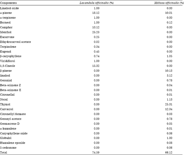 | |
| Total identified constituents (%), Percentages are mean of three replications obtained from electronic measurements using Flame Ionization Detection (FID), 0: Not detected | |
Many researchers have reported that the main components of lemon balm are neral and geranial. Neral and geranial rates in the oil were reported, respectively as 15 and 14.5% by Hefendehl (1970), 19.6-36.1 and 25.3-47.5% by Tittel et al. (1982), 19.5% and 31.6% by Werker et al. (1985). Although, all of discussed studies in above reported that neral and geranial were the main components of the oil, Kirimer et al. (1995) found that the main component of the lemon balm oil, they studied was carvacrol (60%). Essential oil obtained from a few different populations of Melissa officinalis L. cultivated in Poland had been investigated by Patora et al. (2003). In their study, the content of essential oil in the leaves and herb were recorded as 0.08 to 0.25 mL 100 g-1 and 0.06 to 0.167 mL 100 g-1, respectively. However, there were significant differences among the rates of those reported components. The oil showed a significant anti-microbial potential on various tested microorganisms (Table 2) (p = 0.05). The essential oils which showed the best antibacterial activity in disc-diffusion method were Melissa officinalis (20.0-35.0 mm) and Lavandula officinalis (20.0-25.0 mm) (p = 0.05). Streptomycin at 1 μg disc-1 showed inhibition zones in the range of 15.0-20.0 mm (Table 2) (p = 0.05).
| Table 2: | Antibacterial activity of essential oils (1.0 μg mL-1) in disc-diffusion method, inhibition zones (mm) |
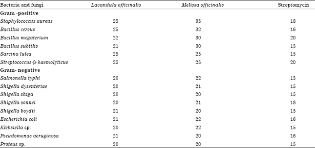 | |
| The data show the diameter of inhibition zone growth in mm. The diameter of paper disc was 6 mm | |
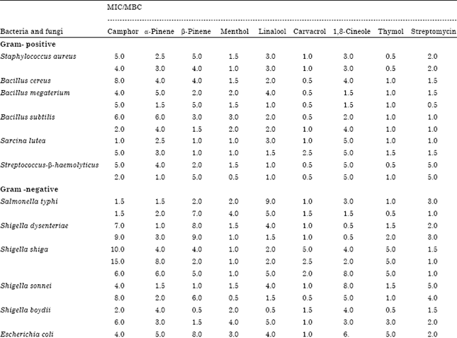
Good inhibition zones were also obtained for Melissa officinalis oils. It can be seen that essential oils from Melissa officinalis and Lavandula officinalis possess a higher antibacterial effect than streptomycin(p = 0.05). Melissa officinalis and Lavandula officinalis oils exhibited much higher antibacterial activity with the same MIC (0.5-0.5 μg mL-1) and MBC (0.5-0.5 μg mL-1) (Table 3) (MIC = 0.02-15.0 μg mL-1). Streptomycin showed MIC at 3.0-1.5 μg mL-1 and MBC at 5.0-1.0 μg mL-1. β-Pinene and linalool showed the lowest of antibacterial activity among the tested components, with inhibition zones 10.0-15.0 mm; α-pinene, Camphor and 1, 8-Cineole possessed almost the same activity, with inhibition zones 9.0-20.0 mm (Table 4) (p = 0.05). Menthol inhibited bacterial growth of all bacteria and inhibition zones were 15.0-25.0 mm, Carvacrol reacted slightly better (inhibition zones 15.0-30.0 mm) while Streptomycin showed activity with inhibition zones 10-20.0 mm (Table 4) (p = 0.05). Thymol showed inhibition with zones of 25.0-35.0 mm. Menthol (MIC at 1.5-1.5 μg mL-1 and MBC at 1.0-1.0 μg mL-1) and carvacrol (MIC at 1.0-1.0 μg mL-1 and MBC at 1.0-1.0 μg mL-1) and thymol (MIC at 0.5-0.5 μg mL-1 and MBC at 0.5-1.0 μg mL-1) possessed much stronger antibacterial activity than streptomycin (Table 5) (MIC concentrations: 0.02-15.0 μg mL-1). Linalool and 1, 8-Cineole showed the lowest antibacterial activity in the microdilution method, MIC at 3.0-2.5 μg mL-1 and MBC at 3-3.0 μg mL-1 (Table 5) (MIC = 0.02-15.0 μg mL-1). The monoterpenic hydrocarbons camphor and β-pinene also showed similar activity with MIC of 5.0-8.0 μg mL-1 and MBC of 4.0-10.0 μg mL-1 while, α-Pinene exhibited inhibitory activity at 2.5-3.0 μg mL-1 and was bactericidal at 3.0-5.0 μg mL-1 (Table 5) (MIC concentrations: 0.02-15.0 μg mL-1). Thymol, carvacrol and menthol showed very strong activity, respectively (Table 5) (MIC concentrations: 0.02-15.0 μg mL-1).. The antibacterial potential of tested oil in both methods can be presented as: Lavandula officinalis<Melissa officinalis. The antibacterial potential of tested essential oils’ components can be presented as: Linalool<1,8-Cineole<β-pinene<α-pinene<camphor<1,8-cineole<menthol<carvacrol<thymol. Among the eight tested essential oil components, thymol, carvacrol and menthol showed the highest activity.
| Table 3: | Antibacterial activity of essential oils (MIC or MBC -μg mL-1), microdilution method |
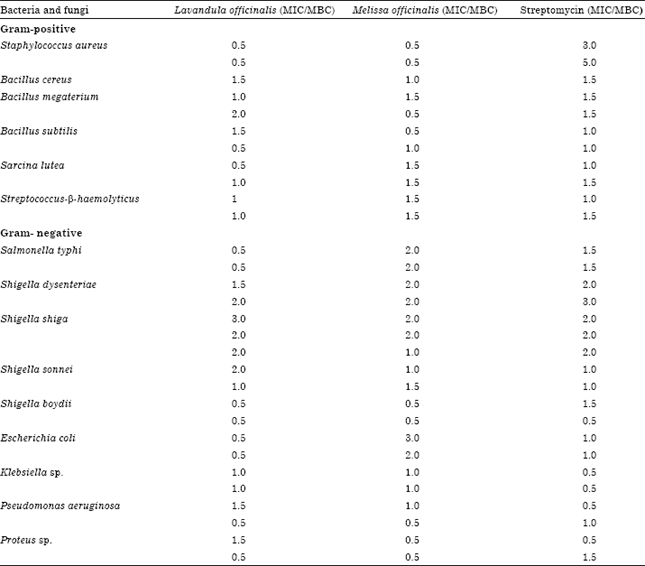 | |
| MBC test: Minimum bactericidal concentration, MIC test: Minimum inhibitory concentration | |
Essential oils which were rich in α-pinene, thymol, carvacrol and menthol demonstrated potential antibacterial activity (Sokovic et al., 2009). Monoterpenes hydrocarbons, terpinenes, have also shown antimicrobial properties that appear to have strong to moderate antibacterial activity against Gram-positive bacteria (Oyedeji and Afolayan, 2005). The antimicrobial activities have been mainly explained through C10 and C15 terpenes with aromatic rings and phenolic hydroxyl groups able to form hydrogen bonds with active sites of the target enzymes, although other active terpenes, as well as alcohols, aldehydes and esters can contribute to the overall antimicrobial effect of essential oils (Belletti et al., 2004). On the other hand, enantiomers of α-pinene, β-pinene, limonene and linalool have a strong antibacterial activity (Magiatis et al., 1999). Essential oils and extracts of Melissa officinalis have been reported to have antiviral (Schnitzler et al., 2008), antimicrobial and antioxidant properties (Dastmalchi et al., 2008). Mimica-Dukic et al. (2003) have described antimicrobial and free Radical Scavenging Capacity (RSC) together with the effects on Lipid Preoxidation (LP) of lemon balm essential oil in their study.
| Table 4: | Antibacterial activity of essential oils components (1.0 μg mL-1) in disc-diffusion method, inhibition zones (mm) |
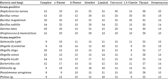 | |
| The data shows the diameter of inhibition zone growth in mm, The diameter of paper disc was 6 mm | |
| Table 5: | Antibacterial activity of essential oils components (MIC and MBC-μg mL-1), microdilution method |
  | |
| MBC test: Minimum bactericidal concentration, MIC test: Minimum inhibitory concentration | |
This high antibacterial activity of Lavandula officinalis essential oil supports the results found by other researchers. Imelouane et al. (2009) showed that Lavandula dentata had a strong antibacterial action against Pseudomonas aeruginosa. Similarly, Lis-Balchin and Deans (1997) showed that lavandin, French lavender, spike lavender, Bulgarian lavender and generic ‘lavender’ (type unspecified) essential oils all have activity against a large number of bacteria and fungi.
CONCLUSION
Present study was conducted to investigate the chemical composition and antibacterial and antifungal activity of essential oil extracted from Lavandula officinalis and Melissa officinalis. The major compounds were α-pinene, Camphor, Menthol, 1, 8-Cineole, β-pinene, linalool, Thymol and Carvacrol. However, further studies are still required to investigate its application in medicine and food industries.
REFERENCES
- Ahmad, N.R., M.A. Hanif and U. Rashid, 2005. Chemical compositional and intra provenance variation for content of essential oil in Eucalyptus crebra. Asian J. Plant Sci., 4: 519-523.
CrossRefDirect Link - Bozin, B., N. Mimica-Dukic, N. Simin and G. Anackov, 2006. Characterization of the volatile composition of essential oils of some lamiaceae spices and the antimicrobial and antioxidant activities of the entire oils. J. Agric. Food Chem., 54: 1822-1828.
CrossRefPubMedDirect Link - Burt, S., 2004. Essential oils: Their antibacterial properties and potential applications in foods-A review. Int. J. Food Microbiol., 94: 223-253.
CrossRefPubMedDirect Link - Cragg, G.M., M.R. Boyd, R. Khanna, R. Kneller and T.D. Mays et al., 1999. International collaboration in drug discovery and development: The NCl experience. Pure Applied Chem., 71: 1619-1633.
Direct Link - Daouk, R.K., S.M. Dagher and E.J. Sattout, 1995. Antifungal activity of the essential oil of Origanum syriacum L. J. Food Prot., 58: 1147-1149.
Direct Link - Dastmalchi, K., H.J.D. Dorman, P.P. Oinonen, Y. Darwis, I. Laakso and R. Hiltunen, 2008. Chemical composition and In vitro antioxidative activity of a lemon balm (Melissa officinalis L.) extract. LWT-Food Sci. Technol., 41: 391-400.
CrossRef - Espinel-Ingroff, A., 2001. Comparison of the E-test with the NCCLS M38-P method for antifungal susceptibility testing of common and emerging pathogenic filamentous fungi. J. Clin. Microbiol., 39: 1360-1367.
CrossRefDirect Link - Ganjewala, D. and R. Luthra, 2007. Essential oil biosynthesis and metabolism of geranyl aceate and geraniol in developing Cymbopogon flexuosus (Nees ex Steud) Wats Mutant cv. GRL-1 leaf. Am. J. Plant Physiol., 2: 269-275.
CrossRefDirect Link - Ganjewala, D. and R. Luthra, 2007. Inhibitors of essential oil biosynthesis in Cymbopogon flexuosus nees ex. steud. Mutant cv. GRL-1 leaves. Am. J. Plant Physiol., 2: 227-232.
CrossRefDirect Link - Fortes, G.A.C., S.S. Naves, F.F.F. Godoi, A.R. Duarte, P.H. Ferri and S.C. Santos, 2011. Assessment of a maturity index in jabuticaba fruit by the evaluation of phenolic compounds, essential oil components, sugar content and total acidity. Am. J. Food Technol., 6: 974-984.
CrossRefDirect Link - Hanel, H. and W. Raether, 1988. A more sophisticated method of determining the fungicidal effect of water-insoluble preparations with a cell harvester, using miconazole as an example. Mycoses, 31: 148-154.
CrossRefPubMedDirect Link - Imelouane, B., H. Amhamdi, J.P. Wathelet, M. Ankit, K. Khedid and A. El Bachiri, 2009. Chemical composition of the essential oil of thyme (Thymus vulgaris) from Eastern Morocco. Int. J. Agric. Biol., 11: 205-208.
Direct Link - Ismail, A., H. Lamia, H. Mohsen and J. Bassem, 2011. Chemical composition of Juniperus oxycedrus L. subsp Macrocarpa essential oil and study of their herbicidal effects on germination and seedling growth of weeds. Asian J. Applied Sci., 4: 771-779.
CrossRefDirect Link - Lis-Balchin, M. and S.G. Deans, 1997. Bioactivity of selected plant essential oils against Listeria monocytogenes. J. Applied Bacteriol., 82: 759-762.
PubMedDirect Link - Louis, B., J. Nguefack and P. Roy, 2011. Evaluation of antifungal potential of Ocimum gratissimum extracts on two seedborne fungi of rice (Oryza sativa L.) in cameroon. Asian J. Biol. Sci., 4: 306-311.
CrossRefDirect Link - Magiatis, P., E. Melliou, A.L. Skaltsounis, I.B. Chinou and S. Mitaku, 1999. Chemical composition and antimicrobial activity of the essential oils of Pistacia lentiscus var. chia. Planta Medica, 65: 749-752.
CrossRefDirect Link - Meftahizade, H., H. Moradkhani, A.F. Barjin and B. Naseri, 2011. Application of Lavandula officinalis L. antioxidant of essential oils in shelf life of confectionary. Afr. J. Biotechnol., 10: 196-200.
Direct Link - Naghibi, F., M. Mosaddegh, M.M. Motamed and A. Ghorbani, 2005. Labiatae family in folk medicine in Iran: From ethnobotany to pharmacology. Iran. J. Pharm. Res., 4: 63-79.
Direct Link - Patra, A.K., 2011. Effects of essential oils on rumen fermentation, microbial ecology and ruminant production. Asian J. Anim. Vet. Adv., 6: 416-428.
CrossRefDirect Link - Sokovic, M.D., J. Vukojevic, P.D. Marin, D.D. Brkic, V. Vajs and L.J.L.D. van Griensven, 2009. Chemical composition of essential oils of Thymus and Mentha speciesand their antifungal activities. Molecules, 14: 238-249.
CrossRefDirect Link - Soltan, M.A.E., R.S. Shewita and S.I. Al-Sultan, 2009. Influence of essential oils supplementation on digestion, rumen fermentation, rumen microbial populations and productive performance of dairy cows. Asian J. Anim. Sci., 3: 1-12.
CrossRefDirect Link - Swamy, K.N. and S.S.R. Rao, 2008. Influence of 28-homobrassinolide on growth, photosynthesis metabolite and essential oil content of geranium [Pelargonium graveolens (L.) Herit]. Am. J. Plant Physiol., 3: 173-179.
CrossRefDirect Link - Tittel, G., H. Wagner and R. Bos, 1982. Chemical composition of the essential oil from Melissa. Planta Med., 46: 91-98.
PubMedDirect Link - Upadhyay, R.K. and D.D. Patra, 2011. Influence of secondary plant nutrients (Ca and Mg) on growth and yield of chamomile (Matricaria recutita L.). Asian J. Crop Sci., 3: 151-157.
CrossRefDirect Link - Verpoorte, R., T.A. van Beek, P.H.A.M. Thomassen, J. Aandewiel and A.B. Svendsen, 1983. Screening of antimicrobial activity of some plants belonging to the Apocynaceae and Loganiaceae. J. Ethnopharmacol., 8: 287-302.
CrossRefDirect Link - Werker, E., U. Ravid and E. Putievsky, 1985. Structure of glandular hairs and identification of the main components of their secreted material in some species of the Labiatae. Israel J. Bot., 34: 31-45.
Direct Link