
M.A. Abd El-Kader
National Research Centre, Department of Biochemistry, 33 El-Tahrir Street, Dokki 12622, Cairo, Egypt
M.M. Ali
National Research Centre, Department of Biochemistry, 33 El-Tahrir Street, Dokki 12622, Cairo, Egypt
N.M. El-Sammad
National Research Centre, Department of Biochemistry, 33 El-Tahrir Street, Dokki 12622, Cairo, Egypt
M.A. El-Shaer
National Research Centre, Department of Pathology, 33 El-Tahrir Street, Dokki 12622, Cairo, Egypt
Asian Journal of Biochemistry
Year: 2011 | Volume: 6 | Issue: 6 | Page No.: 426-438


ABSTRACT
Non steroidal anti-inflammatory drugs are widely used and effective against inflammatory diseases. However, its clinical use is limited by its ulcerogenic effect. This study aimed to investigate the possible gastroprotective effect of alpha-lipoic acid on indomethacin-induced gastric ulcer in rats. Forty adult male Dawley rats were assigned into five groups of eight animals each : group 1 served as control, group 2 was treated intraperitioneally (i.p.) with alpha-lipoic acid (20 mg kg-1 b.w./day) for 7 successive days, group 3: Rats were treated i.p. with indomethacin (20 mg kg-1 b.w./day) for two days, group 4 were treated with indomethacin for 2 days followed by alpha-lipoic acid for 7 days, group 5: Animals were treated with alpha-lipoic acid for 3 days followed by indomethacin for 2 days then alpha-lipoic acid for other 4 days. Alteration of gastric mucosa were analysis. Gastric secretion volume, pH and gastric mucus secretion were determined. In addition, lipid peroxidation and the changes in the antioxidants: reduced glutathione, catalase and superoxide dismutase as markers for antioxidant activity and mucosal defense factors were evaluated. Administration of indomethacin per os induced gastric damage as evidenced by observed ulcers, histological changes, increased gastric secretion volume, decreased pH value and mucus secretion. Alpha lipoic acid were recovered rats from gastric mucosal lesions caused by indomethacin and protected the gastric mucosa covered the stomach wall from injury through decreasing gastric secretion volume, increasing pH value and mucus secretion. All parameters indicating the oxidative injury in gastric mucosa markedly reversed by alpha-lipoic acid treatment due to its potent antioxidant properties where it reduced the level of lipid peroxidation product malondialdehyde, alleviated increases in the catalase activity and ameliorated depressions in reduced glutathione level and the activity of superoxide dismutase caused by indomethacin injection. The results indicate that beneficial effect of alpha-lipoic acid in the protection and treatment of gastric ulcer which result from toxic effect of reactive oxygen species induced by indomethacin.
PDF Abstract XML References Citation
Received: June 01, 2011;
Accepted: July 08, 2011;
Published: October 29, 2011
How to cite this article
M.A. Abd El-Kader, M.M. Ali, N.M. El-Sammad and M.A. El-Shaer, 2011. Antiulcer Effects of Alpha Lipoic Acid on Gastric Acid Secretion and Mucosal Defense Factors in Rats. Asian Journal of Biochemistry, 6: 426-438.
DOI: 10.3923/ajb.2011.426.438
URL: https://scialert.net/abstract/?doi=ajb.2011.426.438
DOI: 10.3923/ajb.2011.426.438
URL: https://scialert.net/abstract/?doi=ajb.2011.426.438
INTRODUCTION
The pathogenesis of gastric ulcer include several factors; mucosal integrity, secretion of gastric acid and pepsin, gastro-duodenal motility, Helicobacter pylori infection and use of nicotine and non-steroidal anti-inflammatory drugs (Huang et al., 2002). Non-steroidal Anti-inflammatory Drugs (NSAID) such as indomethacin are widely used in the treatment of pain, fever and inflammation but the major limitation of their clinical application is serious gastrointestinal side effects (erosions, ulcers and bleeding) (Alsaif, 2004). A number of anti-ulcer drugs like gastric anti-secretory drugs-H2 receptor antagonists, anti-muscarinic agents, proton pump inhibitors, mucosal protective agents-carbenoxolone sodium, sucralfate, thyroxin and prostaglandin analogues are available which are shown to have side effects and limitations. Moreover, various reports have shown that commonly used drugs for gastric ulcers have danger of drug interaction, adverse effect and increased incidence of relapses during ulcer therapy (Oluwole and Saka, 2007; Al-Attar, 2011). Reactive Oxygen Species (ROS) have been implicated in the etiology and pathophysiology of gastrointestinal inflammation and gastric ulcers. (Repetto and Llesuy, 2002). Hence, there is a need for agents to minimize and repair free radical-induced damage. The antioxidants play a key role in these defense mechanisms. Many antioxidant agents; melatonin, Nigella sativa seed, Cucumis melo seeds and vegetable oils have been reported to protect gastric mucosa against indomethacin ulcers (Bandyopadhyay et al., 2000; Rifat-uz-Zaman et al., 2004; Odabasoglu et al., 2008; Gill et al., 2011).
Alpha Lipoic Acid (ALA) is called thiotic acid contain sulfur groups in a dithiol ring structure. It is endogenous synthesized in mitochondria and is covalently bound to specific proteins which function as cofactors for several important mitochondrial enzymes (Cicchillo et al., 2004). Bioavailability studies have reported that 20-40% of lipoic acid from an oral dose appears in circulation and this rapid uptake of lipoic acid in the gastrointestinal system is followed by its transport to different tissues (Chng et al., 2009; Shay et al., 2008). Alpha lipoic acid is reduced by enzymes in mitochondria to its dithiol form known as Dihydrolipoic Acid (DHLA) which enhanced antioxidant activity. It has also a metabolic role in the body by facilitating the production of energy via aiding in the metabolism of glucose. ALA proven to chelate metal ions such as iron, copper and cadmium which catalyze auto oxidation reactions. However, both ALA and DHLA have been found to inhibit copper and iron-mediated oxidative damage (Suh et al., 2004) and to inhibit copper and iron accumulation in animal models (Suh et al., 2005). Several studies have confirmed the beneficial effect of ALA in the treatment of many diseases in which ROS have been implicated such as ischemia-reperfusion injury, diabetes, diabetic neuropathy, atherosclerosis, hypertension, neurodegeneration, disease of joints, radiation injury and acquired immune deficiency syndrome (Song et al., 2004; Bilska and Wlodek, 2005; Winiarska et al., 2008). Because of antioxidant properties mentioned above, this study aimed to verify the gastroprotective and therapeutic effect of ALA in experimental model of gastric ulcer induced by indomethacin.
MATERIALS AND METHODS
Chemicals: Indomethacin was supplied by Arab Co. for pharmaceuticals (Egypt), ALA was obtained from Sigma-Aldrich Chemic (Deisenhofen, Germany), Sigma Chemical, (St. Louis, MO, USA). All other chemicals used were of highest purity and analytical grade and purchased from either Merck (Darmstadt, Germany) or Sigma-Aldrich Chemicals.
Animals: The experiment has been started in the beginning of 2008. Forty adult male Dawley rats weighing 180-200 g were obtained from the animal house of National Research Centre, Cairo, Egypt. Animals were randomly assigned to 5 groups of 8 rats each and housed in plastic cages at an environmentally controlled room (constant temperature 25-27°C, with 12 h light/dark cycle) for one week prior to starting the experiments. All animals received humane care in compliance with the international guiding principles for animal research and they were provided with tap water and standard rat diet. The animals were deprived of food for 24 h before the experiments but had free access to water.
Experimental design: Indomethacin was injected intraperitoneally (i.p.) to animals at a dose of 20 mg kg-1, b.w. day-1 for two consecutive days which is well known to cause significant stomach ulcer in rats (Guidobono et al., 1997; Alsaif, 2004; Jiang et al., 2009). Alpha-lipoic acid was dissolved in saline and administered to animals i.p at a dose of 20 mg kg-1, b.w. day-1. The used dose was selected on the basis of the previous studies (Maritim et al., 2003).
The rats were randomly assigned into five groups containing eight rats each:
| G 1 | : | Served as control received a daily i.p. injection of 1 mL vehicle (saline) for nine days |
| G 2 | : | (ALA- treated group), the rats were administered i.p with ALA at a dose of 20 mg kg-1, b.w. day-1 in 1 mL of vehicle for 7 consecutive days followed by saline for 2 days |
| G 3 | : | (indomethacin- treated group), the rats were administered i.p with 20 mg indomethacin/kg, b.w. day-1 for 2 days |
| G 4 | : | (Indomethacin followed by ALA), rats were received indomethacin as in G 3 followed by ALA for 7 days as in G 2 |
| G 5 | : | (ALA followed by indomethacin then ALA), rats were administered ALA for 3 days and then indomethacin for 2 days followed by ALA for other 4 days |
METHODS
Gastric secretion volume and pH were estimated according to George et al. (1999)
Determination of gastric lesions: After washing with normal saline, the stomachs were checked for the presence of ulceration. Ulcerations were the linear necrohemorrhagic lesions present in the glandular part of the stomach. Photographs for ulcers were taken by digital camera. Stomachs were examined macroscopically for gastric mucosal damage. Each lesion was measured along its greatest diameter (mm). The sum of the lesion lengths was divided by the number of rats and expressed as the mean ulcer scores as described by Ogle et al. (1985).
Measurement of gastric wall mucus: The modified procedure of Corne et al. (1974) was used for the determination of gastric wall mucus.
Biochemical investigation: The stomach tissues were scrapped and homogenized in ice cold phosphate buffer (pH 8). The tissue homogenate was centrifuged at 3.000 rpm for 10 min at 4°C, then the supernatants were used for biochemical investigations.
Malondialdehyde (MDA) assay: Malondialdehyde levels, as the stable product of lipid peroxidation and as a measure of free radical formation, were estimated by the double heating method of Draper and Hadley (1990). The principle of the method is the spectrophotometric measurement of the color generated by the reaction of Thiobarbituric Acid (TBA) with MDA.
Assay of reduced glutathione (GSH) level: The determination of GSH level was carried out according to the method of Ellman (1959).
Assay of catalase (CAT) activity: Catalase activity was assayed according to the procedure of Aebi (1984), based on the principle that the disappearance of hydrogen peroxide was monitored spectrophotometrically at 240 nm.
Assay of superoxide dismutase (SOD )activity: Superoxide dismutase activity was assayed spectrophotometrically as described by Durak et al. (1996).
Histopathological examination were evaluated according to Ross et al. (1989)
Statistical analysis: The statistical significance of the difference between means was estimated by Student’s t-test. The p<0.05 and p<0.01 was selected as the limit of statistical significance.
RESULTS
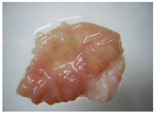
Effect of ALA on gastric ulcer, volume of gastric juice, pH value and mucus secretion: The healing effects and protective of ALA on indomethacin induced gastric ulcer are shown in Fig. 1-5 and in Table 1. Intraperitoneal injection of indomethacin in a dose of 20 mg kg-1 b.w. (G3) induced reddish bands of hemorrhagic erosions in gastric mucosa of rats (Fig. 3) as compared with control group (Fig. 1). The ulcer index was 4.31±0.21 mm (Table 1) while no ulcer formation recorded in rats of the group either post-treated (G4 , Fig. 4) or pre-treated (G5, Fig. 5) with ALA as compared to G3. ALA exert healing effects as well as a preventive effect on formation of gastric ulcer.
Treatment with indomethacin, significantly (p<0.01) increased the gastric juice volume (2.34±0.13 mL) in G3 as compared to control group G1 (1.13±0.01 mL). Either post-treatment (G4) or pre-treatment (G5) with ALA significantly (p<0.05) reduced the gastric juice volume to 1.95±0.04 and 1.72±0.18 mL, respectively as compared to G3. There no recorded changes was found for healthy rats received ALA (1.14±0.07 mL). Indomethacin induced a significant (p<0.01) decrease in the gastric pH value (1.06±0.13) in G3 versus 2.81±0.03 in G1. Administration of ALA attenuated this decrease to 2.31±0.14 and 3.20±0.05 in G4 and G5, respectively as compared to G3.
Also indomethacin produced a reduction in the gastric mucus secretion (p<0.01, mean: 0.279±0.01 mg g-1 tissue) versus 0.370±0.02 mg g-1 tissue in G1.
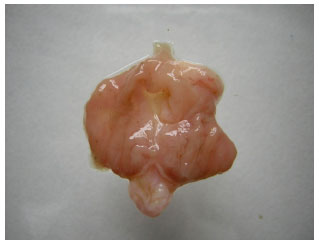 | |
| Fig. 1: | Normal stomach (G1) |
| Table 1: | Effect of ALA on indomethacin-induced gastric damage in rats |
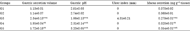 | |
| G1: Served as control, G2: ALA- treated group, G3: Indomethacin- treated group, G4: Indomethacin followed by ALA and G5: ALA (3 days) followed by indomethacin then ALA (4 days). Results are expressed as Mean±SE. a: The indomethacin group was compared to the control group. b: Treated group were compared to indomethacin group. *Significant at p<0.05. **Significant at p<0.01 | |
 | |
| Fig. 2: | Stomach treated with ALA (G 2) |
 | |
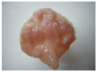
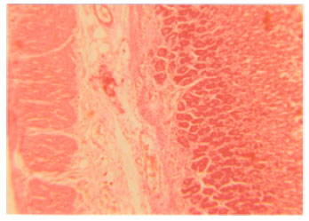
| Fig. 3: | Gastric ulcer induced by indomethacin(G3) |
Treatment with ALA antagonized significantly (p<0.05) the decrease in mucus secretion produced by indomethacin to 0.320±0.01 mg g-1 tissue in G4 and more significant (p<0.01) elevation in mucus secretion was observed in rats pre-treated with ALA (0.354±0.01 mg g-1 tissue) than in G3 group. Treatment with ALA did not exhibit any changes on mucus secretion on healthy rats.
 | |
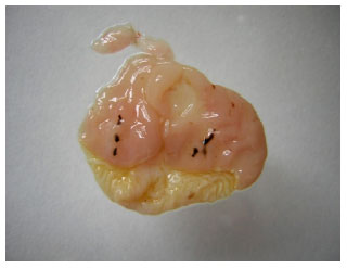
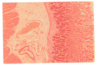
| Fig. 4: | Healing effect of Post-treatment with ALA after ulcer induction by indomethacin (G4) |
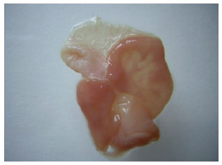 | |
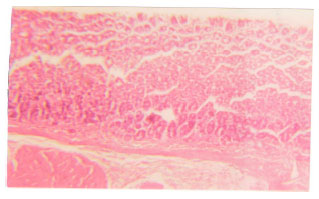
| Fig. 5: | The pre-treatment with ALA followed by indomethacin then ALA (G5) |
Effect of ALA on pro-oxidant/antioxidant parameters
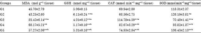
Effect on lipid peroxidation: Administration of indomethacin produced sever changes in the pro-oxidant and antioxidant parameters in gastric tissues of rats as illustrated in Table 2. MDA content in the gastric mucosa as index for lipid peroxidation revealed a significant increment (p<0.01) in group 3 (G3) that received indomethacin (85.42±6.14 mol g-1 tissue) as compared to control group G1 (45.70±2.79 mol g-1 tissue) whereas post-treatment with ALA for 7 days (G4) and treatment with ALA for 3days followed by indomethacin, then ALA for 4days (G5), significantly decreased MDA level (p<0.05, mean: 68.17±5.21 mol g-1 tissue) and (p<0.01, mean: 57.27±2.06 mol g-1 tissue, respectively when compared to G3. No significant change in MDA levels in gastric tissues was observed between healthy rats treated with ALA in G2 (43.25±2.60 mol g-1 tissue) and G1.
Effect on GSH level: The results also showed that GSH level in the gastric tissue was significantly (p<0.01) decreased in G3 (4.01±0.12 nmol mg-1 tissue) in comparison with G1(5.06±0.15 nmol mg-1 tissue). Post-treatment with ALA was significantly (p<0.01) increased GSH level in G4(5.17±0.16 nmol mg-1 tissue) and G5 (5.31±0.16 nmol mg-1 tissue) when compared to G3. Moreover, the treatment of healthy rats with ALA (G2) showed a significant (p<0.01) increase in GSH content (6.11±0.24 nmol mg-1 tissue) as compared to G1 (Table 2).
Effect on CAT activity: CAT activity was significantly (p<0.01) increased in G3 that administrated indomethacin (114.78±4.59 mmol min-1 mg-1 tissue) as compared to G1 (69.94±2.80 mmol min-1 mg-1 tissue). Treatment with ALA caused an opposite changes in the activities of CAT in gastric mucosa of both groups G4 (82.067±3.29 mmol min-1 mg-1 tissue) and G5 (74.93±2.94 mmol min-1 mg-1 tissue) when compared to G3 and insignificant change was found in gastric tissues of healthy animals of G2 that received ALA as regard to G1.
Effect on SOD activity: SOD activity in gastric tissue was reduced significantly (p<0.01) in the G3 (70.48±1.41 mmol min-1 mg-1 tissue) as compared to G1 (118.31±2.37 mmol min-1 mg-1 tissue). However, administration with ALA to the experimentally ulcerated group (G4) increased the SOD activity (93.82±1.87 mmol min-1 mg-1 tissue) while pre-treatment with ALA (G5) significantly (p<0.01) increased SOD activity to (106.43±2.13 mmol min-1 mg-1 tissue) in comparison with G3. The treatment of healthy rats with ALA showed a significant (p<0.05) increase in SOD activity (126.19±3.8 mmol min-1 mg-1 tissue) as regarded to G1.
Histopathological investigation of gastric mucosal lesions: Figure 6 showed a normal histological structure of rat gastric mucosa where normal gastric architecture formed of outer serosa, muscularis layer composed of outer longitudinal and inner circular muscle, submucosa with blood vessels and mucosa composed of muscularis mucosa and gastric glands with connective tissue. The gastric glands are simple tubular, lined by secreting cells of two types, the granular (peptic) cells of polygonal outline and secretory granules with affinity to basic dyes, oxyntic (acidic) cells with rounded outline and affinity to acidic dyes.
The group treated with ALA alone showed preserved gastric architecture free of any pathological changes and retained full thickness as shown in Fig. 7 which revealed a normal appearance of stomach wall as control.
Indomethacin treatment revealed superficial mucosal ulceration, neutrophilic permeation, marked submucosal oedema, congested blood capillaries, marked mucosal changes, sloughing and thinning, detachment of the muscularis mucosa, with glandular necrosis, submucosal hemorrhage and oedema were evident (Fig. 8). The group treated with indomethacin then lipoic acid showing preserved gastric mucosal architecture, mild submucosal congested blood capillaries and oedema, yet glandular epithelium was protected as displayed in Fig. 9.
| Table 2: | Effect of ALA on MDA level as well as CAT and SOD activities in indomethacin -induced gastric damage in rats |
 | |
| G1: Served as control, G2: ALA- treated group, G3: Indomethacin-treated group, G4: Indomethacin followed by ALA and G5: ALA (3 days) followed by indomethacin then ALA (4 days). Results is expressed as Mean±SE. a: The indomethacin group was compared to the control group. b: Treated group were compared to indomethacin group. *Significant at p<0.05. **Significant at p<0.01 | |
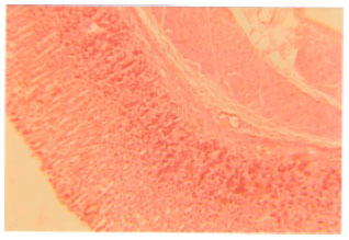 | |
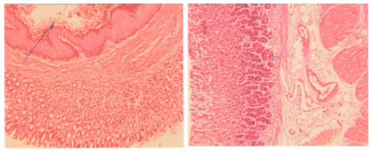
| Fig. 6: | The stomach of a control showing normal appearance (G1). (H and E, X40) |
 | |
| Fig. 7: | The stomach of a rat after treatment with (20 mg kgG1) ALA for 7 day (G2). (H and E, X40) |
 | |
| Fig. 8: | The stomach of a rat after treatment with (20 mg kgG1) indomethacin for 2 days (G3). (H and E, X40) |
 | |
| Fig. 9: | The stomach of a rat after treatment with (20 mg kg-1) indomethacin for 2 days, followed by ALA (20 mg kg-1) for 7 days (G4). (H and E, X40) |
 | |
| Fig. 10: | The stomach of a rat after treatment with ALA for 3 days followed by indomethacin for 2 days then ALA for 4 days (G5). (H and E, X40) |
The group treated with ALA for 3 days then indomethacin for 2 days then ALA for 4 days showed no ulceration, no necrosis, no congestion and preserved gastric mucosal architecture (protected) as shown in Fig. 10.
DISCUSSION
In this study, the antiulcer effect of ALA was investigated in rats using an indomethacin-induced ulcer model. In addition, the effect of ALA on some oxidant and antioxidant parameters in rat stomach tissues was evaluated. ALA was found to significantly inhibit indomethacin- induced ulcer. Indomethacin has been shown to produce higher gastric damage in rats when compared to other NSAIDs (Takeuchi et al., 2005). Gastric epithelium is covered by continuous layer of secreted mucus and bicarbonate which have been widely implicated as important pre-epithelial protective factors against auto-digestion of gastric mucosa by acid and pepsin (Copeman et al., 1994). It has reported that diminished gastric mucus renders the mucosa more susceptible to injury induced by various aggressive factors like indomethacin (Leonard et al., 1994).
In our findings, indomethacin produced visible ulcers in gastric tissues (Fig. 3) and it significantly increased the volume of gastric juice while it caused decrease in both mucus secretion and pH value. Our data were in agreement with reports of several authors who found that indomethacin treatment causes increased gastric secretion volume with significant decreased gastric pH (Rifat-uz-Zaman et al., 2005; Ajeigbe et al., 2008) and markedly decease in mucus content (Olaleye and Ajeigbe, 2009). These gastric changes have been reported due to its lipid peroxidation/ apoptosis activity (Smith and Marnett, 1991).
The present data revealed that ALA protect the gastric mucosa from injury through increasing the mucus secretion in stomach wall and increment of pH value. Goel and Bhattacharya (1991) reported that mucus secretion prevents physical damage and back diffusion of hydrogen ions. The observed increase in mucus secretion in our groups treated with ALA may be due to prostaglandin synthesis. Marnett and Wilcox (1977) reported that ALA stimulate prostaglandin synthesis and release in vitro. Furthermore, various compounds with potential to generate prostaglandins have been reported to protect gastric mucosa against various ulcerogenic agents (Grant et al., 1988).
On the other hand, oxidative stress mediated ROS has been shown to be the important primary factor in indomethacin induced gastric damage (Naito et al., 1998). In the current study, the level of MDA and CAT activity that are indicators of oxidative stress were increased by indomethacin administration. These findings are in accordance with many experimental studies showed that MDA levels and CAT activity in stomach tissue increase in indomethacin- induced gastric damage (Demircan et al., 2005; Bayir et al., 2006; Dengiz et al., 2007). The increase of CAT activity is important in terms of secondarily aiding in gastric protection.
In present study, the gastric GSH levels were depleted in ulcerated rats. Similar observations in ulcer studies were reported by Dengiz et al. (2007) and Bilici et al. (2009) where they found lower concentrations of GSH in indomethacin ulcerated gastric tissues. Such decreases were reversed when treated with ALA in groups 4 and 5. Glutathion has central role in antioxidant network including vitamin C and vitamin E which is continuously recycled by glutathione reductase (Gul et al., 2000). Thus, agents that increase availability of thiols which enhances antioxidant defenses, all reduced NASIDs causing injury (Naito et al., 1998). ALA is potent antioxidant and capable of regenerating several other antioxidants back to their active states, including vitamin C, vitamin E and glutathione. ALA is a small molecule soluble in both water and fat, this allows it to work both inside the cell and at the membrane level, making ALA acts as antioxidant against free radicals by coupling with unpaired electrons and enhancing their cell protection abilities (Packer et al., 1995).
Present results revealed that indomethacin inhibited SOD activity, whereas this activity increased by the administration of ALA in groups (2,4 and 5). Previous studies have also reported that decreased SOD activity and release of ROS produce gastric damage (Basivireddy et al., 2003). SOD plays an important role in eliminating gastric damage by partially preventing oxidative damage and it destroys the highly reactive radical O•-G2 by converting it into the less reactive H2O2 that can be destroyed by CAT. Furthermore, the relation between SOD activity and prostaglandin synthesis may be a possible mechanism to explain indomethacin-induced ulcer (El-Missiry et al., 2001).
Moreover, in vitro and in vivo tests have confirmed that ALA exert potent antioxidant properties not only by ROS scavenging but also through metal chelation, regeneration of endogenous antioxidants and repair of oxdatively damaged biomolecules by react with ROS such as superoxide radicals and hydroxyl radicals, hypochlorous acid, peroxyl radicals and singlet oxygen (Biewenga et al., 1997).
CONCLUSION
The present study suggested that the antioxidant properties of ALA and its mucus protective effect might be the main factors responsible for its strong protective and healing action on indomethacin induced gastric ulcer and also indicating a beneficial therapeutic effect of ALA in cases of gastric disorders induced by NSAIDs.
REFERENCES
- Aebi, H., 1984. Catalase in vitro. In: Methods in Enzymology, Packer, L., Academic Press, Cambridge, Massachusetts, United States, ISBN: 9780121820053, pp: 121-126.
CrossRefDirect Link - Ajeigbe, K.O., E.O. Nwobodo, T.O. Oyesola, D.A. Ofusori and S.B. Olaleye, 2008. Chloroquine phosphate potentiates indomethacin and HCl/Ethanol-induced gastric mucosa injury in rats. Int. J. Pharmacol., 4: 482-486.
CrossRefDirect Link - Al-Attar, A.M., 2011. Protective effect of Avicennia alba leaves extract on gastric mucosal damage induced by ethanol. Res. J. Med. Plant, 5: 477-490.
CrossRefDirect Link - Alsaif, M.A., 2004. Inhibition of gastric mucosal damage by boric acid pretreatment in rats. J. Med. Sci., 4: 102-109.
CrossRefDirect Link - Bandyopadhyay, D., K. Biswas, U. Bandyopadhyay, R.J. Reiter and R.K. Banerjee, 2000. Melatonin protects against stress-induced gastric lesions by scavenging the hydroxyl radical. J. Pineal Res., 29: 143-151.
Direct Link - Bayir, Y., F. Odabasoglu, A. Cakir, A. Aslan, H. Svleyman, M. Halici and C. Kazaz, 2006. The inhibition of gastric mucosal lesion, oxidative stress and neutrophil-infiltration in rats by the lichen constituent diffractaic acid. Phytomedicine, 13: 584-590.
CrossRefPubMedDirect Link - Biewenga, G.P., G.R.M.M. Haenen and A. Bast, 1997. The pharmacology of the antioxidant lipoic acid. Gen. Pharmacol.: Vasc. Syst., 29: 315-331.
CrossRefDirect Link - Bilici, M., C. Ozturk, H. Dursun, F. Albayrak and M.B. Saglam et al., 2009. Protective effect of mirtazapine on indomethacin-induced ulcer in rats and its relationship with oxidant and antioxidant parameters. Digestive Dis. Sci., 54: 1868-1875.
CrossRefPubMedDirect Link - Bilska, A. and L. Wlodek, 2005. Lipoic acid-the drug of the future?. Pharmacol. Rep., 57: 570-577.
PubMed - Chng, H.T., L.S. New, A.H. Neo, C.W. Goh, E.R. Browne and E.C.Y. Chan, 2009. Distribution study of orally administered lipoic acid in rat brain tissues. Brain Res., 1251: 80-86.
CrossRef - Copeman, M., J. Matuz, A.J. Leonard, J.P. Pearson, P.W. Dettmar and A. Allen, 1994. The gastroduodenal mucus barrier and its role in protection against luminal pepsins: The effect of 16, 16 dimethyl prostaglandin E2, carbolpolyacrylate, sucrafate and bismuth salicylate. J. Gastroenterol. Hepatol., 9: 55-59.
Direct Link - Corne, S.J., S.M. Morrissey and R.J. Woods, 1974. Proceedings: A method for the quantitative estimation of gastric barrier mucus. J. Physiol., 242: 116P-117P.
PubMedDirect Link - Dengiz, G.O., F. Odabasoglu, Z. Halici, H. Suleyman, E. Cadirci and Y. Bayir, 2007. Gastroprotective and antioxidant effects of amiodarone on indomethacin-induced gastric ulcers in rats. Arch. Pharm. Res., 30: 1426-1434.
CrossRef - Draper, H.H. and M. Hadley, 1990. Malondialdehyde determination as index of lipid peroxidation. Methods Enzymol., 186: 421-431.
CrossRefPubMedDirect Link - Durak, I., O. Canbolat, M. Kavutcu, H.S. Ozturk and Z. Yurtaslani, 1996. Activities of total, cytoplasmic and mitochondrial superoxide dismutase enzyme in sera and pleural fluids from patients with lung cancer. J. Clin. Lab. Anal., 10: 17-20.
PubMedDirect Link - Ellman, G.L., 1959. Tissue sulfhydryl groups. Arch. Biochem. Biophys., 82: 70-77.
CrossRefPubMedDirect Link - El-Missiry, A.M., H.I. El-Sayed and I.A. Othman, 2001. Protection by metal complexes with SOD-mimetic activity against oxidative gastric injury induced by indometacin and ethanol in rats. Ann. Clin. Biochem., 38: 694-700.
CrossRef - Gill, N.S., J. Bajwa, P. Sharma, K. Dhiman and S. Sood et al., 2011. Evaluation of antioxidant and antiulcer activity of traditionally consumed Cucumis melo seeds. J. Pharmacol. Toxicol., 6: 82-89.
CrossRefDirect Link - Goel, R.K. and S.K. Bhattacharya, 1991. Gastroduodenal mucosal defense and protective agents. Indian J. Exp. Biol., 29: 701-714.
PubMed - Guidobono, F., F. Pagani, C. Ticozzi, V. Sibilia, A. Pecile and C. Netti, 1997. Protection by amylin of gastric erosions induced by indomethacin or ethanol in rats. Br. J. Pharmacol., 120: 581-586.
CrossRef - Gul, M., F.Z. Kutay, S. Temocin and O. Hanninen, 2000. Cellular and clinical implications of glutathione. Indian J. Exp. Biol., 38: 625-634.
PubMed - Huang, J.Q., S. Sridhar and R.H. Hunt, 2002. Role of Helicobacter pylori infection and non-steroidal anti-inflammatory drugs in peptic-ulcer disease: A meta-analysis. Lancet, 359: 14-22.
PubMedDirect Link - Jiang, G.L., W.B. Im, Y. Donde and L.A. Wheeler, 2009. EP4 agonist alleviates indomethacin induced and promotes chronic gastric ulcer healing. World J. Gastroenterol., 15: 5149-5156.
Direct Link - Leonard, A., M.T. Droy-Lefaix and A. Allen, 1994. Pepsin hydrolysis of the adherent mucus barrier and subsequent gastric mucosal damage in rat: Effect of diosmectite and 16, 16-dimethyl prostaglandin E2. Gastroenterol. Clin. Biol., 18: 609-616.
Direct Link - Maritim, A.C., R.A. Sanders and J.B. Watkins, 2003. Effects of alpha-lipoic acid on biomarkers of oxidative stress in streptozotocin-induced diabetic rats. J. Nutr. Biochem., 14: 288-294.
Direct Link - Marnett, L.J. and C.L. Wilcox, 1977. Stimulation of prostaglandin biosynthesis by lipoic acid. Biochem. Biophys. Acta, 487: 222-230.
CrossRef - Naito, Y., T. Yoshikawa, N. Yoshida and M. Kondo, 1998. Role of oxygen radical and lipid peroxidation in indomethacin-induced gastric mucosal injury. Dig. Dis. Sci., 43: 30S-34S.
PubMed - Odabasoglu, F., Z. Halici, A. Cakir, M. Halici and H. Aygun et al., 2008. Beneficial effects of vegetable oils (corn, olive and sunflower oils) and alpha-tocopherol on anti-inflammatory and gastrointestinal profiles of indomethacin in rats. Eur. J. Pharmacol., 591: 300-306.
CrossRef - Ogle, C.W., C.H. Cho, M.C. Tong and M.W.L. Koo, 1985. The influence of verapamil on the gastric effects of stress in rats. Eur. J. Pharmacol., 112: 399-404.
CrossRef - Olaleye, S.B. and K.O. Ajeigbe, 2009. Attenuation of experimental gastric ulceration by sulfadoxine-pyrimethamine in albino rats. J. Med. Sci., 9: 87-92.
CrossRefDirect Link - Oluwole, F.S. and M.T. Saka, 2007. Effect of thyroid hormone on gastric mucus secretion around indomethacin induced gastric ulcers in rats. J. Medical Sci., 7: 678-681.
CrossRefDirect Link - Packer, L., E.H. Witt and H.J. Tritschler, 1995. Alpha-lipoic acid as a biological antioxidant. Free Radic. Biol. Med., 19: 227-250.
CrossRefPubMedDirect Link - Repetto, M.G. and S.F. Llesuy, 2002. Antioxidant properties of natural compounds used in popular medicine for gastric ulcers. Braz. J. Med. Biol. Res., 35: 523-534.
CrossRefDirect Link - Shay, P.K., R.F. Moreau, E.J. Smith and T.M. Hagen, 2008. Is α-lipoic acid a scavenger of reactive oxygen species in vivo? Evidence for its initiation of stress signaling pathways that promote endogenous antioxidant capacity. IUBMB Life, 60: 362-367.
CrossRefPubMedDirect Link - Smith, W.L. and L.J. Marnett, 1991. Prostaglandin endoperoxide synthase: Structure and catalysis. Biochim. Biophys. Acta, 1083: 1-17.
CrossRefPubMedDirect Link - Song, K.H., W.J. Lee, J.M. Koh, H.S. Kim and J.Y. Youn et al., 2004. Alpha-lipoic acid prevents diabetes mellitus in diabetes-prone obese rats. Biochem. Biophys. Res. Commun., 326: 197-202.
CrossRef - Suh, J.H., H. Wang, R.M. Liu, J. Liu and T.M. Hagen, 2004. (R)-alpha-lipoic acid reverses the age-related loss in GSH redox status in post-mitotic tissues: evidence for increased cysteine requirement for GSH synthesis. Arch. Biochem. Biophys., 423: 126-135.
PubMed - Suh, J.H., R. Moreau, S.H. Heath and T.M. Hagen, 2005. Dietary supplementation with (R)- alpha-lipoic acid reverses the age-related accumulation of iron and depletion of antioxidants in the rat cerebral cortex. Redox. Rep., 10: 52-60.
CrossRefPubMedDirect Link - George, S., A. Sathiamoorthy and S.S. Sathimoorthy, 1999. Effect of alpha tocopherol on gastric ulcers induced by pylorus ligation in rats. Indian J. Pharmacol., 31: 431-433.
Direct Link - Takeuchi, K., A. Tanaka, Y. Hayashi and A. Yokota, 2005. COX inhibition and NSAID-induced gastric damage-roles in various pathogenic events. Curr. Top. Med. Chem., 5: 475-486.
PubMed - Winiarska, K., D. Malinska, K. Szymanski, M. Dudziak and J. Bryla, 2008. Lipoic acid ameliorates oxidative stress and renal injury in alloxan diabetic rabbits. Biochimie, 90: 450-459.
CrossRefDirect Link - Rifat-uz-Zaman, M.S. Akhtar and M.S. Khan, 2004. Gastroprotective and anti-secretory effect of Nigella sativa seed and its extracts in indomethacin-treated rats. Pak. J. Biol. Sci., 7: 995-1000.
CrossRefDirect Link - Rifat-uz-Zaman, M.S. Akhtar and M.S. Khan, 2005. Protective effects of Polygonum viviparum L. root and its extracts against lipid-peroxidation induced by Indomethacin in rats. Int. J. Pharmacol., 1: 324-328.
CrossRefDirect Link