
Huseyin Turker
Department of Biology, Faculty of Science, Ankara University, Ankara, Turkey
LiveDNA: 90.23568
Asian Journal of Animal and Veterinary Advances
Year: 2015 | Volume: 10 | Issue: 10 | Page No.: 584-591


ABSTRACT
In general, the Glomerular Basement Membrane (GBM) is the main part of the Glomerular Filtration Barrier (GBF) in living organisms which border the urinary space and capillary lumen. It is thought that, the GBF comprises the endothelium, the basement membrane and the foot processes of podocytes. In this study, the effects of ultraviolet (UV) C radiation on the thickness of GBM in mole rats (Spalax leucodon) kidneys were examined by transmission electron microscope. Thirteen adult mole rats of both sexes, weighing 180-200 g were used in this study. The mole rats were divided into four groups. Group I was separated as the control and did not take any radiation. The other groups were irradiated by artificially produced UVC radiation for 14, 28 and 60 days. After the euthanasia, the kidney samples were taken, dissected out and fixed. Then the specimens were examined on transmission electron microscope. The results showed that the thickening of the GBM was evident at 14 days after UVC radiation exposure and the thickness of GBM increased more than twice of control value at 28 days. But after 60 days of radiation exposure, the thickening decreased slightly. The results showed that UVC radiation caused the thickness of GBM in mole rats according to radiation dose and exposure times.
PDF Abstract XML References Citation
How to cite this article
Huseyin Turker, 2015. Effects of UVC Radiation on the Thickness of Glomerular Basement Membrane in Mole Rats. Asian Journal of Animal and Veterinary Advances, 10: 584-591.
DOI: 10.3923/ajava.2015.584.591
URL: https://scialert.net/abstract/?doi=ajava.2015.584.591
DOI: 10.3923/ajava.2015.584.591
URL: https://scialert.net/abstract/?doi=ajava.2015.584.591
INTRODUCTION
Living organisms are under the effects of radiation coming from the sun, nuclear weapons testing, technical products and medical procedures (NCRP., 2009; Pouget et al., 2015; Sharma et al., 2011). Solar ultraviolet radiation is a part of the spectrum of electromagnetic radiation emitted from the sun. It is arbitrarily divided into 3 categories according to wavelengths: UVA (400-320 nm), UVB (320-290 nm) and UVC (290-200 nm) (Dong et al., 2007; WHO., 1994).
The UVA radiation causes tanning on the epidermis in a short time exposure, due to melanin accumulation in the skin. The UVB radiation causes serious sunburn, associated with intensified erythema and oedema, ache and blister formation in less than one day of exposure. The UVC radiation has sterilization and biocidal properties, but it also has more detrimental effects on the living beings compared to other organisms. Fortunately, majority of this radiation is filtered by the ozone (O3) layer (Corvo et al., 2015; Stolarski et al., 1992; WHO., 1994). The reduction of ozone in the stratosphere occurs as a consequence of human activities such as chlorofluorocarbon and greenhouse gases and besides that the cosmetic sprays leads to an increase in the amount of UVC radiation on the earth. It is estimated that skin cancer, cataract and immune deficiency diseases will increase in the near future (Majumdar et al., 2013; Stolarski et al., 1992; Thiele et al., 2003).
A lot of researchers were interested in the thickening of vascular basal membrane in many pathological diseases and a large number of studies have been carried out. But, these studies were mostly related with aging and variety of diseases, such as diabetes mellitus and rheumatoid arthritis (Carlson et al., 2003; Matsubara et al., 1987; Tyagi et al., 2008).
To our knowledge, plenty of data has been obtained about the effects of radiation on haematological, biochemical or histopathological characteristics of animals (Mansoub and Sarvestani, 2011; Moulder and Cohen, 2007; Santra and Manna, 2009), but similar studies done with UV radiation on the glomerular basement membrane thickness have not been encountered.
The mole rats (Spalax leucodon) are living in underground galleries and nests. Although, the living organisms on earth have a directly exposure to radiation coming from the sun and the other resources, these animals are not under such effects. For this reason, these animals were selected and exposed to artificially produced UVC radiation in the lab and the thickening of Glomerular Basement Membrane (GBM) was compared to the control group values.
MATERIALS AND METHODS
Specimen collection: Thirteen adult mole rats of both sexes, weighing 180-200 g were used in this study. All mole rats were caught within the rural areas of Ankara in Turkey. The mole rats were housed individually in special cages called terrarium and a constant UVC radiation was applied from the upper side (Fig. 1). Before the experiment, dorsal hairs of the animals were shaved. The animals were fed with carrot, potato, plant roots and no special diet was given.
Radiation source: A "Mazda TG" ultraviolet lamp in 30 W powers and a 90 cm length was placed to the cover of the terrarium. The pick value of ultraviolet radiation emitted from the lamp was measured as 254 nm in wavelength and the energy was found to be 0.0014 J cm–2 in 1 sec.
Experimental animals and design: After certain days of acclimatization, the mole rats were divided into four groups. Group I was separated as the control and did not take any radiation. In terms of sunlight period, the other groups were irradiated with artificial UVC radiation for 8 h daily (between 08.00-17.00 h). A feeding interval was given at midday for 1 h. A timer was used to standardize radiation exposure times. Group II was irradiated for 14 days, group III was irradiated for 28 days and group IV was irradiated for 60 days. Experiment groups, exposure times and total dosages applied were shown in Table 1.
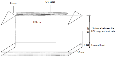 | |
| Fig. 1: | Terrarium where the mole rats were exposed to UVC radiation |
| Table 1: | Experiment groups, exposure times and total dosages applied |
 | |
Sample preparation for electron microscopic studies: Before and at the end of the experiment periods of 14, 28 and 60 days, the animals were sacrificed under ether anaesthesia to detect and compare the GBM thickness induced by irradiation. Anaesthesia of the animals was done with 0.01 mg ketamine injection intramuscularly. After the euthanasia, the kidneys were dissected out and the tissues were cut into small pieces and prepared for electron microscopic examination. The small kidney samples were fixed in glutaraldehyde, washed in buffered salina and post-fixed in 1% osmium tetroxide, dehydrated in ethanol, cleared in propylene oxide and embedded in Araldite CY-212. Ultrathin sections were obtained by an ultramicrotome, picked up onto the finder grids, stained with uranyl acetate and lead citrate. Then, the specimens were examined on Jeol JEM 100 CX-II transmission electron microscope in Gulhane Military Medical Faculty (GATA), Ankara, Turkey.
Statistical analyses: Thickness of Glomerular Basement Membrane (GBM) in control and experimental groups were measured by the direct measurement method and the arithmetic means were calculated. Data were analysed using SPSS for Windows software, Version 18.00 (SPSS Inc., Chicago, IL, USA). The differences between means of GBM in the control and experimental groups were evaluated by ANOVA statistics.
Significant differences in irradiated groups with the control groups are accepted at p<0.05. The study was carried out in accordance with the Ankara University guidelines for the care of experimental animals. In addition, guiding principles and procedures found in Declaration of Helsinki of the World Medical Association regarding animal experimentation were followed in this study.
RESULTS
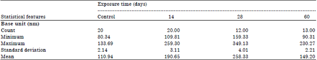
In the control and irradiated animals, the GBM thickness in kidney tissues could be distinguished by the direct measuring technique on TEM micrographs. The measured results and electron microscopic results were given in Table 2, Fig. 2 and 3.
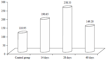
The mean GBM thickness in the control mole rats was found 110.94 nm. The mean GBM thickness in irradiated mole rats were found 190.65 nm after 14 days, 258.33 nm after 28 days and 149.20 nm after 60 days. The results were found significantly different from those of the control group (p<0.05).
DISCUSSION
The UV radiation has both useful and harmful effects on the living organisms. Among the harmful effects of UV radiation are generally such things as an increased damage on DNA and cell functions, blockading of genetic transcription, alteration of cell divisions and so on. Living organisms have well protective and effective cellular repair mechanisms against radiation. However, the excessive radiation exposure may cause a damage on the cells, immune system and blood cells (Stone et al., 2003; WHO., 1994). Accordingly, the present study aimed to elucidate the histopathological changes of GBM in mole rats.
| Table 2: | Mean thickness of GBM in control group and irradiated mole rats |
 | |
GBM: Glomerular basement membrane, Significant differences in irradiated groups with the control groups are accepted at p>0.05 | |
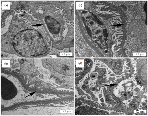 | |
| Fig. 2(a-d): | Electron micrographs of GBM thickness in mole rats, (a) Control, (b) Irradiated for 14 days, (c) Irradiated for 28 days and (d) Irradiated for 60 days, GBM (arrow) |
 | |
| Fig. 3: | Thickness changing of GBM in mole rats according to UVC radiation dose and exposure times |
The pathological effects of radiation begin immediately after radiation exposure, but the histological effects may not become apparent for weeks, months or even years after exposure. According to the appearance of symptoms, radiation injury is classified as early and late effects. Although early effects can be observed within a few weeks, the late effects can emerge from months to years after the exposure (Dorr and Hendry, 2001). The symptoms are developed depending on the dosage and the exposal period and they may be developed gradually or suddenly (Gridley et al., 2001; Stone et al., 2003). While the high dose of radiation destroys the cell structure, the low dose of radiation is no longer considered to be as harmful as once thought, because the pathological effects can be prevented by some antioxidants in cells (Jagetia and Reddy, 2005).
As the kidney is a late-responding organ, the radiation effects may develop for months or years after the exposure. This damage develops slowly and results in nephropathy with arterial hypertension, increased proteinuria and anaemia if the kidneys are treated with high dose of radiation. In contrast with many tissues, there seems to be little regenerative response in kidney cells.
The GBM is the main portion of the Glomerular Filtration Barrier (GBF) that borders the capillary lumen and urinary space (Arkill et al., 2014; Menon et al., 2012). The glomerular filter, through which the infiltration passes, consists of three layers: fenestrated endothelium, intervening GBM and the podocytes layer (Menon et al., 2012; Shirato et al., 1991). This complex membrane is permeable to water and small solutes, but retains most of the proteins and other large molecules as well as whole blood cells (Farquhar, 1991; Solak, 2010).
The GBM is a thick membrane that provides the glomerular cells with a structural support. The endothelial cells in glomerulus and podocytes are responsible for the production of this membrane (Abrahamson, 1985). Thickening of vascular BM has been studied by a lot of researchers into aging and into a variety of diseases, such as diabetes mellitus, rheumatoid arthritis and polymyositis (Carlson et al., 2003; Matsubara et al., 1987; Tyagi et al., 2008). But, no study has been carried out with regard to demonstrating the effects of UVC radiation on the thickness of GBM in mole rats.
In general, the thickness of GBM was 300-350 nm in some animals (Arkill et al., 2014; Solak, 2010), but in the current study, the mean thickness of GBM was measured as 110,94 nm in the control group and it was measured as 190.65 nm for 14 days, 258.33 nm for 28 days and 149.20 nm for 60 days irradiated mole rats.
The mean thickness of the GBM varied with radiation dosage and exposure times. The results showed that the thickening of GBM was more evident at 14 and 28 days of UV radiation exposure compared to the control group value. Thickening of the GBM increased after 14 days exposure and it was twice as high as the control value at 28 days. But the thickening of the GBM decreased a little on 60 days exposure. According to some studies, the expansion or contraction of capillary lumen gives rise to thin or thick basement membrane. These changes of the capillary lumen could be considered as one of the important factors responsible for the variation of the thickness. Similar results were recorded in the studies conducted on some animals and humans (Carlson et al., 2003; Jaggi et al., 2005).
It was also reported that the thickness of GBM occurred possibly due to a decreased amount of heparin sulphate proteoglycan and immunopathological disorders in diabetes (Weber et al., 1984) and accelerated death and replenishment of Endothelial Cells (EC) (Vracko and Benditt, 1970).
A recent investigation showed that interleukin-1 (IL-1) is capable of increasing the production of basement collagen type IV (C(IV)22) in murine mammary epithelial cells. The IL-1 might stimulate the Endothelial Cells (EC) to produce BM components, including collagen type IV and affects the thickening of the GBM (Iguchi et al., 1986). According to other studies, the deposition of immune complexes in the vessel walls of GBM, proteolytic enzymes of leukocytes and macrophages could degrade the basement membrane (Davies et al., 1980).
It was assumed that the pathological changes might result from an increase in the process of lipid peroxidation and a decrease in the activity of antioxidant enzymes of the body with the consequent damage of cellular membranes (Sugiyama et al., 1984). Such degenerative changes occuring in cytoplasm and organelles leading to significant functions in each phase of radiation in order that cell could carry on its crucial activities would certainly decrease its process and distort its structure. These results were in consistent with the studies carried out by local irradiation or high dose radiation on some animals (Gridley et al., 2001; Mansoub and Sarvestani, 2011; Santra and Manna, 2009).
CONCLUSION
The results clearly indicated that the effects of UV radiation on the thickness of GBM in mole rats. It was found that these effects increased depending on the dosage and the exposal period of the radiation. In this respect, further experimental studies are needed to confirm the relation between the radiation effects, exposure time, dosage and cell deformation in the kidney cells.
ACKNOWLEDGMENT
The authors would like to thank the reviewers for their thorough and thoughtful comments and suggestions that improved the overall quality of this study.
REFERENCES
- Abrahamson, D.R., 1985. Origin of the glomerular basement membrane visualized after in vivo labeling of laminin in newborn rat kidneys. J. Cell Biol., 100: 1988-2000.
CrossRefDirect Link - Arkill, K.P., K. Qvortrup, T. Starborg, J.M. Mantell and C. Knupp et al., 2014. Resolution of the three dimensional structure of components of the glomerular filtration barrier. BMC Nephrol., Vol. 15.
CrossRefDirect Link - Carlson, E.C., J.L. Audette, N.J. Veitenheimer, J.A. Risan, D.I. Laturnus and P.N. Epstein, 2003. Ultrastructural morphometry of capillary basement membrane thickness in normal and transgenic diabetic mice. Anat. Record Part A: Discov. Mol. Cell. Evol. Biol., 271: 332-341.
CrossRefDirect Link - Corvo, R., R. Santoni, S.M. Magrini and R.M. Enrici, 2015. Radiobiology as a basic and clinical medical science: What the physicists have forgotten. Tumori J.
CrossRefDirect Link - Davies, M., G.A. Coles and K.T. Hughes, 1980. Glomerular basement membrane injury by neutrophil and monocyte neutral proteinases. Renal Physiol., 3: 106-111.
CrossRefDirect Link - Dong, Q., K. Svoboda, T.R. Tiersch, and W.T. Monroe, 2007. Photobiological effects of UVA and UVB light in zebrafish embryos: Evidence for a competent photorepair system. J. Photochem. Photobiol. B: Biol., 88: 137-146.
CrossRefPubMedDirect Link - Dorr, W. and J.H. Hendry, 2001. Consequential late effects in normal tissues. Radiother. Oncol., 61: 223-231.
CrossRefDirect Link - Gridley, D.S., M.J. Pecaut, G.M. Miller, M.F. Moyers and G.A. Nelson, 2001. Dose and dose rate effects of whole-body gamma-irradiation: II. Hematological variables and cytokines. In vivo, 15: 209-216.
PubMedDirect Link - Iguchi, T., M. Kurosaka and M. Ziff, 1986. Electron microscopic study of HLA‐DR and monocyte/macrophage staining cells in the rheumatoid synovial membrane. Arthritis Rheumatism, 29: 600-613.
CrossRefDirect Link - Jagetia, G.C. and T.K. Reddy, 2005. Modulation of radiation-induced alteration in the antioxidant status of mice by naringin. Life Sci., 77: 780-794.
CrossRefDirect Link - Jaggi, J.S., S.V. Seshan, M.R. McDevitt, K. LaPerle, G. Sgouros and D.A. Scheinberg, 2005. Renal tubulointerstitial changes after internal irradiation with α-particle-emitting actinium daughters. J. Am. Soc. Nephrol., 16: 2677-2689.
CrossRefDirect Link - Majumdar, D., A. Chintada, J. Sahu and C.V.C. Rao, 2013. Emissions of greenhouse and non-greenhouse air pollutants from fuel combustion in restaurant industry. Int. J. Environ. Sci. Technol., 10: 995-1006.
CrossRefDirect Link - Mansoub, N.H. and A.H. Sarvestani, 2011. Effects of gamma irradiation on histomorphology of different organs in rats. Ann. Biol. Res., 2: 431-436.
Direct Link - Matsubara, T., M. Ziff and A. Smith, 1987. Basement membrane thickening of postcapillary venules and capillaries in rheumatoid synovium. Arthritis Rheumatism, 30: 18-30.
CrossRefDirect Link - Menon, M.C., P.Y. Chuang and C.J. He, 2012. The glomerular filtration barrier: Components and crosstalk. Int. J. Nephrol., Vol. 2012.
CrossRefDirect Link - Moulder, J.E. and E.P. Cohen, 2007. Renal dysfunction after total body irradiation: Dose-effect relationship: In regard to Kal and van Kempen-Harteveld (Int J Radiat Oncol Biol Phys 2006;65:1228-1232). Int. J. Radiat. Oncol. Biol. Phys., 67: 319-319.
CrossRefPubMedDirect Link - Pouget, J.P., C. Lozza, E. Deshayes, V. Boudousq and I. Navarro-Teulon, 2015. Introduction to radiobiology of targeted radionuclide therapy. Front. Med., Vol. 2.
CrossRefDirect Link - Santra, K.B. and C.K. Manna, 2009. X-ray induced changes in biochemical and histochemical parameters in the testis of male wild Indian house rat, Rattus rattus. Ceylon J. Sci. (Biol. Sci.), 38: 39-49.
CrossRefDirect Link - Sharma, P., J. Parmar, P. Sharma, P. Verma and P.K. Goyal, 2011. Radiation-induced testicular injury and its amelioration by Tinospora cordifolia (An Indian medicinal plant) extract. Evidence-Based Complement. Altern. Med.
CrossRefDirect Link - Shirato, I., Y. Tomino, H. Koide and T. Sakai, 1991. Fine structure of the glomerular basement membrane of the rat kidney visualized by high-resolution scanning electron microscopy. Cell Tissue Res., 266: 1-10.
CrossRefDirect Link - Solak, Y., 2010. A longitudinal study of kidney structure and function in adults. Nephrol. Dial. Transplant., 25: 3457-3457.
CrossRefDirect Link - Stolarski, R., R. Bojkov, L. Bishop, C. Zerefos, J. Staehelin and J. Zawodny, 1992. Measured trends in stratospheric ozone. Science, 256: 342-349.
CrossRefDirect Link - Stone, H.B., C.N. Coleman, M.S. Anscher and W.H. McBride, 2003. Effects of radiation on normal tissue: Consequences and mechanisms. Lancet Oncol., 4: 529-536.
CrossRefPubMedDirect Link - Sugiyama, M., K. Kajiyama, T. Hidaka, S. Kumano and R. Ogura, 1984. Lipid peroxidation and radical formation in methyl linoleate following ultraviolet light exposure. J. Dermatol., 11: 455-459.
CrossRefDirect Link - Thiele, J.J., F. Dreher, H.I. Maibach and L. Packer, 2003. Impact of ultraviolet radiation and ozone on the transepidermal water loss as a function of skin temperature in hairless mice. Skin Pharmacol. Applied Skin Physiol., 16: 283-290.
CrossRefPubMedDirect Link - Tyagi, I., U. Agrawal, V. Amitabh, A.K. Jain and S. Saxena, 2008. Thickness of glomerular and tubular basement membranes in preclinical and clinical stages of diabetic nephropathy. Indian J. Nephrol., 18: 64-69.
CrossRefDirect Link - Vracko, R. and E.P. Benditt, 1970. Capillary basal lamina thickening. Its relationship to endothelial cell death and replacement. J. Cell Biol., 47: 281-285.
PubMedDirect Link - Weber, L., T. Krieg and R. Timpl, 1984. [Basement membranes-structure, function, pathology]. Hautarzt, 35: 279-286, (In German).
PubMed