
P.L. Lalruatfela
Department of Veterinary Pathology, Post Graduate Institute of Veterinary and Animal Sciences,
Maharashtra Animal and Fishery Sciences University, Akola, 444001, Maharashtra, India
M. Saminathan
Division of Pathology, Indian Veterinary Research Institute, Izatnagar, Bareilly, Uttar Pradesh, 243122, India
R.S. Ingole
Department of Veterinary Pathology, Post Graduate Institute of Veterinary and Animal Sciences,
Maharashtra Animal and Fishery Sciences University, Akola, 444001, Maharashtra, India
K. Dhama
Division of Pathology, Indian Veterinary Research Institute, Izatnagar, Bareilly, Uttar Pradesh, 243122, India
M.V. Joshi
Department of Veterinary Pathology, Post Graduate Institute of Veterinary and Animal Sciences,
Maharashtra Animal and Fishery Sciences University, Akola, 444001, Maharashtra, India
Asian Journal of Animal and Veterinary Advances
Year: 2014 | Volume: 9 | Issue: 9 | Page No.: 523-542


ABSTRACT
Paraquat (PQ) is commonly used in agriculture as a contact herbicide to control the growth of weeds and grasses and is easily accessible to farmers in developing countries. Paraquat is highly toxic to human and is associated with high mortality varying from 35-50%. The main causes for mortality are respiratory and multi organ failure. In this experimental study, twenty four female Wistar rats of 6-7 weeks age were divided into four equal groups. Group-I rats served as control whereas groups-II, III and IV rats were given daily paraquat solution at the dose rate of 10, 15 and 25 mg kg-1 b.wt, respectively, orally by gavage for 28 days. At end of the 2nd week of the experiment, mild diarrhoea, anorexia, polydipsia and reduced locomotional activities were noticed. Hematological observations showed significant decrease in Hb, PCV, TEC and TLC values and biochemical parameters revealed significant increase in AST, ALT and creatinine levels when compared to control group. Gross pathological observations of lungs from all treatment rats showed mild congestion and emphysema. An accidental finding of hydronephrosis was recorded in group-II and atrophy of left kidney and hypertrophy of right kidney was observed in group-III. Few animals from group-IV showed rounded flabby heart. Histopathologically, granular and vacuolar changes were observed in liver and kidneys. Lungs showed prominent congestion and oedema. From the present investigation it was concluded that paraquat toxicity adversely affects general performance, hematological and biochemical parameters in rats. Histopathological changes in liver and kidneys indicated its hepatoxic and nephrotoxic effects.
PDF Abstract XML References Citation
Received: March 08, 2014;
Accepted: July 05, 2014;
Published: August 22, 2014
How to cite this article
P.L. Lalruatfela, M. Saminathan, R.S. Ingole, K. Dhama and M.V. Joshi, 2014. Toxicopathology of Paraquat Herbicide in Female Wistar Rats. Asian Journal of Animal and Veterinary Advances, 9: 523-542.
DOI: 10.3923/ajava.2014.523.542
URL: https://scialert.net/abstract/?doi=ajava.2014.523.542
DOI: 10.3923/ajava.2014.523.542
URL: https://scialert.net/abstract/?doi=ajava.2014.523.542
INTRODUCTION
In traditional India, the entire agriculture was practiced using organic techniques. But during 1950s to 1960s, the ever increasing population and several natural calamities lead to a severe food scarcity in India (Lancaster, 1990; Mehta, 2001). As a result, to increase food security, green revolution programme became erupted in the 1960s (Swaminathan, 2006). In this programme large amount of land was brought under cultivation, hybrid seeds were introduced, natural and organic fertilizers were replaced by chemical fertilizers and locally made pesticides were replaced by chemical pesticides (Wade, 1974; Parayil, 1992). Due to extensive dependence on chemical farming, the land gradually loses its fertility and demand larger quantities of fertilizers to be used. Pests are becoming immune requiring the farmers to use stronger and costlier pesticides (Freebairn, 1995; Sharma, 2000). Acute, sub-acute and chronic toxicity of pesticides to animals and human cases are reported frequently (Gunnell et al., 2007; Dawson et al., 2010). Due to these various reasons, many of the pesticides were banned in India and some are allowed only for restricted use (Vendan, 2011).
Paraquat is one of the most widely used herbicide and held the largest share of the global herbicide market until today. It is a quaternary nitrogen herbicide and brown syrupy liquid belongs to the Bipyridinium compounds and its chemical name is 1,1’-dimethyl 4,4’-bipyridinium (Dasta, 1978; Bismuth et al., 1982, 1990; Raghu et al., 2013). Paraquat is widely used for weed control in fruit orchards and plantation crops, including coffee, cocoa, coconut, oil palms, rubber, bananas, vines, olives and tea, ornamental trees and shrubs and in forestry (Hood et al., 1963; Hood, 1965). Paraquat is classified by WHO as moderately hazardous herbicide and class II poison for acute toxicity (WHO, 2009). In paraquat poisoning, no antidote is available and potential delay in onset of clinical signs by several days differentiates it from organophosphates (Ellenhorn et al., 1997).
It is a highly toxic substance for humans and animals with many cases of acute poisoning and death have been reported (Kelly et al., 1978; Florkowski et al., 1992). The main risks are due to deliberate ingestion and intensity of toxicity depends on dose ingested. Although accidental or occupational poisoning may occur in humans and animals but which is comparatively rare (Hall and Becker, 1995). Other routes of exposure of paraquat are inhalation, ocular route and skin exposure (Bataller et al., 2000; Baharuddin et al., 2011). Skin exposure is common in concentrated forms and causes irritation to the skin. Prolonged contact with skin results in extensive skin damage results in systemic poisoning due to absorption of paraquat through damaged skin and may leads to severe toxicity or even death (Bataller et al., 2000; Marrs and Adjei, 2003).
Paraquat is not metabolized, but is reduced to an unstable free radical which is then re-oxidized to reform the cation and produce a superoxide anion. Redox Cycling is the principal mechanism responsible for the toxicity of paraquat. In anaerobic conditions the paraquat cation can be reduced by NADPH-dependant microsomal flavoprotein reductase to form the reduced radical (Dinis-Oliveira et al., 2008; Mohammadi-Bardbori and Ghazi-Khansari, 2008), which then reacts with molecular oxygen to reform the paraquat cation and the superoxide ion. Paraquat will then continue to cycle from its oxidized to reduced form with the electrons and oxygen. Cell death is caused by free radical induced lipid peroxidation or NADPH depletion and in the lung selective accumulation of paraquat occurs (Eisler, 1990; Garg, 2002; Dinis-Oliveira et al., 2008; Mohammadi-Bardbori and Ghazi-Khansari, 2008).
Clinical effects of paraquat are depending on the dose and route of exposure. Lung is the organ primarily affected in paraquat poisoning, due to the preferential accumulation of paraquat in lung alveolar cells (Eisler, 1990; Dinis-Oliveira et al., 2008; Mohammadi-Bardbori and Ghazi-Khansari, 2008). It causes progressive irreversible pulmonary fibrosis (Garg, 2002). The highest concentrations (0.01-0.02%) being found in the liver, lungs and kidneys (Podprasart et al., 2007). However, some workers reported that highest tissue concentrations were found in the kidney and liver (Sharp et al., 1972; Leahey et al., 1976). Ingestion of large amounts result in multiple organ failure and death (Florkowski et al., 1992). Ingestion of moderate amounts result in renal failure and or massive pulmonary fibrosis, cardiac arrythmias, oesophageal perforation and death. The aim of the present study was to investigate the experimental paraquat toxicity at different dose levels for four weeks periods in female Wistar rats. Also, to evaluate the effects of paraquat on clinical signs, performance, hematology and biochemical parameters, gross and histological alterations in the visceral organs.
MATERIALS AND METHODS
Experimental animals: This experiment was approved by the Institute Animal Ethics Committee (IAEC) and Committee for the Purpose of Control and Supervision of Experiments on Animals (CPCSEA), Ministry of Social Justice and Empowerment, Government of India before its commencement. The guiding principles in the care and use of laboratory animals together with those described in the declaration of Helsinki and Indian standards were strictly adhered to in the conduct of all the experimental procedures. The out bred, female Wistar rats at 6-7 weeks of age were obtained Shree Farm, Bhandara (India), a recognized laboratory animal breeding center. The rats were housed in polypropylene cages in the experimental animal house under environmentally controlled conditions (temperature 25±2°C; relative humidity 30-70%) with a 12/12 h light/dark cycle. The rats were provided with standard rodent pellet feed procured from Swastik Agro Industries Ltd, Maharashtra (India) and water ad libitum. The animals were acclimatized for one week before the commencement of experiments.
Preparation of paraquat solution: Active substance of paraquat (dichloride-1,1-dimethyl-4, 4-bipyridylium) was purchased as 24% w/v solution, with the trade name of ‘Allclear’ (Hindustan Pulverising Mills, India) with properly sealed in opaque plastic container. It was kept at room temperature and during usage caution was taken to avoid spillage, fire and poisoning. The 3 doses of paraquat (1/5LD50, 1/10LD50 and 1/20LD50) was calculated and prepared the solution accordingly by dilution with water as paraquat is readily soluble in water.
Experimental design: A total of 24 rats were randomly divided into 4 groups (Group I, II, III and IV). Group-I (n = 6) didn’t received any paraquat solution and served as control group. Treatment groups-II, III and IV, each group thus comprising of six rats were given daily paraquat solution at the dose rate of 10, 15 and 25 mg kg-1 b.wt., respectively, orally by gavage. The whole experiment duration was of 28 days. The overall experimental design was presented in Table 1.
Clinical studies: Rats of each group were kept under observation for any clinical symptoms for the period of 4 weeks period from the start of experiment. Clinical signs, general performance, average weekly feed consumption, weekly body weight and mortality if any was recorded.
Haematology: Blood was collected from retro-orbital sinus and heart of rats at the time of euthanasia for haematology.
| Table 1: | Details of experimental design |
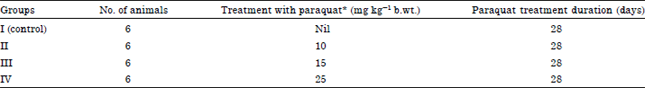 | |
| *All rats were fed with ad-lib feed and drinking water, paraquat solution was administered orally by gavage | |
Blood was collected in dry sterilized vials containing as an Ethylene Diamine Tetra Acetic Acid (EDTA) at 1 mg mL-1 and used for estimation of haemoglobin (Hb), Packed Cell Volume (PCV), Total Erythrocyte Count (TEC), Mean Corpuscular Volume (MCV), Mean Corpuscular Haemoglobin (MCH), Mean Corpuscular Haemoglobin Concentration (MCHC), Total Leukocyte Count (TLC) and absolute leukocyte count was estimated.
Serum biochemistry: Blood samples were collected in heparinized tubes, centrifuged at 2,500 rpm for 15 min and the plasma was separated and stored at -20°C for analysis. Plasma samples were analyzed for Liver Function Test (LFT), biochemical parameters viz., total protein, albumin, globulin, alanine aminotransferases (ALT/SGPT) and aspartate aminotransferases (AST/SGOT) using standard commercial kits (Span diagnostics, India). Kidney function was assessed by estimating using creatinine levels.
Necropsy and gross pathology: After 28 days, all rats from each group were sacrificed using chloroform inhalation anaesthesia to study the gross pathological changes in visceral organs. Thorough necropsy examination of all the animals was carried out and gross lesions were recorded. Lungs, heart, liver, spleen, kidneys, intestine, brain and ovary were collected in 10% neutral buffered formalin for histopathological processing.
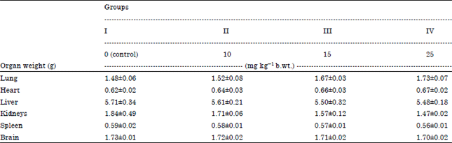
Organ weight: After gross pathological examination, organ weight of lungs, heart, liver, spleen, kidneys, intestine and brain were recorded to evaluate effect of paraquat intoxication on average organ weights.
Histopathological examination: The representative tissue samples i.e., lungs, heart, liver, spleen, kidneys, intestine, brain and ovary were processed for histo-pathological examination. Tissue samples of 1-2 mm thickness were dehydrated in graded alcohol and cleared in xylene and embedded in paraffin blocks. About 4-5 μm thick serial sections were taken with rotator microtome on clean grease free slides and subjected for haematoxylin and eosin staining (Luna, 1968).
Statistical analysis: Statistical analysis was performed using SPSS Advanced Statistics 16.0 software (SPS Inc., Chicago, USA). The one-way ANOVA followed by Tukey’s Post-hoc test was computed to know the effect of paraquat on haematological, serum biochemical parameters and organ weight in different treatment groups. The two-way ANOVA was computed to know the effect of paraquat on average weekly body weight as per Snedecor and Cochran (1980).
RESULTS
Clinical observations: During the 2nd week of the experiment decreased food intake with polydipsia was noticed in all treatment groups-II, III and IV. At the beginning of 3rd week all treatment groups showed weakness, lethargy with reduction in locomotional activities and mild diarrhea when compared with control animals. The groups-IV (25 mg kg-1 b.wt.) rats were showed anorexia, increase rate of respiration and shortness of breath. Cyanosis of visible mucous membrane and dehydration were noticed during the last week of experiment in most of the treatment group animals.
Average weekly body weight (g): The average weekly body weight in different groups were found to be 194.00±7.60 in group-I, 182.60±5.74 in group-II, 182.53±4.84 in group-III, 184.00±3.97 in group-IV. The values of percentage increased on average weight in different groups were calculated on the basis of day 0 average body weight and the pooled mean body weight at the end of experiment. Percentage increased in body weight was found to be up to 12.16% in group-I, 10.88% in group-II, 8.97% in group-III and 7.91% in group-IV. The observations of reduction in percentage increase in body weight suggested the adverse effect of paraquat on weekly body weight of rats.
Hematological observations: Hematological parameters were estimated at the end of 4th week of experiment to assess the effect of paraquat on functional status of hemopoietic and leucopoietic system.
Red blood cells (RBC), hemoglobin (Hb) and packed cell volume (PCV) count: The average mean value of erythrocyte count and hemoglobin was significantly reduced in paraquat treatment groups when compared to control group. The mean packed cell volume was significantly decreased in all treatment groups but in group-III and IV abnormal reduction in PCV was noticed when compared with control group (Table 2).
Erythrocytes indices: The erythrocyte indices values were derived from erythrocyte count, hemoglobin concentration and PCV. Details of erythrocyte indices values are given in Table 2.
MCV, MCH and MCHC: The average MCV, MCH and MCHC values in different treatment groups revealed non-significant decrease when compared with control group. However, the lowest average value was observed in highest dose group-IV (Table 2).
Total Leucocyte Count (TLC): The average total leucocyte count in different groups was depicted in Table 2. Mean average values showed significant decreased in paraquat treatment group when compared with control group.
| Table 2: | Effect of paraquat on hematological values in different treatment groups (Mean±SEM) |
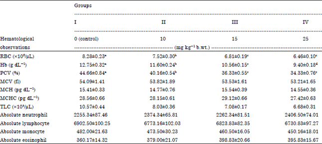 | |
| Mean±SEM values with different superscript letters indicates the significant differences (p<0.05) between paraquat treated and control groups | |
| Table 3: | Effect of praquat on liver and kidney enzyme levels in different treatment groups (Mean±SEM) |
 | |
| Mean±SEM values with different superscript letters indicates the significant differences (p<0.05) between paraquat treated and control groups | |
Absolute leucocyte count: From the total leucocyte count and differential leucocyte count absolute values of neutophil, lymphocyte, monocyte and eosinophil were derived and are given in Table 2. The mean average absolute values of neutrophil, lymphocyte, monocyte and eosinophil in different treatment groups showed non-significant differences when compared to control group. The group-II rats showed decrease in monocyte count whereas, groups-III and IV rats revealed increased count in comparison with the control group-I rats.
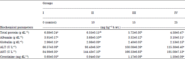
Biochemical parameters: Plasma globulin values were obtained by subtractions of albumin value from total protein values. The average values of plasma total protein, albumin and globulin were found to be significantly decreased in paraquat treated groups-III and IV rats when compared with control group-I and treated group-II. The mean average plasma AST, ALT and creatinine levels in different treated groups showed significant increase when compared with control animals. The highest dose group-IV showed more increased levels. The values of different biochemical parameters were depicted in Table 3.
Gross pathological observation: Most of the paraquat treatment group rats were showed debilitation and pale carcasses. Lungs and kidneys from all treatment group rats showed mild congestion and emphysema. Hydronephrosis was observed as an accidental finding in group-II rat (Fig. 1) and atrophy left kidney and hypertrophy of right kidney were observed in group-III (Fig. 2). Round flabby heart with pin-point hemorrhages and congestion of liver was noticed in highest dose group-IV. Congestion and dark discoloration of spleen and intestinal mucosa showed hyperaemia in all the treatment group rats.
Organ weights: After completion of detailed necropsy examination, visceral organs i.e., liver, lungs, heart, kidneys, spleen and brain were dissected and separated from carcass of each group. The mean average weight of different organs showed non-significant differences when compared with control animals. Liver, kidneys, spleen and brain weights in different group rats showed non-significant decrease in weight in a dose dependent manner. The mean average weight of lungs and heart in different treatment group revealed non-significant increase in weight when compared with the control group. The organ weights in different treatment groups were depicted in Table 4.
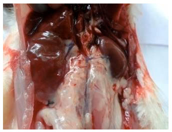 | |
| Fig. 1: | Gross lesion of hydronephrosis in right kidney of group-II rat |
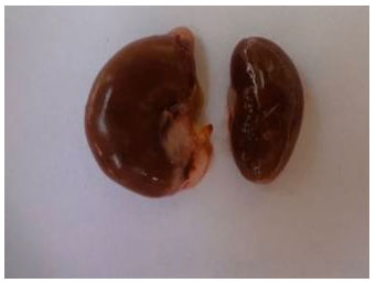 | |
| Fig. 2: | Gross lesions of hypertrophy of right kidney and atrophy of left kidney in group-IV rat |
| Table 4: | Effect of praquat on organ weights in different treatment groups (Mean±SEM) |
 | |
Histopathological observations
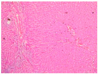
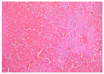
Liver: Histopathological examination of liver section from group-II rats revealed congestion in central vein. Granular and vacuolar changes (Fig. 3) near sub-capsular area with thickening of glisson’s capsule noticed (Fig. 4).
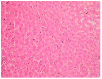 | |
| Fig. 3: | Section of liver from group-II rat showing granular and vacuolar changes and reticular cell hyperplasia. H and E stain 200X |
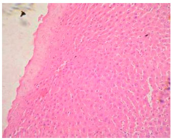 | |
| Fig. 4: | Section of liver from group-II rat showing thickened Glisson’s capsule. H and E stain 200X |
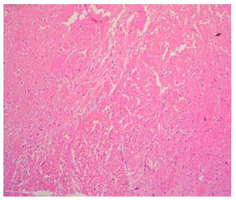
Section also revealed prominent reticular cells in the hepatic lobules and proliferation of bile ducts, suggesting hyperplasia of bile ducts. Sections from group-III rats revealed granular and prominent vacuolar changes in the parenchyma of liver and congestion in central veins (Fig. 5). Focal areas of necrosis around central vein and periportal hyperplasia of bile ducts were noticed. Sections from group-IV animals revealed prominent nuclei with granular cytoplasm of hepatocytes, and hepatic lobule at various places showed dilated sinusoids with stagnated erythrocytes and reticular cells hyperplasia. Most section showed bile duct hyperplasia, granular and degenerative changes in liver parenchyma with infiltration of mononuclear cells (Fig. 6). In few sections, central lobular area revealed atropy of hepatocytes with necrosis.
Lungs: Sections from group-II showed normal parenchyma of lung. However, mild congestion, edema and peribronchiolar round cell infiltration were evident suggesting mild changes of paraquat intoxication. Sections from group-III and IV revealed prominent peribronchiolar round cell infiltration, severe congestion, edema and connective tissue proliferation was noticed (Fig. 7).
 | |
| Fig. 5: | Section of liver from group-IV rat showing congestion in central vein, hemorrhages, degenerative changes and bile duct hyperplasia. H and E stain 200X |
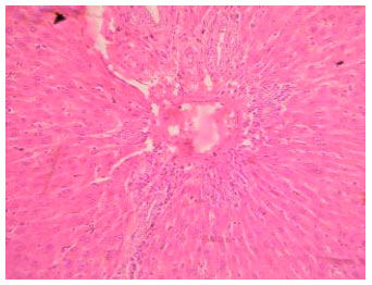 | |
| Fig. 6: | Section of liver from group-IV rat showing massive bile duct hyperplasia around central lobular vein. H and E stain 200X |
 | |
| Fig. 7: | Section of lung from group-IV rat showing congestion and round cell infiltration. H and E stain 200X |
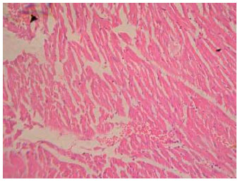 | |
| Fig. 8: | Section of heart from group-III rat showing myocardial hemorrhages. H and E stain 200X |
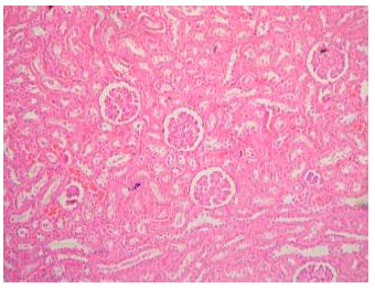 | |
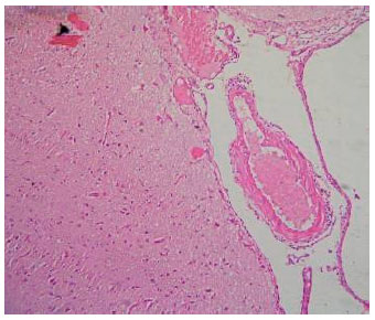
| Fig. 9: | Section of kidney from group-II rat showing severe venous congestion. H and E stain 100X |
Heart: Sections from group-II showed normal myocardial parenchyma and muscles fibres indicating least myocardial damage in lowest dose of paraquat. Sections from group-III rats revealed venous congestion, vacuolar changes, hemorrhages and mild degenerative changes in the myocardium (Fig. 8). Group-IV rats revealed hemorrhages and severe congestion.
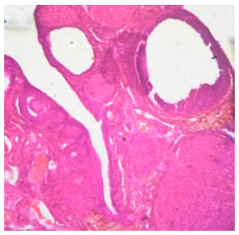
Kidneys: Sections of kidney from group-II rats revealed congestion, focal hemorrhages, desquamation of tubular epithelium in lumen, decrease in glomerular size and tubular nephrosis (Fig. 9). Sections from group-III and IV rats showed severe congestion, decreased in size of the glomeruli, hemorrhages, granular and degenerative changes in the tubular epithelium and desquamation in the lumen. Dilated tubules, cloudy swelling, vacuolar changes and necrosis of tubular epithelium results in occlusion of the lumen were noticed (Fig. 10 and 11).
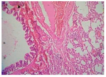
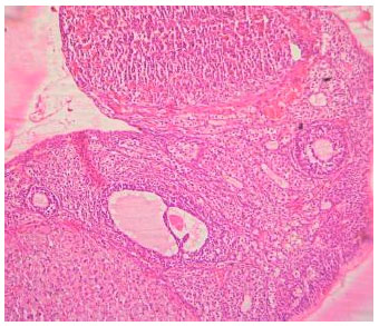
Spleen, intestine, brain and ovary: Sections from spleen of all treatment groups revealed decreased lymphoid population in the parenchyma and prominent stroma (Fig. 12). Sections from intestine of all treatment groups showed distortion and desquamation of epithelium in the lumen of intestine (Fig. 13).
 | |
| Fig. 10: | Section of kidney from group-III rat showing tubular nephrosis and venous congestion. H and E stain 100X |
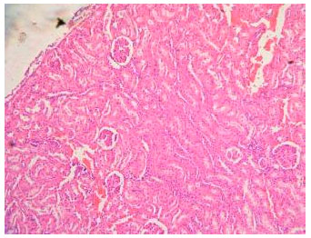 | |
| Fig. 11: | Section of kidney from group-IV rat showing congestion, hemorrhages, tubular nephrosis and decrease in glomerular size. H and E stain 100X |
 | |
| Fig. 12: | Section of spleen from group-IV rat showing decreased lymphoid population. H and E stain 100X |
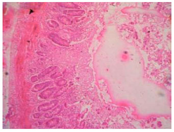 | |
| Fig. 13: | Section of intestine from group-III rat showing desquamation of intestinal mucosa. H and E stain 100X |
 | |
| Fig. 14: | Section of brain from group-IV rat showing meningeal congestion. H and E stain 100X |
Sections from brain of all treatment groups revealed meningeal congestion and edema (Fig. 14 and 15). Sections from ovary of all treatment groups showed degenerating follicles and corpus luteum occupying most of the parenchyma (Fig. 16 and 17).
DISCUSSION
Paraquat is a non-selective contact herbicide and its properties were discovered in 1955, registered as herbicide in 1962 by ICI laboratories. It is easily accessible to farmers in developing countries (Wesseling et al., 2001). Herbicide paraquat is often reported as one the agents that may be used for suicidal attempt, in case of humans the accidental or suicidal intake of paraquat result toxic organ affects with the dose of 4 mg kg-1 b.wt. It is highly toxic to humans and animals, accumulating mainly in the lungs, liver, kidneys and heart. This herbicide has been widely used as an experimental model to study lung injury, especially diffuse alveolar damage, due to its low cost, rapid effect and simplicity of administration (Silva and Saldiva, 1998; Rocco et al., 2003).
 | |
| Fig. 15: | Section of brain from group-IV rat showing cerebral edema. H and E stain 100X |
 | |
| Fig. 16: | Section of ovary from group-I rat showing normal developing follicles. H and E stain 100X |
In the present study, paraquat treated animals showed decreased food intake with polydipsia, weakness, lethargy with reduction in locomotional activities and mild diarrhea when compared with control animals in all treatment groups by the end of 3rd week of the experiment. Groups-IV rats were showed increase rate of respiration and shortness of breath. Cyanosis of visible mucous membrane and dehydration were noticed during the last week of experiment in most of the treatment group animals. These findings were similar to the observations of Gil et al. (2007) who reported anorexia, cyanosis of mucous membrane reported by Dinis-Oliveira et al. (2008), shortness of breath and diarrhea reported by Dinis-Oliveira et al. (2008) and reduction of locomotional activities reported by Chanyachukul et al. (2004) and Prasad et al. (2009). The lower feed consumption could be possibly due to direct adverse effect of paraquat on gastrointestinal tract, which is corrosive and strong irritant to the gastric mucosa and may lead to gastric ulceration and gastritis reported by Garg (2002) and Debe et al. (2007).
 | |
| Fig. 17: | Section of ovary from group-IV rat showing congestion and degenerating follicles. H and E stain 100X |
The observation of decreased weekly body weight in paraquat treatment groups can be correlated with the lower feed intake due to adverse effect of paraquat on gastrointestinal tract. These findings were similar to the observations of Yoshida et al. (1982), Podprasart et al. (2007) and Dinis-Oliveira et al. (2008) who reported rapid and significant weight loss in upto 30% of paraquat treated animals.
Blood is assumed to be the central compartment. Hawksworth et al. (1981) reported a three-compartment model of paraquat distribution suggesting that paraquat toxicity may leads to variable changes in hematological parameters. The average mean value of erythrocyte count, hemoglobin and packed cell volume was significantly reduced in paraquat treatment groups when compared to control group. This finding demonstrates the possibility of paraquat to induce anemia. The erythrocyte indices values define the size and hemoglobin content of erythrocyte. The average MCV, MCH and MCHC values in different treatment groups revealed non-significant decrease when compared with control group. The average total leucocyte count showed significant decreased in paraquat treatment group when compared with control group. The mean average absolute leukocyte count in different treatment groups showed non-significant differences when compared to control group. These results suggested that paraquat has toxic effect on the hemopoietic system. These findings were similar to the observations of Toyoshima et al. (1982), Vuksa et al. (2004) and Akinloye et al. (2011) who reported the reduction in RBC, hemoglobin, PCV, TLC and absolute leucocyte count value in rats due to paraquat toxicity and (Tamuli et al., 2004, 2007) in goat and calves due to hemolysis through lipid peroxidation due to generation of reactive oxygen species. Nagao et al. (1994) reported that paraquat accumulates in the immune and haematopoietic systems (bone marrow) and in various types of blood cells (granulocyte, erythrocyte and megakaryocyte), which may probably the reasons for decreased in leukocyte count indicating adverse effects on leukopoiesis.
Yoshida et al. (1982) reported slight reduction in MCV, MCH and MCHC during paraquat poisoning in male mouse for 13 and 104 weeks study, respectively, at a dose of 35.8 mg kg-1 b.wt. Therefore, it is concluded that the 4 weeks study of paraquat causes only minor changes in MCV possibly due to short duration of experiment.
The average values of plasma total protein, albumin and globulin were found to be significantly decreased in paraquat treated groups-III and IV rats when compared with control group-I and treated group-II. Decreased total protein levels in paraquat toxicity may be due to free radical-mediated membrane damage (Dinis-Oliveira et al., 2008; Mohammadi-Bardbori and Ghazi-Khansari, 2008). The electrophilic free radicals derived from the redox cycling of PQ are capable of abstracting allylic hydrogen atoms from membrane-associated polyunsaturated fatty acids (PUFAs). This results in alterations of membrane structure, ultimately leads to lipid peroxidation (Eisler, 1990; Garg, 2002). Jaiswal et al. (1992) reported the ability of paraquat binding to plasma albumin, thus inducing conformational changes and fragmentation in protein structure. All these mechanisms enhance the understanding of paraquat action on protein degeneration, thus decreasing the protein levels. These findings were similar to the observations of Toyoshima et al. (1982), Wershana (2001) and Podprasart et al. (2007) who reported decreased levels of total protein in paraquat poisoning. Attia and Nasr (2009) and Yoshida et al. (1982) reported reduction in plasma albumin and globulin levels during paraquat toxication in rats.
The mean average plasma AST, ALT and creatinine levels in different treated groups showed significant increase when compared with control animals. These findings were similar to the observations of Hong et al. (2000), Dere and Polat (2001), Vuksa et al. (2004), Attia and Nasr (2009), Samai et al. (2010) and Akinloye et al. (2011). They reported increased ALT, AST and creatinine levels, suggesting possible hepato-renal damage and liver, lung and kidney are the principal target organs for paraquat toxicity.
Gross pathological observation of most of the paraquat treatment group rats showed debilated and pale carcasses. Lungs and kidneys from all treatment group rats showed mild congestion and emphysema. Round flabby heart with pin-point hemorrhages, congestion of liver, spleen and intestinal mucosa were also observed in treated groups. These findings were similar to the observations of Soloukides et al. (2007) and Tamuli et al. (2007) who reported congestion, haemorrhages and emphysema in lungs and spleen.
The weights of organs were recorded to evaluate effect of paraquat toxicity on internal organs. Liver, kidneys, spleen and brain weights in different group rats showed non significant decrease in weight in a dose dependent manner. The mean average weight of lungs and heart in different treatment group revealed non-significant increase in weight when compared with the control group. These findings were similar to the observations of Toyoshima et al. (1982), Florkowski et al. (1992) and Nagao et al. (1994). They reported a decrease in liver, kidney, spleen and brain weight in rats treated with paraquat. Houze et al. (1990) reported an increase in lungs and heart weight during paraquat toxicity in rats and beagle dog.
Histopathological examination is the basis of toxicopathology and is helpful in evaluating adverse effects of toxic substances on morphological alterations at cellular level. Sections from liver revealed congestion, granular and prominent vacuolar changes in the parenchyma of liver. Focal areas of necrosis around central vein and periportal hyperplasia of bile ducts were noticed. Prominent nuclei with granular cytoplasm of hepatocytes and hepatic lobules showed dilated sinusoids with stagnated erythrocytes and reticular cells hyperplasia. Lungs revealed prominent peribronchiolar round cell infiltration, severe congestion, edema and connective tissue proliferation. Sections from heart revealed venous congestion, vacuolar changes, hemorrhages and mild degenerative changes in the myocardium. Spleen revealed decreased lymphoid population and intestine showed distortion and desquamation of epithelium. Brain sections revealed mild venous congestion and edema. These findings were similar to the observations of Akinloye et al. (2011) who reported centrilobular hepatocellular necrosis and proliferation of the bile duct. Lung is the organ mainly affected in paraquat poisoning, largely due to the preferential accumulation of paraquat in lung alveolar cells. Liu et al. (2011), Gil et al. (2007), Gocgeldi et al. (2008) and Lacerda et al. (2009) reported pulmonary edema, infiltration of inflammatory cells and peribronchiolar round cell infiltration. The morphological alterations in heart could be possibly due to hypoxic injury to the myocardium as a result of pulmonary edema and toxic effects of paraquat. Tamuli et al. (2004, 2007) reported the congestion, focal hemorrhages, focal areas of myocardial necrosis and mononuclear cell infiltration in between the myocardial fibres.
In the present study, grossly, hydronephrosis, atrophy of left kidney and hypertrophy of right kidney were observed in paraquat treated groups. Histopathological examination of kidney revealed congestion, focal hemorrhages, desquamation of tubular epithelium in lumen, decreased in glomerular size and tubular nephrosis. Florkowski et al. (1992) reported that paraquat is mainly eliminated through kidneys and acute renal failure is a recognized as complications of paraquat poisoning with both oliguric and non-oliguric symptoms. These findings were similar to the observations of Gil et al. (2005), Lamfon and Al-Rawi (2007) and Zhang et al. (2011). They reported cytoplasmic vacuolation inrenal tubular epithelial cells, glomerular degeneration, congested renal blood vessels and tubular necrosis.
Riahi et al. (2011) reported that the paraquat toxicity on spleen leads to decreased in immunity. Debe et al. (2007) reported that the mucosal ulceration and loss of villi due to intraperitoneal administration of paraquat in rat. Brain lesions were similar to the observations of Grant et al. (1980) and Florkowski et al. (1992). They reported the cerebral edema and hemorrhages due to damaging effect of paraquat on the cerebral blood vessels. Ovarian lesions were similar to the observations of Atkhah et al. (2012) who studied the effects of paraquat on oogenesis and ovary structure in Wistar rats.
ACKNOWLEDGMENT
The authors are very grateful to the staff of PGIVAS, Akola and ICAR, New Delhi for the financial assistance for research programme to the first author.
REFERENCES
- Akinloye, O.A., I. Adamson, O. Ademuyiwa and T.A. Arowolo, 2011. Supplementation of vitamins C, E and its combination on paraquat-intoxicated rats: Effects on some biochemical and markers of oxidative stress parameters. J. Applied Pharm. Sci., 1: 85-91.
Direct Link - Lacerda, A.C., G. Rodrigues-Machado Mda, P.L. Mendes, R.D. Novaes and G.M. Carvalho et al., 2009. Paraquat (PQ)-induced pulmonary fibrosis increases exercise metabolic cost, reducing aerobic performance in rats. J. Toxicol. Sci., 34: 671-679.
PubMed - Atkhah, V.H., M. Najafiyan, M. Farzam and H. Kargar, 2012. Effects of the paraquat herbicide on oogenesis and ovary structure of Wistar rat strain. Adv. Environ. Biol., 6: 1006-1012.
Direct Link - Attia, A.M. and H.M. Nasr, 2009. Evaluation of protective effect of omega- 3 fatty acids and selenium on paraquat intoxicated rats. Slovak J. Anim. Sci., 42: 180-187.
Direct Link - Baharuddin, M.R.B., I.B. Sahid, M.A.B.M. Noora, N. Sulaiman and F. Othman, 2011. Pesticide risk assessment: A study on inhalation and dermal exposure to 2,4-D and paraquat among Malaysian paddy farmers. J. Environ. Sci. Health B., 46: 600-607.
Direct Link - Bataller, R., E. Bragulat, S. Nogue, M.N. Gorbig, M. Bruguera and J. Rodes, 2000. Prolonged cholestasis after acute paraquat poisoning through skin absorption cholestasis due to paraquat. Am. J. Gastroenterol., 95: 1340-1343.
CrossRefDirect Link - Bismuth, C., R. Garnier, F.J. Baud, J. Muszynski and C. Keyes, 1990. Paraquat poisoning. Drug Saf., 5: 243-251.
CrossRef - Bismuth, C., R. Garnier, S. Dally, P.E. Fournier and J.M. Scherrmann, 1982. Prognosis and treatment of paraquat poisoning: A review of 28 cases. J. Toxicol. Clin. Toxicol., 19: 461-474.
CrossRef - Chanyachukul, T., K. Yoovathaworn, W. Thongsaard, S. Chongthammakun, P. Navasumrit and J. Satayavivad, 2004. Attenuation of paraquat-induced motor behavior and neurochemical disturbances by l-valine in vivo. Toxicol. Lett., 150: 259-269.
CrossRef - Dawson, A.H., M. Eddleston, L. Senarathna, F. Mohamed and I. Gawarammana et al., 2010. Acute human lethal toxicity of agricultural pesticides: A prospective cohort study. PLoS med., Vol. 7.
CrossRef - Debe, E.B., B.N. Okolonkwo and A.A. Ngokere, 2007. Toxicological effects of paraquat on the histology of the stomach, small intestine and testis of male albino rat (Rattus norvegicus). Port Harcourt Med. J., 2: 51-55.
Direct Link - Dere, E. and F. Polat, 2001. The effect of paraquat on the activity of some enzymes in different tissues of mice (Mus musculus-Swiss albino). Turk. J. Biol., 25: 323-332.
Direct Link - Dinis-Oliveira, R.J., J.A. Duarte, A. Sanchez-Navarro, F. Remiao, M.L. Bastos and F. Carvalho, 2008. Paraquat poisonings: Mechanisms of lung toxicity, clinical features and treatment. Crit. Rev. Toxicol., 38: 13-71.
PubMedDirect Link - Florkowski, C.M., S.M. Bradberry, G.W. Ching and A.F. Jones, 1992. Acute renal failure in a case of paraquat poisoning with relative absence of pulmonary toxicity. Postgrad. Med. J., 68: 660-662.
Direct Link - Freebairn, D.K., 1995. Did the green revolution concentrate incomes? A quantitative study of research reports. World Dev., 23: 265-279.
CrossRefDirect Link - Gil, H.W., J.O. Yang, E.Y. Lee and S.Y. Hong, 2005. Paraquat-induced fanconi syndrome. Nephrology, 10: 430-432.
CrossRef - Gil, H.W., M.H. Oh, K.M. Woo, E.Y. Lee, M.H. Oh and S.Y. Hong, 2007. Relationship between pulmonary surfactant protein and lipid peroxidation in lung injury due to paraquat intoxication in rats. Korean J. Int. Med., 22: 67-72.
CrossRefDirect Link - Gocgeldi, E., B. Uysal, A. Korkmaz, R. Ogur and R.J. Reiter et al., 2008. Establishing the use of melatonin as an adjuvant therapeutic against Paraquat-induced lung toxicity in rats. Exp. Biol. Med., 233: 1133-1141.
CrossRefDirect Link - Grant, H., P.L. Lantos and C. Parkinson, 1980. Cerebral damage in paraquat poisoning. Histopathology, 4: 185-195.
CrossRefPubMedDirect Link - Gunnell, D., M. Eddleston, M.R. Phillips and F. Konradsen, 2007. The global distribution of fatal pesticide self-poisoning: Systematic review. BMC Public Health, Vol. 7.
CrossRef - Hawksworth, G.M., P.N. Bennett and S. Davies, 1981. Kinetics of paraquat elimination in the dog. Toxicol. Applied Pharmacol., 57: 139-145.
CrossRef - Hong, S.Y., D.H. Yang and K.Y. Hwang, 2000. Associations between laboratory parameters and outcome of paraquat poisoning. Toxicol. Lett., 118: 53-59.
CrossRef - Houze, P., F.J. Baud, R. Mouy, C. Bismuth, R. Bourdon and J.M. Scherrmann, 1990. Toxicokinetics of paraquat in humans. Hum. Exp. Toxicol., 9: 5-12.
CrossRef - Wade, N., 1974. Green revolution (II): Problems of adapting a Western technology. Science, 186: 1186-1192.
PubMed - Jaiswal, R., M.A. Khan and J. Musarrat, 1992. Photosensitized paraquat-induced structural alterations and free radical mediated fragmentation of serum albumin. J. Photochem. Photobiol. B., 67: 163-170.
PubMed - Kelly, D.F., D.G. Morgan, P.G.G. Darke, C. Gibbs, H. Pearson and B.M.Q. Weaver, 1978. Pathology of acute respiratory distress in the dog associated with paraquat poisoning. J. Comput. Pathol., 88: 275-294.
CrossRefPubMedDirect Link - Lamfon, H.A. and M.M. Al-Rawi, 2007. Effect of antox on paraquat-Induced histological and biochemical changes in kidney of albino rats. J. Applied Sci. Res., 3: 988-993.
Direct Link - Liu, S., K. Liu., Q. Sun, W. Liu and W. Xu et al., 2011. Consumption of hydrogen water reduces paraquat-induced acute lung injury in rats. J. Biomed. Biotechnol., Vol. 2011.
CrossRefDirect Link - Mohammadi-Bardbori, A. and M. Ghazi-Khansari, 2008. Alter-native electron acceptors: Proposed mechanism of paraquat mitochondrial toxicity. Environ. Toxicol. Pharmacol., 26: 1-5.
CrossRef - Nagao, M., W.D. Zhang, T. Takatori, Y. Itakura and Y. Yamada et al., 1994. Identification and dynamics of paraquat in the bone marrow, thymus and spleen in rats using immunohistochemical techniques. Nihon Hoigaku Zasshi., 48: 166-168.
PubMed - Parayil, G., 1992. The green revolution in India: A case study of technological change. Technol. Cult., 33: 737-756.
Direct Link - Podprasart, V., J. Satayavivad, S. Riengrojpitak, P. Wilairat and W. Wananukul et al., 2007. No direct hepatotoxic potential following a multiple-low dose paraquat exposure in rat as related to its bioaccumulation. Toxicol. Lett., 170: 193-202.
CrossRefDirect Link - Prasad, K., E. Tarasewicz, J. Mathew, P.A.O. Strickland, B. Buckley, J.R. Richardson and E.K. Richfield, 2009. Toxicokinetics and toxicodynamics of paraquat accumulation in mouse brain. Exp. Neurol., 215: 358-367.
CrossRef - Raghu, K., V. Mahesh, P. Sasidhar, P.R. Reddy, V. Venkataramaniah and A. Agrawal, 2013. Paraquat poisoning: A case report and review of literature. J. Family Community Med., 20: 198-200.
CrossRefDirect Link - Riahi, B., H. Rafatpanah, M. Mahmoudi, B. Memar, A. Fakhr, N. Tabasi and G. Karimi, 2011. Evaluation of suppressive effects of paraquat on innate immunity in Balb/c mice. J. Immunotoxicol., 8: 39-45.
CrossRefDirect Link - Rocco, P.R.M., A.B. Souza, D.S. Faffe, C.P. Passaro and F.B. Santos et al., 2003. Effect of corticosteroid on lung parenchyma remodeling at an early phase of acute lung injury. Am. J. Respir. Crit. Care Med., 168: 677-684.
CrossRef - Sharp, C.W., A. Ottolenghi and H.S. Posner, 1972. Correlation of paraquat toxicity with tissue concentrations and weight loss of the rat. Toxicol. Applied Pharmacol., 22: 241-251.
CrossRef - Silva, M.F.R. and P.H.N. Saldiva, 1998. Paraquat poisoning: An experimental model of dose-dependent acute lung injury due to surfactant dysfunction. Braz. J. Med. Biol. Res., 31: 445-450.
CrossRefDirect Link - Snedecor, G.W. and W.G. Cochran, 1980. Statistical Methods. 7th Edn., Iowa State University Press, Iowa, USA., ISBN-10: 0813815606, Pages: 507.
Direct Link - Soloukides, A., D.A. Moutzouris, T. Kassimatis, G. Metaxatos and V. Hadjiconstantinou, 2007. A fatal case of paraquat poisoning following minimal dermal exposure. Ren Fail., 29: 375-377.
CrossRefDirect Link - Tamuli, S.M., G.K. Baruah and M.K. Tamuli, 2007. Pathology of chronic paraquat toxicity in calves. Indian J. Vet. Pathol., 31: 130-134.
Direct Link - Vendan, S.E., 2011. Endosulfan ban in India: For good or not? Curr. Sci., 101: 1398-1398.
Direct Link - Vuksa, M., N. Neskovic, S. Vitorovic and K. Vesela, 2004. Subacute toxicity of paraquat in rats-biochemical effects. Ecotoxicol. Environ. Safety, 7: 475-483.
CrossRefDirect Link - Wershana, K.Z., 2001. The Influence of vitamin C or selenium on paraquat-induced toxicity in guinea pigs. Pak. J. Biol. Sci., 4: 81-88.
CrossRefDirect Link - Wesseling, C., B. van Wendel de Joode, C. Ruepert, C. Leon, P. Monge, H. Hermosillo and T.J. Partanen, 2001. Paraquat in developing countries. Int. J. Occup. Environ. Health, 7: 275-286.
PubMed - WHO, 2010. The WHO Recommended Classification of Pesticides by Hazard and Guidelines to Classification 2009. World Health Organization, Geneva, Switzerland, ISBN-13: 9789241547963, Pages: 78.
Direct Link - Zhang, Z.J., C.Y. Zhou, Y.J. Luo, L.B. Peng and J. Xia, 2011. Pathologic changes and expression of caspase-3 in kidneys of paraquat poisoned rats. Chi. J. Ind. Med., 24: 3-10.
Direct Link