
T.R. Arun
Indian Veterinary Research Institute, Izatnagar, Bareilly, Uttar Pradesh, 243122, India
R. Rana
Indian Veterinary Research Institute, Izatnagar, Bareilly, Uttar Pradesh, 243122, India
P. Singh
P. Choudhuri
Indian Veterinary Research Institute, Izatnagar, Bareilly, Uttar Pradesh, 243122, India
V.P. Singh
Indian Veterinary Research Institute, Izatnagar, Bareilly, Uttar Pradesh, 243122, India
P. Thomas
Indian Veterinary Research Institute, Izatnagar, Bareilly, Uttar Pradesh, 243122, India
V. Rekha
Indian Veterinary Research Institute, Izatnagar, Bareilly, Uttar Pradesh, 243122, IndiaIndian Veterinary Research Institute, Izatnagar, Bareilly, Uttar Pradesh, 243122, India
K. Nehra
Indian Veterinary Research Institute, Izatnagar, Bareilly, Uttar Pradesh, 243122, India
J. Usharani
Indian Veterinary Research Institute, Izatnagar, Bareilly, Uttar Pradesh, 243122, India
K. Dhama
Indian Veterinary Research Institute, Izatnagar, Bareilly, Uttar Pradesh, 243122, India
Asian Journal of Animal and Veterinary Advances
Year: 2014 | Volume: 9 | Issue: 7 | Page No.: 405-413


ABSTRACT
A gold nanoparticle based lateral flow assay was developed for rapid serodiagnosis of contagious agalactia, an economically important mycoplasmal disease of small ruminants. Sonicated antigen of Mycoplasma agalactiae was used as the test reagent that was immobilized on nitrocellulose membrane along with the control line of goat IgG. The detection reagent, gold nanoparticle conjugated with anti-goat antibody was dried on the conjugate pad. During the assay, specific antibodies against M. agalactiae in the test serum that combined with the detection reagent were captured in the test line and detected visually by the development of a red line on nitrocellulose membrane. The gold conjugate captured in control line produced a red line regardless of the presence of specific antibodies that served as a procedural control. Serum samples collected from an experimentally infected goat were tested with the lateral flow assay and antibodies were detected from 9th day of infection and the assay was also evaluated using 100 goat sera samples. This is the first report regarding development of a gold nanoparticle based lateral flow assay for rapid diagnosis of contagious agalactia in goats. This study suggests that current lateral flow assay can be used as a user friendly diagnostic in laboratories lacking specialized equipments as well as for point of care diagnosis of contagious agalactia.
PDF Abstract XML References Citation
Received: January 12, 2014;
Accepted: April 17, 2014;
Published: June 11, 2014
How to cite this article
T.R. Arun, R. Rana, P. Singh, P. Choudhuri, V.P. Singh, P. Thomas, V. Rekha, K. Nehra, J. Usharani and K. Dhama, 2014. Development of a Gold Nanoparticle Based Lateral Flow Assay for Rapid Diagnosis of Contagious Agalactia in Goats. Asian Journal of Animal and Veterinary Advances, 9: 405-413.
DOI: 10.3923/ajava.2014.405.413
URL: https://scialert.net/abstract/?doi=ajava.2014.405.413
DOI: 10.3923/ajava.2014.405.413
URL: https://scialert.net/abstract/?doi=ajava.2014.405.413
INTRODUCTION
Contagious agalactia is an economically important disease of small ruminants which cause mastitis, agalactia, arthritis, keratoconjunctivitis, pneumonia and neonatal mortality (Bergonier et al., 1997). The classical etiological agent is Mycoplasma agalactiae although some other mycoplasmas also produce similar type of disease conditions (Gomez-Martin et al., 2013). The disease has worldwide prevalence, being enzootic in the Mediterranean area, Africa, USA and in some Asian countries (Campos et al., 2009; Kumar et al., 2014). Gomez-Martin et al. (2013) conducted a detailed study screening of 300 bulk tank milk and 381 milk samples from goats with clinical mastitis in Spain. The presence of mycoplasmas was detected in 66.7% of the herds and M. agalactiae was identified in 95.45% of these positives herds. The disease been reported from many Indian states (Vihan, 1989; Srivastava et al., 1996; Mondal et al., 2004; Kumar et al., 2009) but the prevalence of disease is largely under estimated since a very few laboratories are routinely working on mycoplasmas and due to lack of a point of care diagnostic.
The laboratory diagnosis of contagious agalactia is mainly based on the isolation and identification of M. agalactiae from clinical samples. The isolation procedure requires specialized technical personnel, high investment and long time for conclusion. The diagnosis can also be done by detection of M. agalactiae antibodies in serum by serological techniques such as Serum Agglutination Test (SAT), Indirect Haemagglutination Assay (IHA), Complement Fixation Test (CFT), Indirect Fluorescent Antibody Test (IFT), Enzyme Linked Immunosorbant Assay (ELISA) and immunoblotting (Bergonier et al., 1997; Poveda and Nicholas, 1998; Kumar et al., 2014). The Office International des Epizooties (OIE) recommends western blotting, complement fixation and ELISA tests as diagnostic tests (World Organisation for Animal Health, 2008). The CFT is limited in sensitivity due to false positives and cross reactions in comparison to other diagnostic methods. For this reason, ELISA tests have been used as reliable diagnostics for contagious agalactia (Lambert et al., 1998; Pepin et al., 2003; Kittelberger et al., 2006; Fusco et al., 2007). An indirect ELISA utilizing total antigen (ELISA-Gt) and sonicated antigen (ELISA-Gs) of M. agalactiae was standardized for the detection of M. agalactiae antibodies in goats (Campos et al., 2009). Relative sensitivity of the ELISA-Gt and ELISA-Gs was 77.27 and 88.63%, respectively, while both had specificity of 95.24%. The results of this study were similar to those recorded by Fusco et al. (2007) using a recombinant antigen based ELISA.
A rapid pen-side test would help field veterinarians in the early diagnosis of contagious agalactia in suspect animals. Gold nanoparticle based lateral-flow assays have been developed recently for rapid serodiagnosis of many bacterial diseases like anthrax, leptospirosis, brucellosis, tuberculosis, scrub typhus etc. (Ching et al., 2001; Smits et al., 2001; Smits et al., 2003; Lyashchenko et al., 2007). It has several advantages over traditional serological tests, such as the simplicity of the procedure, low cost and ease of use in the field. At present, rapid serodiagnostic test for contagious agalactia is not available in India. Therefore, the aim of the present study was to develop a novel gold nanoparticle based lateral flow assay platform for rapid diagnosis of contagious agalactia in goats.
MATERIALS AND METHODS
Growth and characterization of M. agalactia isolates: Indian isolate VP20-L15/02 and standard strain of M. agalactiae (NCTC 10123), obtained from National Referral Laboratory on Mycoplasma, Division of Bacteriology and Mycology, Indian Veterinary Research Institute, Izatnagar (U.P), were employed in the study. The bacterial growth was accomplished in solid and liquid modified pleuropneumonia like organism (PPLO) medium (pH 7.6) supplemented with 15% horse serum. The genomic DNA of isolates was extracted as per the protocol of Wilson (1987) with a slight modification. Mycoplasma genus specific PCR was performed employing the primers based on 16S rRNA gene (Van Kuppeveld et al., 1992) and species specific PCR was performed employing the primers specific for M. agalactiae (Tola et al., 1996). The primer sequences used and the expected product sizes are presented in Table 1.
Antigen preparation: Sonicated antigen of standard strain of M. agalactiae was prepared according to the method of Bhanuprakash and Srivastava (1996). The cell pellet obtained from 1 L M. agalactiae culture was resuspended in 3 mL sterile Phosphate Buffered Saline (PBS) and treated ultra sonically at 15 amp for 15 sec with 15 sec break for 10 cycles (MSE-soniprep 150) for production of sonicated antigen.
| Table 1: | Details of PCR primers used |
 | |
Protein concentration of antigen was estimated by Bradford method using Bovine Serum Albumin (BSA) as standard (Bradford, 1976) and 0.01 M PBS (pH 7.4) was added to make the final protein concentration to 2 mg mL-1.
Synthesis of gold nanoparticles: Colloidal gold nanoparticles (GNPs) were synthesized by citrate reduction method (Frens, 1973). An aqueous chloroauric acid solution (100 mL of 0.01% HAuCl4, Sigma) was boiled with vigorous stirring, followed by the rapid addition of 2 mL of 1% sodium citrate to the solution. Boiling was continued for 10 min, the heat source was then removed and stirring was continued for an additional 15 min. After the solution reached at Room Temperature (RT), colloidal gold was supplemented with 0.02% (w/v) sodium azide and stored at 4°C. The absorption spectrum of synthesized gold nanoparticles at λ 350-700 was checked using an UV-visible spectrophotometer.
Salt agglomeration test for determining the minimal protective amount: Minimal Protective Amount (MPA) is the minimum amount of protein required to protect the gold nanoparticles against salt agglomeration and pH changes during the experiment procedures. For determining the minimal protective amount of anti-goat IgG (2.5 mg mL-1 Sigma-Aldrich) for conjugation, 1, 2.5, 5, 10, 20 and 40 μg of anti-goat IgG was added to 1 mL of gold nanoparticles (GNPs) (pH adjusted to 8.5). After incubation for 20-30 min at room temperature in a shaker, 100 μL of 10% NaCl in distilled water was added to each tube and incubated for 10 min at room temperature. All the tubes were observed for any change in color from red to blue due to agglomeration of GNPs.
Conjugation of anti goat IgG with gold nanoparticles: Conjugation of anti goat IgG with gold nanoparticles was performed in 20 mM Tris buffer (pH 8.5) as described by Thobhani et al. (2010). The pH value of the colloidal gold solution (1%, w/v) was adjusted to 8.5 by addition of 0.02 M K2CO3. The 10 μL of anti-goat antibody (2.5 mg mL-1 Sigma) was added to 1 mL pH adjusted colloidal gold solution. The mixture was gently mixed for 2 h in a shaker and then centrifuged at 12,000xg at 4°C for 30 min to remove unconjugated antibodies. After centrifugation, the colloidal gold pellets were redispersed in 1 mL 20 mM Tris buffer, pH 8.5, containing 2% Bovine Serum Albumin (BSA) and gently mixed for 1 h in a shaker to block any uncoated sites on the gold colloids and centrifuged at 12,000xg at 4°C for 30 min to remove unconjugated BSA. The pellets were then redispersed in 0.01 M PBS (pH adjusted to 7.4) that contained 0.01% BSA and 0.02% sodium azide. The anti goat IgG coated colloidal gold nanoparticles were ready for use without further preparation and were stored at 4°C.
Spotting of test and control lines on the nitrocellulose membrane: Goat IgG (Sigma) at a concentration of 1 mg mL-1 was dispensed as control line and sonicated antigen at a concentration of 2 mg mL-1 were spotted on the nitrocellulose (NC) membrane as a test line. Approximately 2 μL cm-1 of protein was applied on the NC membrane using an Easy printer. Membranes were dried at room temperature, blocked with 1% (w/v) BSA in 0.01 M PBS for 30 min and washed three times with 0.01 M PBS for 5 min, dried at RT and stored at 4°C.
Preparation of lateral flow device: The sample pad, conjugate pad, NC membrane and absorption pad (MDI, India) were assembled on the support board sequentially with a 1-2 mm overlap. The assembled laminates were then cut into 5 mm wide pieces and 2.5 μL of anti goat antibody-gold nanoparticle conjugate was applied onto the conjugate matrix. The lateral flow devices were dried at room temperature and sealed in a plastic bag and stored at 4°C.
Lateral flow assay procedure: The serum samples were diluted 1:50 with 0.01 M PBS and 100 μL was applied on the sample pad. The assembly was placed horizontally for 5-10 min to observe the result. For standardization of the assay, hyper immune serum raised in goat and goat serum purchased from Sigma-Aldrich were used as the known positive and negative samples, respectively. Serum samples collected from an experimentally infected adult goat at regular intervals and 100 goat serum samples obtained from National Referral Laboratory on Mycoplasma, IVRI were tested using the lateral flow assay.
RESULTS
Indian isolate VP20-L15/02 and standard strain of M. agalactiae were confirmed by biochemical tests and growth inhibition test. Further confirmation was done by genus specific Polymerase Chain Reaction (PCR) that amplified fragment of 715 bp and species specific PCR produced an amplicon size of 375 bp (Fig. 1). Sonicated antigen was prepared from standard and Indian strains of M. agalactiae and protein concentration of antigen was adjusted to 2 mg mL-1 using 0.01 M PBS.
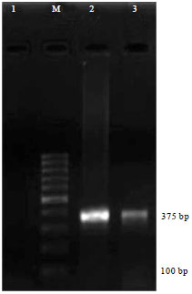 | |
| Fig. 1: | Mycoplasma agalactiae species specific PCR. Lane 1: No. template control, Lane M: 100 bp ladder, Lane 2: Mycoplasma agalactiae standard strain NCTC 10123, Lane 3: Indian isolate VP20-L15/02 |
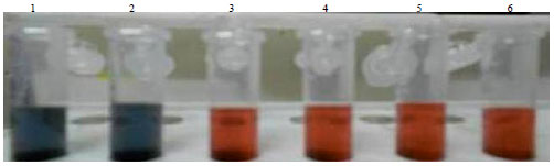 | |
| Fig. 2: | Salt agglomeration test results determining the minimal protective amount of anti-goat IgG. Concentrations of anti-goat IgG added in 1 mL of gold nanoparticles, tubes No. (1) 1, (2) 2.5, (3) 5, (4) 10, (5) 20 and (6) 40 μg. Tube 1 and 2 showed salt agglomeration, while tubes 3 to 6 revealed no salt agglomeration |
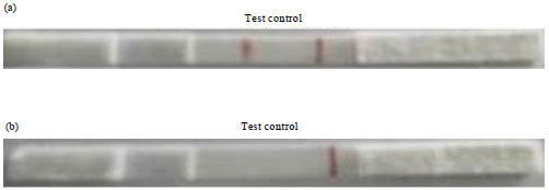 | |
| Fig. 3(a-b): | Lateral flow assay with Mycoplasma agalactiae sonicated antigen and goat IgG on the test and control lines, respectively. (a) Positive serum showed red colored lines on both test and control zones, whereas (b) Negative serum showed red colored line only on control zone |
Colloidal gold nanoparticles were prepared from an aqueous chloroauric acid solution (0.01%) by citrate reduction method (Frens, 1973). Salt agglomeration test was performed for determining the minimal protective amount and 10 μg of protein antigen mL was found to be protecting GNPs from salt agglomeration (Fig. 2). The absorption spectra of synthesized gold nanoparticles at λ 350-700 was checked using an UV-visible spectrophotometer and a peak was observed at 525 nm in accordance with the earlier workers who used the similar method for synthesis of gold nanoparticles of 40 nm size (Jiang et al., 2011). Further, the conjugation and blocking steps were also confirmed spectrophotometrically as increase in the absorbance of gold nanoparticles was observed after each step.
Test serum sample, diluted in 0.01 M PBS (1:50) was applied on the sample pad and assembly was placed horizontally for 5-10 min to observe the result. Both test and control lines appeared red when assay was conducted using hyper immune sera against standard strain of M. agalactiae and only the control line appeared red when the assay was performed using PBS and known negative sera (Fig. 3).
The lateral flow assay was standardized using the positive sera obtained from experimentally infected goat and known negative sera (goat sera purchased from Sigma Aldrich). Dilutions of the infected sera prepared in 0.01 M PBS (1:10, 1:20, 1:50, 1:100, 1:150, 1:200) were also tested and serum sample diluted up to 1:150 gave positive results in the assay. Serum samples collected from the experimentally infected goat at three days interval (day 3 to 45) were also tested with the lateral flow assay and antibodies were detected from 9th day of infection. The lateral flow assay was evaluated using 100 goat serum samples, wherein 9 samples were found to positive for antibodies against M. agalactiae that was in accordance with results obtained with serum agglutination test. The cross-reactivity of lateral flow assay strip was examined using hyper immune and infected serum for Mycoplasma mycoides ssp. capri, a Mycoplasma spp. prevalent in small ruminants in India and no cross reactivity was observed.
DISCUSSION
Contagious agalactia is a notifiable disease listed by World Organisation for Animal Health (WOAH/OIE) and has been responsible for causing severe economic losses to goat and sheep industries. The disease has been reported from India (Vihan, 1989; Srivastava et al., 1996; Mondal et al., 2004; Kumar et al., 2009, 2014) but the prevalence of disease is overlooked due to lack of a rapid field diagnostic test. The isolation and biochemical identification of the organism is more tedious and time consuming (Aluotto et al., 1970; Poveda, 1998). Serological techniques such IHA, CFT, IFT, ELISA and immunoblotting assays have been considered to be most suitable for screening the herds (Bergonier et al., 1997; Poveda and Nicholas, 1998; Kumar et al., 2014). Despite these advancements in diagnostic techniques, most of these methods have limitations, including bulky instrumentation, laborious sample preparation and slow data readout.
The lateral flow assays are user friendly diagnostics without the need for specific equipment, training or electricity. Moreover, the assay components are highly stable and devices can be stored for a prolonged time without the need for refrigeration (Posthuma-Trumpie et al., 2009). Recently, lateral-flow assays have been developed for rapid serodiagnosis of many bacterial diseases like anthrax, leptospirosis, brucellosis, tuberculosis, scrub typhus etc. (Ching et al., 2001; Smits et al., 2001, 2003; Lyashchenko et al., 2007). In the current study, a lateral flow assay platform was adapted for rapid detection of M. agalactiae as described by Smits et al. (2003) for serodiagnosis of human brucellosis, in which Brucella lipopolysaccharide (LPS) was used as the capture reagent and colloidal gold-conjugated anti-human IgG/IgM as the detection reagent. During the current lateral flow assay procedure, the sample (serum) is allowed to react with the colloidal gold-anti goat IgG conjugate. For a positive serum sample, the conjugate binds to the antibody forming a gold nanoparticle-anti-goat antibody-antibody complex that binds to antigen immobilized on test line and forms a red color. Absence of this test line suggests a negative result. The excess gold conjugate will continue to move by capillary action and encounter a control line composed of goat IgG. Function as a procedural control, a red line will always appear at the control zone as the gold conjugate binds to goat IgG regardless of the presence of specific antibodies against M. agalactiae (Fig. 4).
Gold nanoparticles of 40 nm size were prepared by citrate reduction method (Frens, 1973) that produced a peak at 525 nm in the absorption spectra in accordance with the previous workers who used the same method for synthesis of gold nanoparticles of similar size (Jiang et al., 2011). The red color was obvious and easy to distinguish which showed that the size of the synthesized gold nanoparticles was suitable for the assay. The minimal protective amount was determined to be 10 μg of anti goat IgG mL-1 by salt agglomeration test. Minimal Protective Amount (MPA) is the minimal amount of protein required to protect the gold nanoparticles against salt agglomeration and pH changes during the assay.
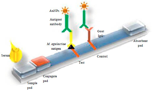 | |
| Fig. 4: | Pictorial representation of lateral flow assay format used. Test line: M. agalactiae sonicated antigen, control line: Goat IgG, detection reagent: Gold nanoparticle-anti goat IgG conjugate, test sample: Serum |
Gold colloid maintained the red color in the tubes that contain enough protein to stabilize the particles even in the presence of electrolytes, while those tubes which contain less protein, agglomeration takes place and the color changes from red to dark purple/black (Karthik et al., 2011). Even though the minimal protective amount was determined to be 10 μg, 2.5 times MPA of anti goat IgG, used for effective conjugation with gold nanoparticles.
Sonicated antigen prepared from standard and Indian strains was used as the capture probe in the assay. Recently, an indirect ELISA assay utilizing sonicated antigen (ELISA-Gs) of M. agalactiae was standardized for the serodiagnosis of contagious agalactia in goats (Campos et al., 2009). The sonicated antigen based ELISA showed sensitivity and specificity of 88.63 and 95.24%, respectively. The current lateral flow assay using sonicated antigen do not show any cross reactions with Mycoplasma mycoides ssp., capri a Mycoplasma spp., that produce symptoms similar to that produced by M. agalactia (Kumar et al., 2013). The assay could detect M. agalactiae antibodies in serum from 9th day of infection in accordance with results obtained by Fusco et al. (2007) using immunoblotting.
CONCLUSION
In conclusion, a gold nanoparticle based lateral flow assay was developed for the first time for rapid detection of contagious agalactia in goats which is an easy-to-perform diagnostic method. The lateral flow assay using sonicated antigen of M. agalctiae requires only 5-10 min of handling time to perform, does not require special equipment or electricity and can be performed by modestly trained personnel. Due to its robustness and simplicity, the test is highly suitable for application under field conditions. It can also be used as an important tool for the seroepidemiological screening of goats in small laboratory settings in developing countries which in turn would contribute significantly to the control of this economically important disease. Wider application of this novel test developed for rapid detection contagious agalactia in goats with screening of larger number of field serum samples is suggested. Besides this, evaluating the stability of antigen and gold conjugate used in the lateral flow device under different storage conditions would strengthen the field applicability of the developed test.
REFERENCES
- Aluotto, B.B., R.G. Wittler, C.O. Williams and J.E. Faber, 1970. Standardized bacteriologic techniques for the characterization of Mycoplasma species. Int. J. Syst. Bacteriol., 20: 35-58.
CrossRef - Bergonier, D., X. Berthelot and F. Poumarat, 1997. Contagious agalactia of small ruminants: Current knowledge concerning epidemiology, diagnosis and control. Rev. Sci. Tech. Office Int. Epizoot., 16: 848-873.
Direct Link - Bhanuprakash, V. and N.C. Srivastava, 1996. Partial purification and electrophoretic profile of Mycoplasma arginini antigenic preparations. Indian J. Anim. Sci., 66: 542-544.
Direct Link - Bradford, M.M., 1976. A rapid and sensitive method for the quantitation of microgram quantities of protein utilizing the principle of protein-dye binding. Anal. Biochem., 72: 248-254.
CrossRefPubMedDirect Link - Campos, A.C., J.A.A. Teles, E.O. Azevedo, E.R. Nascimento, M.M.M. Oliveira, S.A. Nascimento and R.S. Castro, 2009. ELISA protein G for the diagnosis of contagious agalactia in small ruminants. Small Ruminant Res., 84: 70-75.
CrossRefDirect Link - Ching, W.M., D. Rowland, Z. Zhang, A.L. Bourgeois, D. Kelly, G.A. Dasch and P.L. Devine, 2001. Early diagnosis of scrub typhus with a rapid flow assay using recombinant major outer membrane protein antigen (r56) of Orientia tsutsugamushi. Clin. Diagnostic Lab. Immunol., 8: 409-414.
CrossRef - Frens, G., 1973. Controlled nucleation for the regulation of the particle size in monodisperse gold suspensions. Nature, 241: 20-22.
CrossRefDirect Link - Fusco, M., L. Corona, T. Onni, E. Marras, C. Longheu, G. Idini and S. Tola, 2007. Development of a sensitive and specific Enzyme-linked immunosorbent assay based on recombinant antigens for rapid detection of antibodies against Mycoplasma agalactiae in sheep. Clin. Vaccine Immunol., 14: 420-425.
CrossRef - Gomez-Martin, A., J. Amores, A. Paterna and C. De la Fe, 2013. Contagious agalactia due to Mycoplasma spp. in small dairy ruminants: Epidemiology and prospects for diagnosis and control. Vet. J., 198: 48-56.
CrossRef - Jiang, T., Z. Liang, W. Ren, J. Chen and X. Zhi et al., 2011. Development and validation of a lateral flow immunoassay using colloidal gold for the identification of Serotype-specific Foot-and-mouth disease virus O, A and Asia 1. J. Virol. Methods, 171: 74-80.
CrossRef - Karthik, K., P. Singh and P. Das, 2011. Dipstick immunoassay for rapid diagnosis of paratuberculosis in small ruminants. Small Ruminant Res., 99: 214-221.
CrossRefDirect Link - Kittelberger, R., J.S. O'Keefe, R. Meynell, M. Sewell and S. Rosati et al., 2006. Comparison of four diagnostic tests for the identification of serum antibodies in small ruminants infected with Mycoplasma agalactiae. N. Z. Vet. J., 54: 10-15.
CrossRef - Kumar, V., R. Rana, S. Mehra and P.K. Rout, 2013. Isolation and characterization of Mycoplasma mycoides subspecies capri from milk of natural goat mastitis cases. ISRN Vet. Sci.
CrossRef - Van Kuppeveld, F.J., J.T. van der Logt, A.F. Angulo, M.J. van Zoest and W.G. Quint et al., 1992. Genus-and Species-specific identification of mycoplasmas by 16S rRNA amplification. Applied Environ. Microbiol., 58: 2606-2615.
Direct Link - Lambert, M., M. Calamel, P. Dufour, E. Cabasse, C. Vitu and M. Pepin, 1998. Detection of False-positive sera in contagious agalactia with a multiantigen ELISA and their elimination with a protein G conjugate. J. Vet. Diagnostic Invest., 10: 326-330.
CrossRef - Mondal, D., A.K. Pramanik and D.K. Basak, 2004. Clinico-haematology and pathology of caprine mycoplasmal pneumonia in rain fed tropics of West Bengal. Small Ruminant Res., 51: 285-295.
CrossRef - Lyashchenko, K.P., R. Greenwald, J. Esfandiari, D. Greenwald and C.A. Nacy et al., 2007. PrimaTB STAT-PAK assay, a novel, rapid Lateral-flow test for tuberculosis in nonhuman primates. Clin. Vaccine Immunol., 14: 1158-1164.
CrossRef - World Organisation for Animal Health, 2008. Contagious Agalactia. In: Manual of Diagnostic Tests and Vaccines for Terrestrial Animals (Mammals, Birds and Bees), OIE (Ed.). 6th Edn., Vol. 2, Chapter 2.7.5, Office International Des Epizooties (OIE), Paris, France, pp: 992-999.
Direct Link - Pepin, M., P. Dufour, M. Lambert, M. Aubert and A. Valognes et al., 2003. Comparison of three enzyme-linked immunosorbent assays serologic diagnosis of contagious agalactiae in sheep. J. Vet. Diagn. Invest., 15: 281-285.
Direct Link - Posthuma-Trumpie, G.A., J. Korf and A. Van Amerongen, 2009. Lateral flow (immuno)assay: Its strengths, weaknesses, opportunities and threats. A literature survey. Anal. Bioanal. Chem., 393: 569-582.
CrossRef - Smits, H.L., C.K. Eapen, S. Sugathan, M. Kuriakose and M.H. Gasem et al., 2001. Lateral-flow assay for rapid serodiagnosis of human leptospirosis. Clin. Diagn. Lab. Immunol., 8: 166-169.
CrossRef - Smits, H.L., T.H. Abdoel, J. Solera, E. Clavijo and R. Diaz, 2003. Immunochromatographic Brucella-specific immunoglobulin M and G lateral flow assays for rapid serodiagnosis of human brucellosis. Clin. Diagn. Lab. Immunol., 10: 1141-1146.
CrossRef - Thobhani, S., S. Attree, R. Boyd, N. Kumarswami, J. Noble, M. Szymanski and R.A. Porter, 2010. Bioconjugation and characterisation of gold Colloid-labelled proteins. J. Immunol. Methods, 356: 60-69.
CrossRef - Tola, S., G. Idini, D. Manunta, G. Galleri, A. Angioi, A.M. Rocchigiani and G. Leori, 1996. Rapid and specific detection of Mycoplasma agalactiae by polymerase chain reaction. Vet. Microbiol., 51: 77-84.
CrossRef