
P.K. Nanda
Indian Veterinary Research Institute, Eastern Regional Station, Belgachia, Kolkata, 700037, India
P. Swain
Central Institute of Freshwater Aquaculture, Kausalyaganga, Bhubaneswar, 751002, India
S.K. Nayak
Central Institute of Freshwater Aquaculture, Kausalyaganga, Bhubaneswar, 751002, India
T. Behera
Central Institute of Freshwater Aquaculture, Kausalyaganga, Bhubaneswar, 751002, India
P. Jayasankar
Central Institute of Freshwater Aquaculture, Kausalyaganga, Bhubaneswar, 751002, India
K. Dhama
Division of Veterinary Pathology, Indian Veterinary Research Institute, Izatnagar, 243122, India
Asian Journal of Animal and Veterinary Advances
Year: 2014 | Volume: 9 | Issue: 7 | Page No.: 395-404


ABSTRACT
The success in establishing in vitro culture from anchorage dependent tissue explants depends upon their ability to attach to culture substratum. In this study, evaluation of different coating factors on the attachment and subsequent monolayer formation from various tissue explants of Indian Major Carp, Mrigal, Cirrhinus mrigala was investigated to establish cell culture. Explants from heart, liver, gills, testis, ovary, kidney and fin were taken in respective culture flasks coated either with type I collagen, gelatin or fibronectin to know their ability on attachment and proliferation of cells resulting in attaining confluency. The percentage attachment of explants from same tissue, when cultured in different coated flasks, varied greatly. Explants (40-65%) from liver, ovary, testis and fin were found to attach well in fibronectin coated flasks, whereas maximum attachment (>65%) from heart tissue was recorded in gelatin coated flasks. This difference in percentage attachment of explants to substratum significantly affected growth, proliferation of cells and subsequent formation of confluent monolayer. Overall, fibronectin as a coating factor was found to be good for attachment of explants from most of the tissues of C. mrigala. Contrary to this, attachment percentage of explants from fin, gills, ovary and testis in type I collagen coated flasks was less which eventually failed to form monolayer. The findings indicate that coating factor(s) has a major role in establishing cell culture which not only influences attachment but also growth and proliferation of cells from explants obtained from different tissues of C. mrigala. Further investigations are suggested to find out suitable coating factors for each cell types of different fish species so as to establish cell cultures.
PDF Abstract XML References Citation
Received: January 13, 2014;
Accepted: April 17, 2014;
Published: June 11, 2014
How to cite this article
P.K. Nanda, P. Swain, S.K. Nayak, T. Behera, P. Jayasankar and K. Dhama, 2014. Evaluation of Different Coating Factors to Establish Cell Culture from Tissue Explants of Indian Major Carp, Cirrhinus mrigala. Asian Journal of Animal and Veterinary Advances, 9: 395-404.
DOI: 10.3923/ajava.2014.395.404
URL: https://scialert.net/abstract/?doi=ajava.2014.395.404
DOI: 10.3923/ajava.2014.395.404
URL: https://scialert.net/abstract/?doi=ajava.2014.395.404
INTRODUCTION
The availability of continuous cell lines is limited in aquaculture for which short term primary and explant cultures from a broad range of tissues and organs of fish are obtained to be utilized as tools in many fields of biological research (Babich et al., 1993; Ostrander et al., 1993; Hightower and Renfro, 1988; Chen et al., 2004; Ye et al., 2006). The primary cell cultures in fish, like any other higher animals are done either by explant or cells derived through tissue dissociation (mechanical and enzymatic) methods (Bols and Lee, 1991; Swain et al., 2014). In later case, certain fish cells are reported to be sensitive to tissue dissociation, especially enzymatic method using proteolytic enzymes like trypsin (Nanda et al., 2014a). In such cases, explant method to obtain primary cell culture is the way out. However, one of the major constraints of anchorage dependent fish cells is their inability to attach firmly to culture substrate (Khan et al., 1997). It is because, most of the normal fish cells and explants are anchorage dependent and they need a substrate or culture substratum to attach and spread out (Folkman and Moscona, 1978; Ireland et al., 1989; Freshney, 2010; Danen and Yamada, 2001). To overcome this problem, coating culture flasks with natural (collagen, gelatin, fibronectin) and/or with synthetic biodegradable polymers blended with natural polymers is often done (Freshney, 2010; Nanda et al., 2014b). This improves the adhesive properties by allowing the negatively charged cells to attach and interact with the well bottom for optimal growth of a particular cell type (Barnes, 1984; Cooke et al., 2008). Further, this allows the cells to produce an extra cellular matrix (ECM) that promote and/or influence in vitro cell attachment to the culture surface facilitating proliferation and further rapid spreading of cells (Damsky et al., 1984; Kleinman et al., 1987). Besides, these attachment or spreading factors also play important role in influencing the cell behavior, cellular functions such as differentiation and gene expression, as well as in maintaining cell viability (Folkman and Mascona, 1978; Ben-Ze’ev et al., 1980; Cooke et al., 2008). Although, cell culture reports from different tissues and organs are available (Sathe et al., 1995; Rao et al., 1997; Joseph et al., 1998; Rathore et al., 2007), no attempts have been made to compare various types of attachment or coating factors to find their suitability or efficacy in establishing primary culture from different species of Indian Major Carps (IMC). Keeping this in view, the present study was undertaken to investigate the suitability of different coating factors on attachment efficiency of tissue explants and study their growth and subsequent proliferation to establish primary cell culture from IMC, Mrigal, Cirrhinus mrigala.
MATERIALS AND METHODS
Fish: Cirrhinus mrigala juveniles (40-50 g) and mature fish (800-1000 g) were brought from nearby farms and maintained in wet laboratory of Fish Health Management Division, Central Institute of Freshwater Aquaculture, Bhubaneswar, India. Fish were acclimatized for 15 days before start of the experiment. During that period, fish were fed with commercial carp diet with one third water exchange on every alternate day.
Serum: Goat serum was collected and used in this study based on the procedure of our previous study (Nanda et al., 2009). In brief, blood was aseptically collected from jugular vein of goat (Capra indica) and allowed to clot at room temperature. After which, serum was collected by centrifuging at 1500xrpm for 10 min at 4°C. The supernatant obtained was inactivated at 56°C for 30 min and pre-filtered through 0.45 μm filter (Millipore, India) followed by refiltration with 0.2 μm filter. The serum, thus obtained was stored at -20°C until further use.
Coating of tissue culture flasks: The effect of coating factors on the attachment of explants and subsequent monolayer formation was investigated by coating the bottom surface of tissue culture flasks (25 cm2, Nunc, Roskilde, Denmark) with coating factors following the procedure described by Mazia et al. (1975). In brief, 0.5 mL of the solution of type I collagen (Sigma, USA), gelatin (Sigma, USA), or fibronectin (Sigma, USA) was separately spread uniformly over the culture surface of the flask by the help of scrapper and incubated for 3 h at 37°C. Before the cell culture, individual flasks were washed with Dulbecco’s phosphate buffered saline (pH, 7.2) followed by one time washing with Dulbecco’s Modified Eagle’s Medium (DMEM, Sigma, USA).
Preparation of explants from different tissues of C. mrigala: Different tissues like fin, liver, kidney, heart, gills, testis and ovary were collected from C. mrigala by sacrificing the fish following standard ethical protocols of CIFA. Briefly, fish (juveniles and matured) were first killed by an overdose of the anaesthetic MS-222 (Tricaine methane sulphonate) and then the entire body was swabbed with 70% alcohol. Processing of fish was done aseptically one at a time using sterile instruments under laminar air flow. Testis and ovary were only collected from matured fish whereas fin, liver, kidney, heart and gills were excised in succession from juveniles and immersed immediately in DMEM. Prior to cultivation, the tissues were minced using surgical scalpel blades to a size of 1-2 mm3 and repeatedly washed with DMEM containing 1% antibiotics to remove excess blood cells and cellular debris.
Primary cell culture using explant method: Approximately 30 (thirty) explants, obtained from different tissues like fin, liver, kidney, heart, gills, testis and ovary were distributed uniformly in each coated culture flasks and then added with 5 mL of DMEM supplemented with glutamine (0.3% w/v) and non-essential amino acids solution (100x). To this, goat serum at 10% and antibiotic-antimycotic solution (1%) containing 10,000 units penicillin, 10 μg streptomycin and 25 μg amphotericin B per mL (Sigma, USA) were added. Each coated culture flask, incubated at 27±1°C in CO2 incubator with 5% CO2 tension was routinely observed under phase-contrast microscope (Olympus, Japan) for percentage attachment of explants (upto 72 h), proliferation of cells and duration for formation of confluent monolayer of cells from different tissue explants of C. mrigala. Further, photographs were taken to know the morphological characteristic of the proliferated cells obtained from explants of different tissues using a digital camera attached to the microscope.
RESULTS
The effect of coating material on percentage attachment of tissue explants obtained from C. mrigala is presented in Table 1. Coating of culture flasks with different coating factors (Type I collagen, gelatin or fibronectin) affected the attachment of different tissue explants of C. mrigala to the culture substratum.
| Table 1: | Effect of various coating material on the percentage attachment and monolayer formation from different tissue explants of Indian Major Carp (Cirrhinus mrigala) |
 | |
| *-+ <15%, +15-25%, ++25-40%, +++40-65%, ++++>65% | |
 | |
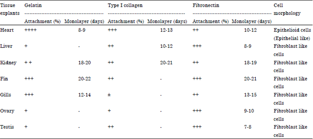
| Fig. 1(a-b): | (a) Outgrowth of epithelioid (epithelial-like cells) from attached heart explants of Cirrhinus mrigala in gelatin coated culture flask (x100) and (b) Formation of complete monolayer by epithelial like cells from heart explants of Cirrhinus mrigala in gelatin coated culture flask (x200) |
The percentage attachment of explants varied not only between tissue explants but also in the same tissue explant cultured in different coated flask. Out of the three coating factors evaluated, fibronectin was found to be the best as far as attachment of tissue explants is concerned in which the percentage attachment of explants from liver, ovary, testis and fin was 40-65% while that of heart, gills and kidney was in between 25-40%.
On the other hand, with gelatin as a coating material, best attachment was recorded in heart explants (>65%) followed by in gills and fin explants (40-65%) coated flasks. However, the attachment of tissue explants of liver, ovary and testis was found to be moderate to poor (15-25%) in gelatin coated flasks.
 | |
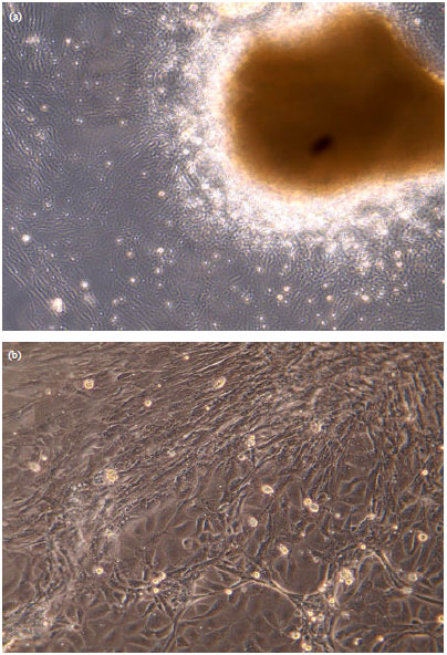
| Fig. 2(a-b): | (a) Outgrowth of fibroblast like cells from the attached liver explants of Cirrhinus mrigala in fibronectin coated culture flask (x100) and (b) Formation of complete monolayer by fibroblast like cells from liver explants of Cirrhinus mrigala in fibronectin coated culture flask (x200) |
In comparison, type I collagen coated flask performed poorly where in attachment of gills explants was less than 15%. Although, 40-65% of tissue explants from heart tissues found attached in type I collagen coated flasks, the attachment percentage varied between 25-40% in explants from liver, kidney, fin and testis.
The percentage attachment of explants to the tissue culture flasks had a direct bearing on subsequent proliferation of cells from explants. Cells proliferated from the explants that strongly adhered to the culture flask and generally grew as a monolayer. But the degree of attaining confluency (monolayer) depended upon the number of attached explants to the flask that proliferated as compared to flasks with less explants, except in type I collagen coated flasks. Further, poor proliferation and delay in formation of confluent monolayer was recorded in explants of same tissue or organ, when cultured in different coated flasks. Cells from heart explants started proliferating and attained confluency within 8-9 days in gelatin coated flasks while it was prolonged upto 13 days in type I collagen coated flasks. In case of liver, monolayer formation was observed within 8-9 days in fibronectin coated culture flasks whereas type I collagen coated flask took 10-12 days to achieve this.
No other coating factor was found to be good, except gelatin and fibronectin for formation of monolayer from explants of gills. Likewise, monolayer formation from liver (8-9 days), ovary (9-10 days), testis (7-8 days) and fin (20-21 days) was found to be good in fibronectin coated flasks. On the contrary to this, no confluency or monolayer formation could be recorded from explants of fin, gills, ovary and testis cultured in type I collagen and for explants of liver, ovary and testis in gelatin coated flasks. However, culture of tissue explants of C. mrigala in different coated flasks did not have effect on their cell morphology. The morphology of attached explants and proliferating cells, through microscopic examination of different tissue explants, exhibited either epithelioid (epithelial-like) (Fig. 1a, b) or fibroblast-like (Fig. 2a, b) pattern of growth. Explants from heart tissues exhibited epithelial like cells, whereas rest of the cultured explants exhibited fibroblast like growth pattern.
DISCUSSION
The success in establishing primary culture from anchorage dependent tissue explants depends upon their ability to attach to culture substratum. To overcome the problem, considerable progress has also been made for identification and characterization of specific matrix proteins as coating factors which induces adhesion and rapid spreading of cells (Damsky et al., 1984). Now-a-days, coated flasks and numerous attachment factors are available to coat plastic surfaces to enhance cell attachment in vitro (Swain et al., 2014). In this aspect, fibronectin, type I collagen and gelatin are well known cell adhesion mediators that improve the adhesive properties of the culture flask bottom to which negatively charged cells attach (Guruvenket et al., 2003; Freshney, 2010). Moreover, these coating factor(s) helps in producing an ECM facilitating attachment, proliferation and further spreading of the cells (Kleinman et al., 1987).
Like animal cell culture, some progress has also been made in establishing cell culture from fish in general and IMC in particular. However, many times fish cell culture practice suffers serious setbacks due to several reasons. Recently, we have also demonstrated that explant method of culture is better as compared to dissociation method for several fish tissues due to their sensitivity towards proteolytic enzyme like trypsin (Nanda et al., 2014a). Therefore, looking into the anchorage dependent nature of most of the normal fish cells and explants, we have evaluated the effect of different coating factors such as gelatin, type I collagen and fibronectin to know their ability on attachment, proliferation and growth of explants from the tissues like fin, heart, liver, kidney, testis, ovary and gills of C. mrigala.
In this study, we found that the attachment percentage of explants, obtained from same tissue, varied greatly among different coated culture flasks. This could be due to the fact that different coating factors can affect differently on the attachment of explants or isolated cells to the substrate as reported by earlier workers (Bols et al., 1994; Butler and Nowak, 2004). However, different attachment factors did not affect the cell morphology. Proliferated cells from heart explants exhibited epithelial like cells while all other cultured tissue explants exhibited fibroblast like growth pattern. Out of the three coating factors used, fibronectin coated flasks were found to be better for attachment of most of the C. mrigala tissue explants with fibroblast like cells. In culture flasks coated with fibronectin, 40-65% attachment of tissue explants from liver, ovary, testis and fin was recorded whereas the percentage of attachment was in between 25-40% for heart, gills and kidney explants. The better efficacy of fibronectin is probably due to the fact that fibronectin which is produced by fibroblasts (Ruoslahti and Vaheri, 1974; Yamada and Weston, 1974; Baum et al., 1977), could have mediated the attachment of the explants to the tissue culture plastic surface as demonstrated by earlier workers (Klebe, 1974; Grinnell, 1978; Yamada and Kennedy, 1979; Loir, 1988).
On the contrary, type I collagen was found to poorly support the attachment of most of the tissue explants and even tissues like fin, gill, ovary and testis failed to form monolayer. Earlier Part et al. (1993) also failed to demonstrate the significant improvement in attachment of primary gill cells of rainbow trout by coating the culture dishes with rat-tail collagen, trout skin extract, laminin or a mixture of human basement proteins (matrigel). Similarly, 25-40% of attachment with 10-12 days time to form monolayer was recorded in case of liver explants. The findings are also in the agreement with earlier reports on the low attachment efficiency of trout hepatocytes on collagen substrates (Lipsky et al., 1986; Rabergh et al., 1995). Furthermore, Buchanan et al. (1999) also demonstrated poor attachment and spreading performances of collagen I in eastern oyster, Crassostrea virginica. The findings are however, contradictory to the improved attachment of rainbow trout hepatocytes on collagen-coated culture dishes as observed by Kocal et al. (1988) and better hepatocytes attachment of neotropical fish, Haplias malabaricus by Filipak Neto et al. (2006). This difference in attachment and spreading performances could be attributed to the type of collagen used and target tissue from different species.
On the other hand, attachment of heart explants of C. mrigala was highest (>65%) in gelatin coated flasks. The fact that glycoproteins which are different from fibronectin, actively support the attachment of epithelial cells (Murray et al., 1979; Terranova et al., 1980). Therefore, this could be the reason for better attachment of heart explants with epithelial like cells in gelatin and also in collagen coated flasks. Earlier, gelatin has also been used for the attachment and spreading for trout myosatellite cells (Powell et al., 1989). But explants of liver, ovary and testis not only failed in attaining the confluency but also attached poorly using the same coating factor. Although, difficult to conclude, they might require separate attachment proteins to provide specific interaction of the cell surface with its matrix.
Generally, good growth of cells is associated with good tissue attachment to the flask. This was observed in case of explants from heart, liver, ovary and testis where emergence of cells was relatively faster resulting in attaining confluency (monolayer formation), even though cultured in different coated flasks. However, explants from few tissues like fin, gills and kidney showed comparatively slower growth rate in attaining confluency. This could perhaps be due to the nature of the tissue as also observed by Joseph et al. (1998) while evaluating the tissues of IMC for development of cell lines by explant method.
CONCLUSION
This study indicates that coating factors influence the attachment and spreading of different cell types obtained tissue explants of C. mrigala. Further, the role and success of using different coating factors not only varied in between tissue explants but also in the same tissue explants obtained from same species. It was also found that the components of the extracellular matrices of fibroblasts and epithelial cells differ as they require different type of coating factor for better attachment and proliferation of cells. Investigations may, therefore, be required to find the suitable coating factor(s) for each and every cell types from different fish species to establish cell culture and definitely, there is scope for further research in this area.
REFERENCES
- Babich, H., M.R. Palace and A. Stern, 1993. Oxidative stress in fish cells: In vitro studies. Arch. Environ. Contam. Toxicol., 24: 173-178.
CrossRef - Baum, B.J., J.A. McDonald and R.G. Crystal, 1977. Metabolic fate of the major cell surface protein of normal human fibroblasts. Biochem. Biophys. Res. Commun., 79: 8-15.
CrossRef - Ben-Ze'ev, A., S.R. Farmer and S. Penman, 1980. Protein synthesis requires cell-surface contact while nuclear events respond to cell shape in anchorage-dependent fibroblasts. Cell, 2: 365-372.
CrossRef - Bols, N.C. and L.E.J. Lee, 1991. Technology and uses of cell cultures from the tissues and organs of bony fish. Cytotechnology, 6: 163-187.
CrossRef - Bols, N.C., R.C. Ganassin, D.J. Tom and L.E.J. Lee, 1994. Growth of fish cell lines in glutamine-free media. Cytotechnology, 16: 159-166.
CrossRef - Buchanan, J.T., J.F. La Peyre, R.K. Cooper and T.R. Tiersch, 1999. Improved attachment and spreading in primary cell cultures of the eastern oyster, Crassostrea virginica. In vitro Cell. Dev. Biol. Anim., 35: 593-598.
CrossRef - Butler, R. and B.F. Nowak, 2004. A dual enzyme method for the establishment of long- and medium-term primary cultures of epithelial and fibroblastic cells from Atlantic salmon gills. J. Fish Biol., 65: 1108-1125.
CrossRef - Chen, S.L., G.C. Ren, Z.X. Sha and C.Y. Shi, 2004. Establishment of a continuous embryonic cell line from Japanese flounder Paralichthys olivaceus for virus isolation. Dis. Aquat. Org., 60: 241-246.
CrossRef - Cooke, M.J., S.R. Phillips, D.S.H. Shah, D. Athey, J.H. Lakey and S.A. Przyborski, 2008. Enhanced cell attachment using a novel cell culture surface presenting functional domains from extracellular matrix proteins. Cytotechnology, 56: 71-79.
CrossRef - Danen, E.H.J. and K.M. Yamada, 2001. Fibronectin, integrins and growth control. J. Cell. Physiol., 189: 1-13.
CrossRef - Folkman, J. and A. Moscona, 1978. Role of cell shape in growth control. Nature, 273: 345-349.
CrossRef - Grinnell, F., 1978. Cellular adhesiveness and extracellular substrata. Int. Rev. Cytol., 53: 65-144.
PubMed - Guruvenket, S., M. Komath, S.P. Vijayalakshmi, A.M. Raichur and G. Mohan Rao, 2003. Wettability enhancement of polystyrene with electron cyclotron resonance plasma with argon. J. Applied Polym. Sci., 90: 1618-1623.
CrossRef - Hightower, L.E. and J.L. Renfro, 1988. Recent applications of fish cell culture to biomedical research. J. Exp. Zool., 248: 290-302.
CrossRef - Ireland, G.W., P.J.C. Dopping-Hepenstal, P.W. Jordan and C.H. O'Neill, 1989. Limitation of substratum size alters cytoskeletal organization and behaviour of Swiss 3T3 fibroblasts. Cell Biol. Int. Rep., 13: 781-790.
CrossRef - Joseph, M.A., R.K. Sushmitha, C.V. Mohan and K.M. Shankar, 1998. Evaluation of tissues of Indian major carps for development of cell lines by explant method. Curr. Sci., 75: 1403-1406.
Direct Link - Khan, E.A., A.K. Dasmahapatra and R. Ghosh, 1997. Evaluation of EDTA and fish skin extract in primary culture of fish liver cells. Methods Cell Sci., 19: 153-159.
CrossRef - Klebe, R.J., 1974. Isolation of a collagen-dependent cell attachment factor. Nature, 250: 248-251.
CrossRef - Kleinman, H.K., L. Luckenbill-Edds, F.W. Cannon and G.C. Sephel, 1987. Use of extracellular matrix components for cell culture. Anal. Biochem., 166: 1-13.
CrossRefDirect Link - Kocal, T., B.A. Quinn, I.R. Smith, H.W. Ferguson and M.A. Hayes, 1988. Use of trout serum to prepare primary attached monolayer cultures of hepatocytes from rainbow trout (Salmo gairdneri). In Vitro Cell. Dev. Biol., 24: 304-308.
CrossRef - Lipsky, M.M., T.R. Sheridan, R.O. Bennett and E.B. May, 1986. Comparison of trout hepatocyte culture on different substrates. In Vitro Cell. Dev. Biol., 22: 360-362.
CrossRef - Loir, M., 1988. Trout sertoli and leydig cells: Isolation, separation and culture. Gamete Res., 20: 437-458.
CrossRef - Nanda, P.K., P. Swain, S.K. Nayak, T. Behera and K. Dhama, 2014. A comparative study on enzymatic and explant method in establishing primary culture from different cultivable cells of Indian Major Carp, Cirrhinus mrigala. Asian J. Anim. Vet. Adv., 9: 281-291.
CrossRef - Nanda, P.K., P. Swain, S.K. Nayak, S. Dash, P. Routray, S.K. Swain and B.C. Patra, 2009. Goat serum as an alternative to establish cell culture from Indian major carp, Cirrhinus mrigala. In vitro Cell. Dev. Biol. Anim., 45: 148-151.
CrossRef - Filipak Neto, F.F., S.M. Zanata, M.A.F. Randi, E. Pelletier and C.A. Oliveira Ribeiro, 2006. Hepatocytes primary culture from the Neotropical fish, trahira Hoplias malabaricus (Bloch). J. Fish Biol., 69: 1524-1532.
CrossRef - Ostrander, G.K., J.B. Blair, B.A. Stark, J.G. Hurst, 1993. Response of rainbow trout liver to partial hepatectomy. Aquat. Toxicol., 25: 31-42.
CrossRef - Part, P., L. Norrgren, E. Bergstrom and P. Sjoberg, 1993. Primary cultures of epithelial cells from rainbow trout gills. J. Exp. Biol., 175: 219-232.
Direct Link - Powell, R.E., M.V. Dodson and J.G. Cloud, 1989. Cultivation and differentiation of satellite cells from skeletal muscle of the rainbow trout Salmo gairdneri. J. Exp. Zool., 250: 333-338.
CrossRef - Rabergh, C.M., A.S. Kane, R. Reimschuessel and M.M. Lipsky, 1995. Viability and induction of tyrosine aminotransferase in rainbow trout hepatocytes cultured on laminin and polylysine in a serum-free medium. Methods Cell Sci., 17: 207-215.
CrossRef - Rathore, G., G. Kumar, T.R. Swaminathan, N. Sood, V. Singh, R. Abidi and W.S. Lakra, 2007. Primary cell culture from fin explants of Labeo rohita (Ham.). Indian J. Fish., 541: 93-97.
Direct Link - Ruoslahti, E. and A. Vaheri, 1974. Novel human serum protein from fibroblast plasma membrane. Nature, 248: 789-791.
CrossRef - Terranova, V.P., D.H. Rohrbach and G.R. Martin, 1980. Role of laminin in the attachment of PAM 212 (epithelial) cells to basement membrane collagen. Cell, 22: 719-726.
CrossRef - Yamada, K.M. and D.W. Kennedy, 1979. Fibroblast cellular and plasma fibronectins are similar but not identical. J. Cell Biol., 80: 492-498.
CrossRef - Yamada, K.M. and J.A. Weston, 1974. Isolation of a major cell surface glycoprotein from fibroblasts. Proc. Natl. Acad. Sci., 71: 3492-3496.
Direct Link - Ye, H.Q., S.L. Chen, Z.X. Sha and M.Y. Xu, 2006. Development and characterization of cell lines from heart, liver, spleen and head kidney of sea perch Lateolabrax japonicas. J. Fish Biol., 69: 115-126.
CrossRef - Nanda, P.K., P. Swain, S.K. Nayak, T. Behera and K. Dhama, 2014. A comparative study on enzymatic and explant method in establishing primary culture from different cultivable cells of Indian Major Carp, Cirrhinus mrigala. Asian J. Anim. Vet. Adv., 9: 281-291.
CrossRef