
A.R. Golchin Manshadi
Department of Aquatic Animal Health, Faculty of Veterinary Medicine, Kazerun Branch, Islamic Azad University, Kazerun, Iran
M. Masoumian
Department of Aquatic Animal Health, Fisheries Research Institute of Iran, Tehran, Iran
B. Jalali Jafari
Department of Aquatic Animal Health, Faculty of Specialized Veterinary Medicine, Islamic Azad University, Science and Research Branch, Tehran, Iran
M. Barzegar Dowlatabadi
MS in Fisheries Engineering, Tehran, Iran
Asian Journal of Animal and Veterinary Advances
Year: 2012 | Volume: 7 | Issue: 9 | Page No.: 842-850


ABSTRACT
In parasitological investigations on some fishes of Parishan Lake in 2008, totally 76 numbers of fish from 6 species were examined. They were as follows: Cyprinus carpio, Barbus luteus, Barbus grypus, Carassius carassius, Liza abu and Mastacembelus mastacembelus. The fishes were collected seasonally and were transported alive to the aquatic laboratory in Islamic Azad University of Kazerun. According to the results, there were eight different parasites collected; five species of Myxozoa including: Myxobolus karuni, Myxobolus persicus, Myxobolus mulleri, Myxobolus bramae, Myxidium pfeiffer and three species of protozoa including: Ichthyophthirius multifiliis, Trichodina nigra, Trichodina puytoraci. During this study, Barbus grypus is introduced as new host for Myxobolus bramae and also Barbus luteus for Myxobolus mulleri, Myxidium pfeiffer and Trichodina nigra; The Trichodinads were recorded for the first time from Iranian freshwater fishes. No parasites were seen from examined Mastacembelus mastacembelus.
PDF Abstract XML References Citation
Received: November 04, 2011;
Accepted: February 17, 2012;
Published: May 10, 2012
How to cite this article
A.R. Golchin Manshadi, M. Masoumian, B. Jalali Jafari and M. Barzegar Dowlatabadi, 2012. Protozoan and Myxozoan Infections in Some Fishes of Parishan Lake. Asian Journal of Animal and Veterinary Advances, 7: 842-850.
DOI: 10.3923/ajava.2012.842.850
URL: https://scialert.net/abstract/?doi=ajava.2012.842.850
DOI: 10.3923/ajava.2012.842.850
URL: https://scialert.net/abstract/?doi=ajava.2012.842.850
INTRODUCTION
Parishan Lake lays at a distance of 12 km to the southeast of Kazeroon city, southern area of Famur’s mountain, in Fars province, Iran. As water of the lake is supplied from around springs’ comes, its area varies in different seasons according to the amount of rainfall. This lake’s water is brackish and a small amount is used for agricultural uses. Native fishermen fish strongly and native residents feed from them with a passion, because of the good quality of the fish (Takamy et al., 1979). Other reports from the Fars province are related to studies on Kaftar’s Lake (Barzegar and Jalali, 2000). Mentioned studies show Trichodina sp. from skin and gills of fish silver carp, grass carp, common carp, Capoeta aculeata and Ichthyophthirius multifiliis from the gills of Chondrostoma orientalis and Leuciscus persidis and Dermocystidium sp. from the gills of common carp were isolated.
In the case of Myxozoa, comprehensive studies have been done in the species level of fishes: Masoumian et al. (1994, 1996a, b), Molnar et al. (1996), Masoumian et al. (2005), Pazooki et al. (2007) and Masoumian et al. (2004, 2003). In this studies about 50 species of Myxozoa from fish has been identified, classified and reported.
| Table 1: | Fish identification keys |
 | |
About Trichodina parasite, in the genus level there are many reports of fish from several areas of Iran and in the species level, T. domerguei and T. perforata from sturgeon and carp fish and T. polycolpus from Capoeta capoeta have been reported by Mortezai and Abbasi (2001). The purpose of this study was identification and introduction of Myxozoa and Protozoa parasites of fish in Parishan Lake.
MATERIALS AND METHODS
During studies on fish’s parasite of the Parishan Lake in 2007, total number of 76 fish were caught and studied. Sampling was done during the year. The fish were caught by fixed net in different areas, including areas of deep lakes, high vegetation areas or deep zone and immediately transferred alive by containers specialized for transporting fish contain the lake’s water to the laboratory of parasitology in Azad University, where they were kept in the aquarium. Fish identification was performed by using keys of Berg (1965), Coad (1995), Abdoli (1999) and Vossoughi and Mostajir (2004) (Table 1). All internal and external organs of fish were put in Petri dishes and after adding normal saline were studied with stereo microscope and light microscope. Obtained protozoa and spores of myxozoa fixed in gelatin glycerin and were identified and categorized by keys of Shulman (1990) and Lom and Dykova (1992).
RESULTS
During this study, eight parasites were identified totally which is consist of: Myxobolus karuni, Myxobolus persicus, Myxobolus mulleri, Myxobolus bramae, Myxidium pfeifferi, Ichthyophthirius multifiliis, Trichodina nigra and Trichodina puytoraci. In freshwater eel no Protozoa and Myxozoa were observed. Parasites isolated from the studied fish, the hosts and the infected organs are shown in Table 2.
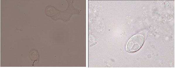
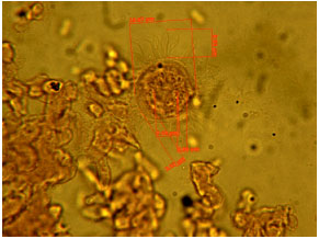
Myxobolus karuni (Masoumian et al., 1994): This parasite were isolated and identified from primary gill filaments of Barbus grypus and Barbus luteus. Spores are relatively large and ovoid in frontal view, lemon shaped in side view, with distinct sutural line. The ends of the anterior part of polar capsules are separated from each other and there is a distinct intercapsular appendix. Spores valves are symmetrical and smooth, relatively thin surface and the end of the anterior part of them is flattened. Spores are 14.1 (13-14.9) μm long, 10.2 (9.7-10.4) μm wide and 7.2 (6.5-7.8) μm thick. Spores have two equal polar and elongated oval capsules, 6.2 (6.5-7.5) μm long and 3.4 (3.2-3.9) μm wide and are tapering at the discharging canals of the polar filament. The polar capsules are a bit longer than semilength of the spore. Polar filament has 11-10 turns. Spores have a large iodinophilous vacuole in the sporoplasm (Fig. 1).
| Table 2: | Protozoa and myxozoa isolated from fish of Parishan lake |
 | |
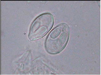 | |
| Fig. 1: | Extracted spores from cyst of Myxobolus karuni of Barbus grypus’s gill (X 1710) |
Myxobolus persicus (Masoumian et al., 1994): This parasite was isolated and identified from secondary filaments of Barbus grypus. Spores are ovoid in frontal view, lemon shaped in side view, with distinct sutural line. The ends of the anterior part of polar capsules are separated from each other and there is a distinct intercapsular appendix. Spores valves are symmetrical and smooth, relatively thin surface. Spores are 10(9.1-10.4) μm long, 7.3(6.5-7.8) μm wide and 6/3(5.2-6.5) μm thick. The two polar capsules are pear-shaped in form and unequal (occasionally equal)in size. The larger is 5.1(4.5-5.8) μm long and 2.7(2.6-3.2) μm wide. The smaller is 4.8(3-4.5) μm long. The larger polar capsule is longer than semi length of the spore with 6-7 turns in larger and 7-8 turns in the smaller one. Sporoplasm does not have iodinophilous vacuole (Fig. 2).
Myxobolus bramae (Reuss, 1906): This parasite was isolated and identified from secondary filaments of Barbus grypus. The spores are ovoid, rarely narrow in the anterior part. Spore valves are seen distinctly in frontal view. The two polar capsules are pear-shaped and their lengths are half or occasionally more than half of the length of the spores which have largely occupied the anterior part of spore. The anterior ends of polar capsules are set apart to each other and intercapsular appendix is triangular shaped, large and visible. Spores are 9-15 μm long, 8-12 μm wide and 4.5-8 μm thick. Polar capsules are 4-7.2 long and 2.5-3 μm wide (Fig. 3). This parasite produces milky white and circular cysts in gills of infected fish.
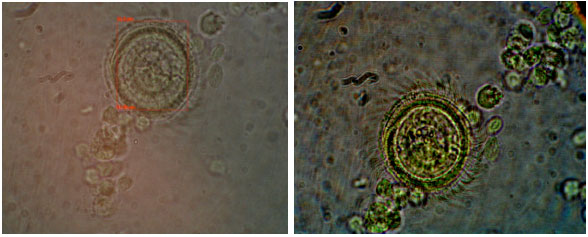
Myxobolus mulleri (Butschli, 1882): This parasite were isolated and identified from bile fluid of Barbus luteus. Spores are ovoid and seem to be enlarged at the anterior part. In some cases, are seen ellipsoidal in shape.
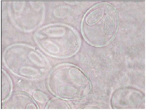 | |
| Fig. 2: | Extracted spores from cyst of Myxobolus persicus of Barbus grypus’s gill (X 1960) |
 | |
| Fig. 3: | Extracted spores from cyst of Myxobolus bramae of Barbus grypus’s gill (X 810) |
The pear-shaped polar capsules are longer than the half length of spore. The anterior ends of polar capsules are closed to each other and intercapsular appendix is triangular shaped, hardly visible. Spores are 8-13 μm long, 7-10 μm wide and 5.4-6 μm thick. The polar capsules are 3.6-5.5 long and 2.5-3 μm wide (Fig. 4).
Myxidium pfeifferi (Auerbach, 1908): This parasite was observed as a Ceolozoit form and isolated from bile fluid of Barbus luteus (Fig. 5). Spores are spindle in shape and in some cases the wide of middle part are less than beginning and end of them. Spores are 12-18 μm long, 5-6 μm wide. Polar capsule is 5-6 μm long.
Ichthyophthirius multifiliis (Fouquet, 1876): Parasite is around to ovoid in shape, 0.05 to 1 mm diameter and has a cytostome in the third of anterior part of body. The Parasite was isolated from gills and skin’s moisture spread of Carassius carassius, Barbus luteus and Cyprinus carpio (mirror carp) (Fig. 6).
Genus Trichodina (Ehrenberg, 1838): The other external protozoan parasite is Trichodina sp. cause of Trichodiniasis. The genus characterized by teeth with central part of dense and cone-shaped and by outside semicircle blades and inside straight thorn.
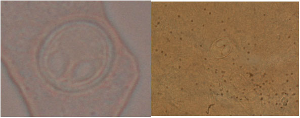 | |
| Fig. 4: | Spores of Myxobolus mulleri isolated from bile fluid of Barbus grypus (left picture X1920 and right picture X 410) |
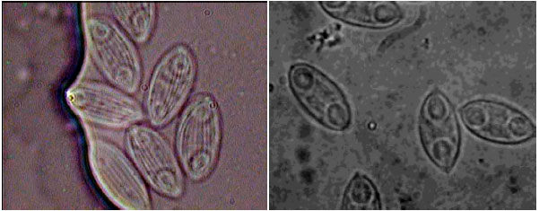 | |
| Fig. 5: | Spores of Myxidium pfeifferi isolated from bile fluid of Barbus luteus, Ceolozoit form, X 2200 |
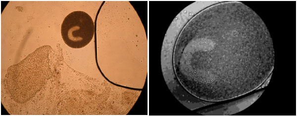 | |
| Fig. 6: | Ictyophthirius multifiliis isolated from gills of Barbus luteus |
 | |
| Fig. 7: | View of Trichodina nigra isolated from skin of Barbus luteus |
 | |
| Fig. 8: | View of Trichodina puytoraci isolated from skin of Liza abu |
Diameter of adhesive disk and dental ring, size and number of teeth and diameter of the macro and micronucleus and their ratio together are considered criteria for differentiation of species of this genus.
Trichodina nigra (Lom, 1960): This protozoa was isolated from the skin and gills of common carp and Barbus luteus. Adhesive disk diameter (da) is 19.87 μm and of tooth ring’s diameter (dd) is 31.9 μm (Fig. 7).
Trichodina puytoraci (Lom, 1962): Trichodina puytoraci was isolated from the skin and gills of Liza abu. Adhesive disk diameter (da) is 31.36 μm and of tooth ring’s diameter (dd) is 14.56 μm (Fig. 8).
DISCUSSION
Among the protozoan parasites obtained in this study Ichthyophthirius multifiliis is considered as one of the important pathogenic ciliated protozoan of freshwater’s fish. This parasite is a factor of white spot disease and lead to severe losses and can reduce fish growth. The parasites have been reported by Moghinemyi et al. (1992) and Molnar and Baska (1993) from large numbers of wild fish and rearing farm of Iran. In this study Ichthyophthirius multifiliis was isolated from gills and skin of Carassius carassius, Barbus luteus and Cyprinus carpio (mirror carp) of water basin’s Parishan for the first time.
Genus Trichodina is another important protozoa. Moghinemyi et al. (1992) reported as the parasites of Barbus sharpeyi, Aspius vorax and Barbus grypus and Liza abu of Hoor-Elazim and Mortezai and Abbasi (1997) reported from Barbus sharpeyi of Shadgan Marsh. Also Mokhayyer (1980) reported from sturgeon and rain bow trout. As other reports, it could be pointed to Mortezai et al. (2009) in study on Barboid fish of Khuzestan province that it was reported from Barbus sharpeyi, B. barbulus and B. grypus and also Trichodina perforata was reported from C. capoeta by Masoumian et al. (2005). In this study Trichodina nigra from skin and gills of common carp and also B. luteus and Trichodina puytoraci from skin of Liza abu were isolated for the first time in Iran.
Phylum of Myxozoa is a controversial group of parasites that are known in the fish. New theory about the life cycle of these parasites have been confirmed with new experiments: (El-Mansy et al., 1998; Szekely et al., 1998; El-Mansy and Molnar, 1997) and finally with the molecular experiments as well as position classification phylum of Myxozoa between metazoan parasites were proposed to belong to Cnidaria, however, parasites belonging to this phylum are most important parasites in fish (Smother et al., 1994; Bush et al., 2001; Kent et al., 2001).
In this study, four species of Myxobolus were reported including Myxobolus karuni and Myxobolus bramae from gills of B. grypus, Myxobolus mulleri from bile of B. luteus and Myxobolus persicus from gills of B. grypus and B. luteus. Also Myxidium pfeifferi were isolated from bile of B. luteus. This is the first time that five species of myxozoa are reported, among the reports has been done in other water basins of Iran.
M. karuni from gills of B. luteus and B. sharpeyi and M. persicus from gills of B. grypus by and M. persicus from gills of B. luteus and B. sharpeyi and M. karuni from gills of B. grypus by Masoumian et al. (1994) were reported. Also Masoumian et al. (1994) in a study on some of the fish in the provinces of Gilan and Mazandaran, isolated M. bramae from gills of Rutilus frisii Kutum and M. mulleri from muscle of Leuciscus cephalus and Myxidium pfeifferi from the gallbladder of Scardinius erythrophthalmus.
Estimating the pathogenicity and economic importance of the parasites is very difficult and the exact amount of loss and mortality in fish, due to our lack of information is not available. In addition, in the aquaculture system comparing data of different incidence of parasite and infection rate of Myxosporea is very difficult because the data varies, comparing the type and extent of rearing system, fish species and ultimately climate. All of the parasites has been studied had no sign of the disease.
Parishan Lake is one of the important water basin of Fars province and supplies fish protein source, especially for people in the region and the fish economically can also be selected for fish farms.
REFERENCES
- Kent, M.L., K.B. Andree, J.L. Bartholomew, M. El-Matbouli and S.S. Desser et al., 2001. Recent advences in our knowledge of the Myxozoa. J. Eukaryotic Microbiol., 84: 395-413.
CrossRef - Masoumian, M., F. Baska and K. Molnar, 1994. Description of Myxobolus karuni sp. n. and Myxobolus persicus sp. n. (Myxosporea, Myxozoa) from Barbus grypus of the River Karun, Iran. Hung. Nat. Hist. Museum Parasit. Hung., 27: 21-26.
Direct Link - Masoumian, M., F. Baska and K. Molnar, 1996. Description of Myxobolus bulbocordis sp. nov. (Myxosporea: Myxobolidae) from the heart of Barbus sharpeyi (Gunther) and histopathological changes produced by the parasite. J. Fish Dis., 19: 19-21.
CrossRefDirect Link - Molnar, K., M. Masoumian and S. Abasi, 1996. Four New Myxobolus spp (Myxosporea: Myxobolidae) from Iranian barboid fishes. Arch. Protistenkunde, 741: 115-123.
CrossRef